آنتی بادیهای فلوسایتومتری
-
 آنتی بادیهای فلوسایتومتری
آنتی بادی مونوکلونال فلوسایتومتری CD117-PE ، کلون 104D2
اطلاعات بیشترName: Flow Cytometry Antibody CD117-PE Clone 104D2
Antibody CD117 is a mAb designed for flow cytometry use as a direct immunofluorescence reagent in the identification and enumeration of CD117 antigen-expressing cells. CD117 is a specific marker to detect leukemic cells committed to the myeloid lineage and therefore it represents a useful reagent in the characterization of acute leukaemias. C-kit expression would be particularly relevant for the diagnosis of biphenotypic acute leukaemias as well since in these cases its association with myeloid lineage is greater than that of the CD13 and CD33 antigens.
CD117 reacts specifically with human c-kit gene product (SCF receptor). The c-kit proto-oncogen (CD117) has been shown to be present in several cell types including normal and neoplastic haemopoietic cells. The majority of CD117+ bone marrow (BM) cells (50-70%), which mainly correspond to myeloid precursors, coexpress the progenitor associated CD34 antigen. The c-kit+/CD34- cells mainly consist of immature cells, mast cells and CD34- erythroid precursors. In acute leukaemias CD117 expression was initially associated with AML. Nevertheless, at present it is well established that CD117 expression may also be found in a relatively important proportion of T-ALL while it is usually absent in B-lineage ALL. The analysis of CD117 could be relevant for the investigation of minimal residual disease (MRD) since in combination with other antigens may be useful for the identification of leukaemia associated phenotypes in patients who achieved morphological complete remission. The combination of CD117 and other myeloid-associated antigens such as CD11b and CD15 may be of great help to monitoring MRD in AML patients although it defines a subpopulation of myeloid cells which are either absent or present at very low frequencies in normal human BM . Additionally, some acute leukaemias express CD117 over-expression which is not detected in normal cells. Moreover, different studies have shown that in around one-third of both multiple myeloma cases and patients with monoclonal gammopathy of undetermined significance myelomatous plasma cells display reactivity for CD117 .BM mast cells are clearly identifiable on the basis of their light scatter properties and strong CD117 expression. BM mast cells studies result interesting for the diagnosis of adult indolent systemic mast cell disease. and mast cell leukaemia.
REAGENT COMPOSITION
The purified monoclonal CD117 antibody (mAb) conjugated with one fluorochrome (see table above) and supplied in phosphate buffered saline with <0,1% (m/v) sodium azide.
- Clone: 104D2
- Isotype: IgG1
- Amount per 1 ml vial: 100 tests (10 µl mAb per determination)
- Reagent is considered non-sterile.
-
 آنتی بادیهای فلوسایتومتری
آنتی بادی مونوکلونال فلوسایتومتری IgG1-FITC ، کلون SAG1
اطلاعات بیشترName: Flow Cytometry Antibody IgG1-FITC Clone SAG1
Antibody IgG1-FITC is designed for flow cytometry use as a direct immunofluorescence reagent in the identification and enumeration of IgG1 expressing cells. IgG1-FITC reacts with the Fc region of human IgG1.
IgG is the most abundant immunoglobulin in human serum (approximately 80% of total). Soluble IgG (sIgG) antibodies are distributed equally between the intra- and extravascular pools.
There are four different IgG subclasses responsible of protection against microorganisms: IgG1 (67% overall), IgG2 (22% overall), IgG3 (7% overall) and IgG4 (4% overall). These molecules have different biological properties as distinct ability to fix complement proteins and interact with phagocytic cell receptors. The IgG1 and IgG3 subclasses are particularly good opsonins and mediators of antibodydependent cell-mediated cytotoxicity (ADCC).
sIgG antibodies are important activators of complement but not as efficient as sIgM antibodies. IgG3 is the most efficient complementfixing IgG subclass, while IgG1 is somewhat less efficient, and IgG2 is even less so. IgG4 is unable to bind C1q and so cannot fix complement at all in the classical pathway.
Only IgG antibodies can cross the mammalian placenta, and maternal IgG1, IgG3, and IgG4 molecules have been found to be crucial for conferring the passive immunity that protects the developing human fetus and newborn in the first few months of life. IgG2 crosses the placenta with much lower efficiency.
CYT-IGG1F should be used with BulkLysisTM (CYT-BL) buffer that allows you to stain a great number of cells and analyze lowfrequency subsets, as IgG1+ memory B cells and plasma cells. If another lysing protocol is used, two pre-staining washing steps using PBS+1% (m/v) BSA + 0,09% (m/v) sodium azide must be performed.
This reagent must be used by flow cytometry qualified personnel.
REAGENT COMPOSITION
Purified monoclonal anti-IgG1 antibody (mAb) conjugated with fluorescein isothiocyanate (FITC), supplied in phosphate buffered saline with 0,09% (m/v) sodium azide and BSA 1% (m/v).
- Clone: SAG1.
- Isotype: Mouse IgG2b.
- Amount per vial: 25 tests using 3µL of mAb to 106 cells
-
آنتی بادیهای فلوسایتومتری
آنتی بادی مونوکلونال فلوسایتومتری IgG1-PE ، کلون SAG1
اطلاعات بیشترName: Flow Cytometry Antibody IgG1-PE, Clone SAG1
Antibody IgG1-PE is designed for flow cytometry use as a direct immunofluorescence reagent in the identification and enumeration of IgG1 expressing cells. IgG1-PE reacts with the Fc region of human IgG1.
IgG is the most abundant immunoglobulin in human serum (approximately 80% of total). Soluble IgG (sIgG) antibodies are distributed equally between the intra- and extravascular pools.
There are four different IgG subclasses responsible of protection against microorganisms: IgG1 (67% overall), IgG2 (22% overall), IgG3 (7% overall) and IgG4 (4% overall). These molecules have different biological properties as distinct ability to fix complement proteins and interact with phagocytic cell receptors. The IgG1 and IgG3 subclasses are particularly good opsonins and mediators of antibody dependent cell-mediated cytotoxicity (ADCC) (1).
sIgG antibodies are important activators of complement but not as efficient as sIgM antibodies. IgG3 is the most efficient complement fixing IgG subclass, while IgG1 is somewhat less efficient, and IgG2 is even less so. IgG4 is unable to bind C1q and so cannot fix complement at all in the classical pathway (1).
Only IgG antibodies can cross the mammalian placenta, and maternal IgG1, IgG3, and IgG4 molecules have been found to be crucial for conferring the passive immunity that protects the developing human fetus and newborn in the first few months of life. IgG2 crosses the placenta with much lower efficiency (1).
CYT-IGG1PE should be used with BulkLysisTM (CYT-BL) buffer that allows you to stain a great number of cells and analyze low frequency subsets, as IgG1+ memory B cells and plasma cells. If another lysing protocol is used, two pre-staining washing steps using PBS + 1% (m/v) BSA + 0,09% (m/v) sodium azide must be performed.
This reagent must be used by flow cytometry qualified personnel.
REAGENT COMPOSITION
Purified monoclonal anti-IgG1 antibody (mAb) conjugated with R-phycoerythrin (PE), supplied in phosphate buffered saline with 0,09% (m/v) sodium azide and BSA 1% (m/v).
- Clone: SAG1.
- Isotype: Mouse IgG2b.
- Amount per vial: 25 tests using 3µL of mAb to 106 cells.
-
آنتی بادیهای فلوسایتومتری
آنتی بادی مونوکلونال فلوسایتومتری IgG2-FITC ، کلون SAG2
اطلاعات بیشترName: Flow Cytometry Antibody IgG2-FITC, Clone SAG2
Antibody IgG2-FITC is designed for flow cytometry use as a direct immunofluorescence reagent in the identification and enumeration of IgG2 expressing cells. IgG2-FITC reacts with the Fc region of human IgG2.
IgG is the most abundant immunoglobulin in human serum (approximately 80% of total). Soluble IgG (sIgG) antibodies are distributed equally between the intra- and extravascular pools.
There are four different IgG subclasses responsible of protection against microorganisms: IgG1 (67% overall), IgG2 (22% overall), IgG3 (7% overall) and IgG4 (4% overall). These molecules have different biological properties as distinct ability to fix complement proteins and interact with phagocytic cell receptors. The IgG1 and IgG3 subclasses are particularly good opsonins and mediators of antibodydependent cell-mediated cytotoxicity (ADCC).
sIgG antibodies are important activators of complement but not as efficient as sIgM antibodies. IgG3 is the most efficient complement fixing IgG subclass, while IgG1 is somewhat less efficient, and IgG2 is even less so. IgG4 is unable to bind C1q and so cannot fix complement at all in the classical pathway.
Only IgG antibodies can cross the mammalian placenta, and maternal IgG1, IgG3, and IgG4 molecules have been found to be crucial for conferring the passive immunity that protects the developing human fetus and newborn in the first few months of life. IgG2 crosses the placenta with much lower efficiency.
CYT-IGG2F should be used with BulkLysisTM (CYT-BL) buffer that allows you to stain a great number of cells and analyze lowfrequency subsets, as IgG2+ memory B cells and plasma cells. If another lysing protocol is used, two pre-staining washing steps using PBS + 1% (m/v) BSA + 0,09% (m/v) sodium azide must be performed. This reagent must be used by flow cytometry qualified personnel.
REAGENT COMPOSITION
Purified monoclonal anti-IgG2 antibody (mAb) conjugated with fluorescein isothiocyanate (FITC), supplied in phosphate buffered saline with 0,09% (m/v) sodium azide and BSA 1% (m/v).
- Clone: SAG2.
- Isotype: Mouse IgG1.
- Amount per vial: 25 tests using 3µL of mAb to 106 cells.
-
آنتی بادیهای فلوسایتومتری
آنتی بادی مونوکلونال فلوسایتومتری IgG2-PE ، کلون SAG2
اطلاعات بیشترName: Flow Cytometry Antibody IgG2-PE, Clone SAG2
Antibody IgG2-PE is designed for flow cytometry use as a direct immunofluorescence reagent in the identification and enumeration of IgG2 expressing cells. IgG2-PE reacts with the Fc region of human IgG2.
IgG is the most abundant immunoglobulin in human serum (approximately 80% of total). Soluble IgG (sIgG) antibodies are distributed equally between the intra- and extravascular pools.
There are four different IgG subclasses responsible of protection against microorganisms: IgG1 (67% overall), IgG2 (22% overall), IgG3 (7% overall) and IgG4 (4% overall). These molecules have different biological properties as distinct ability to fix complement proteins and interact with phagocytic cell receptors. The IgG1 and IgG3 subclasses are particularly good opsonins and mediators of antibody dependent cell-mediated cytotoxicity (ADCC).
sIgG antibodies are important activators of complement but not as efficient as sIgM antibodies. IgG3 is the most efficient complement fixing IgG subclass, while IgG1 is somewhat less efficient, and IgG2 is even less so. IgG4 is unable to bind C1q and so cannot fix complement at all in the classical pathway.
Only IgG antibodies can cross the mammalian placenta, and maternal IgG1, IgG3, and IgG4 molecules have been found to be crucial for conferring the passive immunity that protects the developing human fetus and newborn in the first few months of life. IgG2 crosses the placenta with much lower efficiency.
CYT-IGG2PE should be used with BulklysisTM (CYT-BL) buffer that allows you to stain a great number of cells and analyze low frequency subsets, as IgG2+ memory B cells and plasma cells. If another lysing protocol is used, two pre-staining washing steps using PBS + 1% (m/v) BSA + 0,09% (m/v) sodium azide must be performed.
This reagent must be used by flow cytometry qualified personnel
REAGENT COMPOSITION
Purified monoclonal anti-IgG2 antibody (mAb) conjugated with R-phycoerythrin (PE), supplied in phosphate buffered saline with 0,09% (m/v) sodium azide and BSA 1% (m/v).
- Clone: SAG2.
- Isotype: Mouse IgG1.
- Amount per vial: 25 tests using 3µL of mAb to 106 cells.
-
آنتی بادیهای فلوسایتومتری
آنتی بادی مونوکلونال فلوسایتومتری IgG3-FITC ، کلون SAG3
اطلاعات بیشترName: Flow Cytometry Antibody IgG3-FITC, Clone SAG3
Antibody IgG3-FITC is designed for flow cytometry use as a direct immunofluorescence reagent in the identification and enumeration of IgG3 expressing cells. IgG3-FITC reacts with the hinge region of the heavy chain of human IgG3.
IgG is the most abundant immunoglobulin in human serum (approximately 80% of total). Soluble IgG (sIgG) antibodies are distributed equally between the intra- and extravascular pools.
There are four different IgG subclasses responsible of protection against microorganisms: IgG1 (67% overall), IgG2 (22% overall), IgG3 (7% overall) and IgG4 (4% overall). These molecules have different biological properties as distinct ability to fix complement proteins and interact with phagocytic cell receptors. The IgG1 and IgG3 subclasses are particularly good opsonins and mediators of antibody dependent cell-mediated cytotoxicity (ADCC) (1).
sIgG antibodies are important activators of complement but not as efficient as sIgM antibodies. IgG3 is the most efficient complement fixing IgG subclass, while IgG1 is somewhat less efficient, and IgG2 is even less so. IgG4 is unable to bind C1q and so cannot fix complement at all in the classical pathway.
Only IgG antibodies can cross the mammalian placenta, and maternal IgG1, IgG3, and IgG4 molecules have been found to be crucial for conferring the passive immunity that protects the developing human fetus and newborn in the first few months of life. IgG2 crosses the placenta with much lower efficiency.
CYT-IGG3F should be used with BulkLysisTM (CYT-BL) buffer that allows you to stain a great number of cells and analyze low frequency subsets, as IgG3+ memory B cells and plasma cells. If another lysing protocol is used, two pre-staining washing steps using PBS + 1% (m/v) BSA + 0,09% (m/v) sodium azide must be performed.
This reagent must be used by flow cytometry qualified personnel. This reagent must be used by flow cytometry qualified personnel
REAGENT COMPOSITION
Purified monoclonal anti-IgG3 antibody (mAb) conjugated with fluorescein isothiocyanate (FITC), supplied in phosphate buffered saline with 0,09% (m/v) sodium azide and BSA 1% (m/v).
- Clone: SAG3.
- Isotype: Mouse IgG1.
- Amount per vial: 25 tests using 3µL of mAb to 106 cells.
-
آنتی بادیهای فلوسایتومتری
آنتی بادی مونوکلونال فلوسایتومتری IgG4-APC ، کلون SAG4
اطلاعات بیشترName: Flow Cytometry Antibody IgG4-APC, Clone SAG4
Antibody IgG4-APC is designed for flow cytometry use as a direct immunofluorescence reagent in the identification and enumeration of IgG4 expressing cells. IgG4-APC recognizes an epitope within Fc region (CH2 domain) of human IgG4.
IgG is the most abundant immunoglobulin in human serum (approximately 80% of total). Soluble IgG (sIgG) antibodies are distributed equally between the intra- and extravascular pools.
There are four different IgG subclasses responsible of protection against microorganisms: IgG1 (67% overall), IgG2 (22% overall), IgG3 (7% overall) and IgG4 (4% overall). These molecules have different biological properties as distinct ability to fix complement proteins and interact with phagocytic cell receptors. The IgG1 and IgG3 subclasses are particularly good opsonins and mediators of antibody dependent cell-mediated cytotoxicity (ADCC).
sIgG antibodies are important activators of complement but not as efficient as sIgM antibodies. IgG3 is the most efficient complement fixing IgG subclass, while IgG1 is somewhat less efficient, and IgG2 is even less so. IgG4 is unable to bind C1q and so cannot fix complement at all in the classical pathway.
Only IgG antibodies can cross the mammalian placenta, and maternal IgG1, IgG3, and IgG4 molecules have been found to be crucial for conferring the passive immunity that protects the developing human fetus and newborn in the first few months of life. IgG2 crosses the placenta with much lower efficiency.
CYT-IGG4AP should be used with BulkLysisTM (CYT-BL) buffer that allows you to stain a great number of cells and analyze low frequency subsets, as IgG4+ memory B cells and plasma cells. If another lysing protocol is used, two pre-staining washing steps using PBS + 1% (m/v) BSA + 0,09% (m/v) sodium azide must be performed.
This reagent must be used by flow cytometry qualified personnel.
REAGENT COMPOSITION
Purified monoclonal anti-IgG4 antibody (mAb) conjugated with allophycocyanin (APC), supplied in phosphate buffered saline with 0,09% (m/v) sodium azide and BSA 1% (m/v).
- Clone: SAG4.
- Isotype: Mouse IgG1.
- Amount per vial: 25 tests using 3µL of mAb to 106 cells
-
آنتی بادیهای فلوسایتومتری
آنتی بادی مونوکلونال فلوسایتومتری IgG4-FITC ، کلون SAG4
اطلاعات بیشترName: Flow Cytometry Antibody IgG4-FITC, Clone SAG4
Antibody IgG4-FITC is designed for flow cytometry use as a direct immunofluorescence reagent in the identification and enumeration of IgG4 expressing cells. IgG4-FITC recognizes an epitope within Fc region (CH2 domain) of human IgG4.
IgG is the most abundant immunoglobulin in human serum (approximately 80% of total). Soluble IgG (sIgG) antibodies are distributed equally between the intra- and extravascular pools.
There are four different IgG subclasses responsible of protection against microorganisms: IgG1 (67% overall), IgG2 (22% overall), IgG3 (7% overall) and IgG4 (4% overall). These molecules have different biological properties as distinct ability to fix complement proteins and interact with phagocytic cell receptors. The IgG1 and IgG3 subclasses are particularly good opsonins and mediators of antibody dependent cell-mediated cytotoxicity (ADCC).
sIgG antibodies are important activators of complement but not as efficient as sIgM antibodies. IgG3 is the most efficient complement fixing IgG subclass, while IgG1 is somewhat less efficient, and IgG2 is even less so. IgG4 is unable to bind C1q and so cannot fix complement at all in the classical pathway.
Only IgG antibodies can cross the mammalian placenta, and maternal IgG1, IgG3, and IgG4 molecules have been found to be crucial for conferring the passive immunity that protects the developing human fetus and newborn in the first few months of life. IgG2 crosses the placenta with much lower efficiency.
CYT-IGG4F should be used with BulkLysisTM (CYT-BL) buffer that allows you to stain a great number of cells and analyze low frequency subsets, as IgG4+ memory B cells and plasma cells. If another lysing protocol is used, two pre-staining washing steps using PBS + 1% (m/v) BSA + 0,09% (m/v) sodium azide must be performed.
This reagent must be used by flow cytometry qualified personnel.
REAGENT COMPOSITION
Purified monoclonal anti-IgG4 antibody (mAb) conjugated with fluorescein isothiocyanate (FITC), supplied in phosphate buffered saline with 0,09% (m/v) sodium azide and BSA 1% (m/v).
- Clone: SAG4.
- Isotype: Mouse IgG1.
- Amount per vial: 25 tests using 3µL of mAb to 106 cells.
-
آنتی بادیهای فلوسایتومتری
آنتی بادی مونوکلونال فلوسایتومتری IgM-APC ، کلون SADA4
اطلاعات بیشترName: Flow Cytometry Antibody IgM-APC, Clone SADA4
Antibody IgM-APC is designed for flow cytometry use as a direct immunofluorescence reagent in the identification and enumeration of IgM expressing cells.
Monomeric IgM is always the first form and isotype of Ig generated by naive B cells. Following its initial activation by antigen, the naive B cell proliferates and differentiates, and its progeny produce the pentameric secreted form of IgM. Thus, it is IgM antibodies that are expressed first in any primary immune response. IgM antibodies comprise only about 5–10% of normal serum Ig. Although the bulk of IgM is found in the blood, if vascular permeability has been increased in a local area by the release of vasoactive compounds during an inflammatory response, IgM antibodies can exit the blood and enter the tissues to reach sites of infection. Although the concentration of IgM antibodies in the external secretions is very low compared to that of IgA, secretory IgM antibodies do play an important role in mucosal humoral immunity.
CYT-IGMAP should be used with BulklysisTM (CYT-BL) buffer that allows you to stain a great number of cells and analyze low-frequency subsets, as IgM+ memory B cells and plasma cells. If another lysing protocol is used, two pre-staining washing steps using PBS+1% (m/v) BSA + ≤0.09% (m/v) sodium azide must be performed. This reagent must be used by flow cytometry qualified personal. This reagent must be used by flow cytometry qualified personnel
REAGENT COMPOSITION
Purified monoclonal anti-IgM antibody (mAb) conjugated with Allophycocyanin (APC), supplied in phosphate buffered saline with <0,1% (m/v) sodium azide.
- Clone: SA-DA4
- Isotype: Mouse IgG1
- Amount per vial: sufficient volume for 25 tests using 5µl per 106 cells
-
آنتی بادیهای فلوسایتومتری
آنتی بادی مونوکلونال فلوسایتومتری IgM-PerCP-Cyanine5.5 ، کلون MHM-88.
اطلاعات بیشترName: Flow Cytometry Antibody IgM-PerCP-Cyanine5.5, Clone MHM-88.
Antibody IgM-PerCP-Cyanine5.5 is designed for flow cytometry use as a direct immunofluorescence reagent in the identification and enumeration of IgM expressing cells.
Monomeric IgM is always the first form and isotype of Ig generated by naive B cells. Following its initial activation by antigen, the naive B cell proliferates and differentiates, and its progeny produce the pentameric secreted form of IgM. Thus, it is IgM antibodies that are expressed first in any primary immune response. IgM antibodies comprise only about 5–10% of normal serum Ig.
Although the bulk of IgM is found in the blood, if vascular permeability has been increased in a local area by the release of vasoactive compounds during an inflammatory response, IgM antibodies can exit the blood and enter the tissues to reach sites of infection. Although the concentration of IgM antibodies in the external secretions is very low compared to that of IgA, secretory IgM antibodies do play an important role in mucosal humoral immunity.
CYT-IGMC2 should be used with BulkLysisTM (CYT-BL) buffer that allows you to stain a great number of cells and analyze low frequency subsets, as IgM+ memory B cells and plasma cells. If another lysing protocol is used, two pre-staining washing steps using PBS + 1% (m/v) BSA + 0,09% (m/v) sodium azide must be performed.
This reagent must be used by flow cytometry qualified personnel.
REAGENT COMPOSITION
Purified monoclonal anti-IgM antibody (mAb) conjugated with Peridin chlorophyll protein-Cyanine 5.5 (PerCP-Cyanine5.5), supplied in phosphate buffered saline with 0,09% (m/v) sodium azide and BSA 1% (m/v).
- Clone: MHM-88.
- Isotype: Mouse IgG1.
- Amount per vial: 25 tests using 3µL per 106 cells
-
 آنتی بادیهای فلوسایتومتری
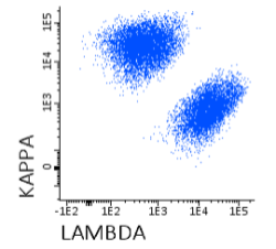
آنتی بادی مونوکلونال فلوسایتومتری KAPPA-PE ، کلون Polyclonal
اطلاعات بیشترName: Flow Cytometry Antibody KAPPA-PE, Clone Polyclonal
- Antibody Kappa-PE is a polyclonal antibody (pAb) labelled with R-Phycoerythrin (PE) and designed for use as a direct immunofluorescence reagent in the identification and enumeration of cells that express human Kappa immunoglobulin Light Chains by flow cytometry. Antibodies to Kappa Light Chains are useful for the identification of clonal excess in B-cell lymphoproliferative disorders together with a panel of other antibodies. Anti-Kappa Light Chains reacts with free Kappa chains as well as Kappa chains in intact immunoglobulin molecules. This reagent must be used by flow cytometry qualified personal.
REAGENT COMPOSITION
Purified polyclonal antibody Anti-human Kappa Light Chains, Goat F(ab’)2, conjugated with R-Phycoerythrin (PE), supplied in phosphate buffered saline with <0,1% (m/v) sodium azide.
- Amount per 0,5 ml
- vial: 100 tests (5 µl pAb to 106 cells).
- Reagents are not considered sterile.
-
 آنتی بادیهای فلوسایتومتری
آنتی بادی فلوسایتومتری 7-AAD Viability Staining Solution
اطلاعات بیشتر7-AAD Viability Staining Solution
Company: BioLegend
Catalog: 420403
Size: 200 tests
Storage & Handling: Protect from light. Store between 2°C and 8°C
Description:
7-AAD (7-amino-actinomycin D) has a high DNA binding constant and is efficiently excluded by intact cells. It is useful for DNA analysis and dead cell discrimination during flow cytometric analysis. When excited by 488 laser light, 7- AAD fluorescence is detected in the far red range of the spectrum (650 nm longpass filter).
-
 آنتی بادیهای فلوسایتومتری
آنتی بادی فلوسایتومتری APC anti-human CD3 Antibody
اطلاعات بیشترAPC anti-human CD3
Company: BioLegend
Catalog : 300311, 300312
Size: 25 tests / 100 tests
Clone: HIT3a
Isotype : Mouse IgG2a, κ
Reactivity: Human
Description:
CD3ε is a 20 kD chain of the CD3/T-cell receptor (TCR) complex which is composed of two CD3ε, one CD3γ, one CD3δ, one CD3ζ (CD247), and a T-cell receptor T-cell heterodimer. It is found on all mature T lymphocytes, NK-T cells, and some thymocytes. CD3, also known as T3, is a member of the immunoglobulin superfamily that plays a role in antigen recognition, signal transduction, and T cell activation.
-
 آنتی بادیهای فلوسایتومتری
آنتی بادی فلوسایتومتری APC anti-mouse CD8a Antibody
اطلاعات بیشترAPC anti-mouse CD8a Antibody
Company:BioLegend
Catalog Number: 100711
Size : 25 µg
Clone : 53-6.7
Isotype : Rat IgG2a, κ
Reactivity : Mouse
Description :
CD8, also known as Lyt-2, Ly-2, or T8, consists of disulfide-linked α and β chains that form the α(CD8a)/β(CD8b) heterodimer and α/α homodimer. CD8a is a 34 kD protein that belongs to the immunoglobulin family. The CD8 α/β heterodimer is expressed on the surface of most thymocytes and a subset of mature TCR α/β T cells. CD8 expression on mature T cells is non-overlapping with CD4. The CD8 α/α homodimer is expressed on a subset of γ/δ TCR-bearing T cells, NK cells, intestinal intraepithelial lymphocytes, and lymphoid dendritic cells. CD8 is an antigen co-receptor on T cells that interacts with MHC class I on antigenpresenting cells or epithelial cells. CD8 promotes T cell activation through its association with the TCR complex and protein tyrosine kinase lck.
-
 آنتی بادیهای فلوسایتومتری
آنتی بادی فلوسایتومتری FITC anti-human CD2 Antibody
اطلاعات بیشترFITC anti-human CD2 Antibody
Company:BioLegend
Catalog : 300206
Size : 100 tests
Clone : RPA-2.10
Isotype : Mouse IgG1, κ
Reactivity : Human, African Green, Baboon, Capuchin Monkey, Chimpanzee, Cynomolgus, Pigtailed Macaque, Rhesus, Swine (Pig, Porcine)
Description:
CD2 is a 50 kD type I transmembrane glycoprotein also known as LFA-2, T11, and sheep red blood cell receptor (SRBC-R). This immunoglobulin superfamily member is expressed on thymocytes, T lymphocytes, NK cells, and thymic B cell subsets. The major ligand for CD2 is CD58 (also known as LFA-3). CD2 has also been reported to bind CD48, CD59, and CD15. CD2 plays a critical role in alternative T cell activation, T cell signaling, and cell-cell adhesion.
-
 آنتی بادیهای فلوسایتومتری
آنتی بادی فلوسایتومتری FITC anti-human CD16 Antibody
اطلاعات بیشترFITC anti-human CD16 Antibody
Company:BioLegend
Clone: B73.1
Isotype: Mouse IgG1, κ
Reactivity: Human
Cat Number: 360716
Antibody Type: Monoclonal
Description:
CD16 is known as low affinity IgG receptor III (FcγRIII). It is expressed as two distinct forms (CD16a and CD16b). CD16a (FcγRIIIA) is a 50-65 kD polypeptide-anchored transmembrane protein. It is expressed on the surface of NK cells, activated monocytes, macrophages, a subset of T cells and placental trophoblasts in humans. CD16b (FcγRIIIB) is a 48 kD glycosylphosphatidylinositol (GPI)-anchored protein. Its extracellular domain is over 95% homologous to that of CD16a, and it is expressed specifically on neutrophils. CD16 binds aggregated IgG or IgG-antigen complex which functions in NK cell activation, phagocytosis, and antibody-dependent cell-mediated cytotoxicity (ADCC).
-
 آنتی بادیهای فلوسایتومتری
آنتی بادی فلوسایتومتری FITC anti-human CD33 Antibody
اطلاعات بیشترFITC anti-human CD33 Antibody
Company:BioLegend
Catalog : 303303
Size: 25 tests
Isotype: Mouse IgG1, κ
Reactivity: Human, Cross-Reactivity : Chimpanzee
Description:
CD33 is a 67 kD type I transmembrane glycoprotein also known as Siglec-3, gp67, and p67. It is a sialoadhesion immunoglobulin superfamily member expressed on myeloid progenitors, monocytes, granulocytes, dendritic cells and mast cells. CD33 is absent on normal platelets, lymphocytes, erythrocytes and hematopoietic stem cells. CD33 functions as a sialic acid-dependent cell adhesion molecule with carbohydrate/lectin binding activity.
-
 آنتی بادیهای فلوسایتومتری
آنتی بادی فلوسایتومتری FITC anti-mouse CD4 Antibody
اطلاعات بیشترFITC anti-mouse CD4 Antibody
Company:BioLegend
Catalog Number : 100405
Size : 50 µg
Clone : GK1.5
Isotype: Rat IgG2b, κ
Reactivity : Mouse
Description :
CD4 is a 55 kD protein also known as L3T4 or T4. It is a member of the Ig superfamily, primarily expressed on most thymocytes, a subset of T cells, and weakly on macrophages and dendritic cells. It acts as a coreceptor with the TCR during T cell activation and thymic differentiation by binding MHC class II and associating with the protein tyrosin kinase, lck.
-
 آنتی بادیهای فلوسایتومتری
آنتی بادی فلوسایتومتری FITC anti-mouse CD8a Antibody
اطلاعات بیشترFITC anti-mouse CD8a Antibody
Company:BioLegend
Catalog Number : 100705
Size : 50 µg
Clone : 53-6.7
Isotype : Rat IgG2a, κ
Reactivity : Mouse
Description :
CD8, also known as Lyt-2, Ly-2, or T8, consists of disulfide-linked α and β chains that form the α(CD8a)/β(CD8b) heterodimer and α/α homodimer. CD8a is a 34 kD protein that belongs to the immunoglobulin family. The CD8 α/β heterodimer is expressed on the surface of most thymocytes and a subset of mature TCR α/β T cells. CD8 expression on mature T cells is non-overlapping with CD4. The CD8 α/α homodimer is expressed on a subset of γ/δ TCR-bearing T cells, NK cells, intestinal intraepithelial lymphocytes, and lymphoid dendritic cells. CD8 is an antigen co-receptor on T cells that interacts with MHC class I on antigenpresenting cells or epithelial cells. CD8 promotes T cell activation through its association with the TCR complex and protein tyrosine kinase lck.
-
 آنتی بادیهای فلوسایتومتری
آنتی بادی فلوسایتومتری FITC anti-mouse CD49b (pan-NK cells) Antibody
اطلاعات بیشترFITC anti-mouse CD49b (pan-NK cells) Antibody
Company:BioLegend
Catalog Number : 108905
Size : 50 µg
Clone : DX5
Isotype : Rat IgM, κ
Reactivity : Mouse
Description :
DX5 antigen has been recently characterized as CD49b. It is a 150 kD integrin α chain also known as α integrin, VLA-2 α chain, and integrin α chain. CD49b non-covalently associates with CD29 (β integrin) to form the CD49b/CD29 complex known as VLA-2, a receptor for collagen and laminin. CD49b is expressed on platelets, the majority of NK cells, NKT cells, and a small subset of CD8+ T cells (this population can be significantly increased following viral infection). DX5 is used for the identification and isolation of NK cells, and is especially useful for identifying NK cells in mice lacking the NK1.1 antigen.