مونوکلونال
-
 آنتی بادیهای ایمونوهیستوشیمی
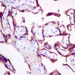
آنتیبادی Pan-Cadherin (CH-19)
اطلاعات بیشترName:Pan-cadherin Antibody clone CH-19
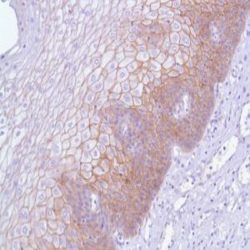
Description and applications: Cadherins are a broad family of cell-cell adhesion molecules with varying degrees of tissue expression. E-cadherin is found in all normal epithelia, whereas Pcadherin is expressed in trophoblastic cells and shows limited expression in epithelia, being predominantly expressed in bronchial and mammary basal epithelia. Cadherins are calcium-dependent adhesion molecules; intracellularly, E-cadherin binds to members of the catenin family (β, γ or α-catenins), which in turn are connected to the cytoskeleton’s actin filaments. Cadherins play an important role in cell growth and development, maintaining tissue integrity and architecture. Loss of cadherin expression plays an important role in neoplasms, facilitating local invasion and metastases generation. Immunohistochemical studies on carcinomas show decreased cadherin expression compared to normal tissues, especially in highly invasive tumours such as gastric adenocarcinomas.
Composition: Anti-human Pan-Cadherin mouse monoclonal antibody purified from serum and prepared in 10mM PBS, pH 7.4, with 0.2% BSA and 0.09% sodium azide
-
 آنتی بادیهای ایمونوهیستوشیمی
آنتی بادی Pancreatic lipase (EPR6275)
اطلاعات بیشترName: Pancreatic lipase Antibody clone EPR6275
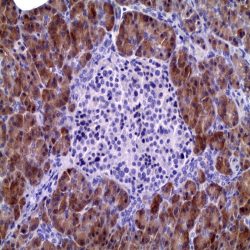
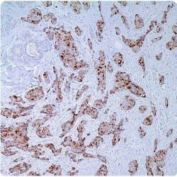
Description and applications: Pancreatic lipase is a 56kDa molecular mass enzyme actively secreted in the duodenum by the pancreas in the presence of bile salts and colipase. It belongs to the family of triglyceride lipases, of which there are at least three tissue-specific isoenzymes (pancreatic, hepatic and gastric and lingual) that break down dietary fats by converting triglycerides into monoglycerides and free fatty acids. Pancreatic lipase together with bile salts favors the absorption into the lymphatic system of the monomers resulting from digestion. Human pancreatic lipase is encoded by the PNLIP gene of 13 exons and more than 20 kb, located on the chromosomal region 10q25.3. In normal tissues this antibody identifies pancreatic acinar cells and in neoplasms it recognizes acinary cell carcinoma of the pancreas, which expresses this marker in more than 50% of cases. For this reason and integrated in a panel of suitable antibodies, pancreatic lipase allows the differentiation of acinar adenocarcinoma of pancreas from other malignancies with similar morphology (especially neuroendocrine carcinoma with acinary architecture) and establish the pancreatic origin of distant metastases, that on numerous occasions may be the first manifestation of the tumor.
Composition: Anti-human Pancreatic lipase mouse monoclonal antibody purified from serum and prepared in 10mM PBS, pH 7.4, with 0.2% BSA and 0.09% sodium azide
-
 آنتی بادیهای ایمونوهیستوشیمی
آنتی بادی Cytokeratin 14 (LL002)
اطلاعات بیشترName: Cytokeratin 14 antibody clone LL002
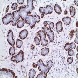
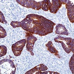
Description and aplications: Cytokeratin 14 belongs to the type A (acidic) subfamily of low molecular weight keratins and exists in combination with keratin 5. Cytokeratin 14 has been studied as a prognostic marker in breast cancer. Here it is used as a marker for the myoepithelial cells and for the identification of basaloid histotype of breast carcinoma.
The antibody also distinguishes stratified epithelial cells from simple epithelial cells and has been reported useful in the identification of squamous cell carcinomas.Composition: anti-human Cytokeratin 14 mouse monoclonal antibody purified from ascites fluid by Protein G chromatography. Prepared in 10mM PBS, pH 7.4, with 0.2% BSA and 0.09% sodium azide
Immunogen: A synthetic peptide of 15 amino acid residues from the C-terminus of human keratin 14.
-
 آنتی بادیهای ایمونوهیستوشیمی
آنتی بادی Cytokeratin 5/6/8/18 (EP24/EP67/B22.1&B23.1)
اطلاعات بیشترName:Cytokeratin Antibody clone EP24/EP67/B22.1&B23.1
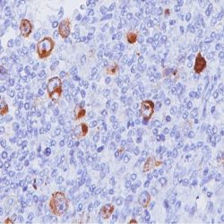
Description and applications: This antibody recognises cytokeratins 5, 6, 8, and 18. Proteins generically called intermediate filaments, because they measure between 7 and 22 nm in diameter (i.e., with a size between actin, 5-7 nm, and tubulin, 22-25 nm), are part, along with actin and tubulin, of the vertebrate cytoskeleton. This superfamily is composed of six subfamilies of molecules with different tissue expression patterns. Cytokeratins constitute homology groups I and II, and in humans they are encoded by more than 49 different genes located in chromosomes 17 (I) and 12 (II). The nomenclature chosen in 1982 by Moll and Franke assigns numbers from 1 to 8 to type II cytokeratins (neutral or basic) and from 9 to 21 to type I cytokeratins (acidic). An analogous nomenclature for hair keratins has now been defined, with the addition of letters Ha and Hb to distinguish type I from type II. Structurally, cytokeratins share with the rest of intermediate filaments a central axis of 310 amino acid residues consisting of four highly preserved α-helical domains (1A, 1B, 2A, and 2B) that define the type of intermediate filament that will be formed after assembly; these domains are separated by three non-helical linking regions (L1, L12, and L2) and two end domains which are highly different in size and sequence (head, 1, and tail, 2), each with constant (E1/E2), variable (V1/V2), and homology (H1/H2) regions, the latter characteristic of type II keratins and absent in type I keratins. The major immunogenic properties and the main differences between each keratin class are found in the variable domains. Keratins are usually assembled into I/II heterodimers and are specifically co-expressed in pairs in each tissue. Due to the high homology among the different molecules, it is common for a monoclonal antibody to react with different types of cytokeratins; for example, anti-cytokeratin AE1 labels cytokeratins 10, 14, 15, 16, and 19. The anti-cytokeratin monoclonal antibody cocktail is a broad-spectrum antibodt for general use. This antibody mixture reacts with carcinomas in general, but as with other anti-cytokeratin antibodies, it is possible that poorly differentiated carcinomas occasionally exhibit poor immunostaining. Its most common application is the differential diagnosis of malignant tumours, in which case it can be used within a panel of antibodies, such as anti-ALC, which recognises lymphoma, or anti-S100 or HMB45, which labels malignant melanoma. The staining of other non-carcinoma tumours is uncommon, although as with other anti-cytokeratin 8 antibodies, smooth muscle and leiomyoma labelling may occasionally be observed.
Composition: Anti-human Cytokeratin 5/6/8/18 rabbit/mouse monoclonal antibody purified from serum and prepared in 10mM PBS, pH 7.4, with 0.2% BSA and 0.09% sodium azide
-
 آنتی بادیهای ایمونوهیستوشیمی
آنتی بادی Calponin (EP63)
اطلاعات بیشترName:Antibody Calponin clone EP63
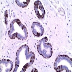
Description and applications: Calponin is a smooth muscle specific, actin-, tropomyosin- and calmodulin-binding protein thought to be involved in regulation of actomyosin as well as the regulation or modulation of contraction. It is expressed on smooth muscle cells and myoepithelial cells. Calponin antibody has been used to identify invasion of breast lesion. Additionally, Calponin is expressed on malignant fibrous histiocytoma of bone and adenoid cystic carcinoma of salivary gland. The consistently positive staining pattern in adenoid cystic carcinomas may be useful in discriminating
Composition: Anti-human Calponin rabbit monoclonal antibody purified from serum and prepared in 10mM PBS, pH 7.4, with 0.2% BSA and 0.09% sodium azide
-
 آنتی بادیهای ایمونوهیستوشیمی
آنتی بادی Delta Catenin-1 (p120) (EP66)
اطلاعات بیشترName: Delta Catenin-1 (p120) (Clone EP66)
Description and applications:Catenins are proteins that are linked to the cytoplasmic domain of transmembrane cadherins. p120 Catenin is a member of this Armadillo gene family of junctional plaque proteins. The association of catenins to cadherins produces a complex which is linked to the actin filament network, and which seems to be important for cadherins cell-adhesion properties. Cytoplasmic accumulation of p120 Catenin has been observed in lung cancer, pancreatic cancer, gastric cancer and colon cancers and is associated with poor progress in colon cancer patients. In breast lobular neoplasia, anti p120 Catenin shows a diffuse cytoplasmic immunostaining pattern, while breast ductal neoplasia retains the membrane immunostaining pattern. p120 Catenin antibody is useful in differentiation of lobular carcinoma from ductal carcinoma of the breast and in identifying early lesions of lobular neoplasia.
Composition:Anti-human Delta Catenin-1 (p120) rabbit monoclonal antibody purified from serum and prepared in 10mM PBS, pH 7.4, with 0.2% BSA and 0.09% sodium azide.
Immunogen: A synthetic peptide corresponding to residues in human p120 Catenin protein.
-
 آنتی بادیهای ایمونوهیستوشیمی
آنتی بادی Epithelial Membrane Antigen (EMA) (E29)
اطلاعات بیشترName: Epithelial Membrane Antigen(EMA)-CloneE29
Description and applications: Anti-EMA antibody is a useful marker for staining many carcinomas. It stains normal and neoplastic cells from various tissues, including mammary epithelium, sweat glands and squamous epithelium. Hepatocellular carcinoma, adrenal carcinoma and embryonal carcinomas are consistently EMA negative, so keratin positivity with negative EMA favours one of these tumours. EMA is frequently positive in meningioma, which can be useful when distinguishing it from other intracranial neoplasms, e.g. Schwannomas. The absence of EMA can also be of value since negative EMA staining is characteristic of some tumours including adrenal carcinoma, seminomas, paraganglioma and hepatoma. Many mesotheliomas, epithelioid and synovial sarcomas, chordomas, choroid plexus tumors, nodular lymphocyte-predominant Hodgkin lymphoma, anaplastic CD30 / ALK positive lymphomas and myelomas are positive; occasionally some small round cell tumours and small cell sarcomas, T null phenotype or CD56 / CD57 + T / NK lymphomas are positive. Other lymphomas, basal cell carcinomas, hepatocellular carcinomas, melanomas, endocrine neoplasms and soft tissue tumours are consistently negative.
Composition: anti-human EMA mouse monoclonal antibody purified from serum and prepared in 10mM PBS, pH 7.4, with 0.2% BSA and 0.09% sodium azide.
-
 آنتی بادیهای ایمونوهیستوشیمی
آنتی بادی Epithelial Specific Antigen (BER-EP4)
اطلاعات بیشترName: Epithelial Specific Antigen-Clone Ber-EP4
Description and aplications: This antibody reacts with two glycoproteins of 34 and 49 kD molecular weight present on the surface and in the cytoplasm of all epithelial cells except the surface layer of the squamous epithelium, hepatocytes and parietal cells of the stomach. This antibody is specific to HEA125 equivalent. The antibody shows a broad pattern of reactivity with human epithelial tissues like simple epithelium, basal layer of the stratified squamous non-keratinized epithelia and epidermis. In contrast to other known anti-epithelial antibodies, this antibody does not mark the mesothelial cells and only a small number of mesotheliomas stained positively. This antibody does not react with nervous, glial, muscle, mesenchymal or lymphoid tissue. Due to the labile nature of the epitope in tissues fixed in buffered formalin and embedded in paraffin, negative staining results should be considered with caution.
Composition: anti-Ber-EP4 mouse monoclonal antibody obtained from supernatant culture and prediluted in a tris buffered solution pH 7.4 containing 0.375mM sodium azide solution as bacteriostatic and bactericidal.
Immunogen: MCF-7 human breast carcinoma cell line.
-
 آنتی بادیهای ایمونوهیستوشیمی
آنتی بادی Epstein-Barr Virus / LMP1 (CS1-4)
اطلاعات بیشترName: Epstein-Barr Virus / LMP1 (Clone CS1-4)
Description and applications:The antibody strongly reacts with EBV-positive lymphoblastoid cell lines and EBV infected B cell immunoblasts in infectious mononucleosis. It also reacts with 25 to 50 per cent of EBV associated undifferentiated nasopharyngeal carcinomas and with Reed Sternberg cells in approximately 90% of EBVassociated Hodgkin’s disease cases. The cocktail recognizes distinct epitopes on the hydrophilic carboxyl region of LMP which is exposed to the cytosol.
Composition:Anti-human Epstein-Barr Virus / LMP1 mouse monoclonal antibody purified from serum and prepared in 10mM PBS, pH 7.4, with 0.2% BSA and 0.09% sodium azide.
Immunogen: EBV-encoded recombinant latent membrane protein.
-
 آنتی بادیهای ایمونوهیستوشیمی
آنتی بادی Fascin (FCN01)
اطلاعات بیشترName: Fascin (Clone FCN01)
Description and applications:Anti-Fascin is a very sensitive marker for ReedSternberg cells and variants in nodular sclerosis, mixed cellularity, and lymphocyte depletion Hodgkin’s disease. It is uniformly negative in lymphoid cells, plasma cells and myeloid cells. Anti-Fascin is positive in dendritic cells. This marker may be helpful in distinguishing between Hodgkin’s disease and nonHodgkin’s lymphoma in difficult cases. Also, the lack of expression of Fascin in the neoplastic follicles in follicular lymphoma can be helpful in distinguishing these lymphomas from reactive follicular hyperplasia in which the number of follicular dendritic cells is normal or increased. Anti-Fascin has been suggested as a prognostic marker in neuroendocrine neoplasms of the lung, as well as ovarian cancer.
Composition: Anti-human Fascin mouse monoclonal antibody purified from serum and prepared in 10mM PBS, pH 7.4, with 0.2% BSA and 0.09% sodium azide.
-
 آنتی بادیهای ایمونوهیستوشیمی
آنتی بادی GH (Growth Hormone) (EP267)
اطلاعات بیشترName: Rabbit anti-human GH (Growth Hormone) Monoclonal Antibody clone EP267
Description and applications: Growth hormone (GH or hGH), also known as somatotropin or somatropin, is a peptide hormone that is produced and secreted by somatotrophs of the anterior pituitary gland. GH exerts a wide variety of biological actions in many different tissues and cell types. The actions of GH at the cellular level can be divided into three categories: those affecting mitogenesis, differentiation, and metabolism.The GH antibody specifically labels somatotrophs in pituitary in normal tissues. It is useful in classification of pituitary tumor.
Composition: Anti-human GH (Growth Hormone) rabbit monoclonal antibody purified from serum and prepared in 10mM PBS, pH 7.4, with 0.2% BSA and 0.09% sodium azide
Immunogen: A synthetic peptide corresponding to residues of human Growth Hormone protein
-
 آنتی بادیهای ایمونوهیستوشیمی
آنتی بادی Glutamine Synthetase (GS-6)
اطلاعات بیشترName: Glutamine Synthetase (Clone GS-6)
Description and applications:Glutamine synthetase (GS) catalyzes the synthesis of glutamine from glutamate and ammonia in the mammalian liver. In normal liver, GS expression is seen in pericentral hepatocytes, but not in mid-zonal or periportal hepatocytes. Glutamine, the end product of GS activity, is the major energy source of tumor cells. Based on findings from experimental hepatocarcinogenesis, GS positive tumor cells are believed to be derived from GS positive hepatocytes. Thus, anti-GS has been suggested as a marker for hepatocellular carcinoma (HCC). GS immunoreactivity has been seen in a majority of HCC (37 of 53 cases, 70%), including 7 of 10 cases of early HCC (70%) and 12 of 22 (59%) for low grade HCC. In nonmalignant nodules, GS overexpression was only seen in 3 high grade dysplastic nodules (HGDN,13.6%). In these cases, GS overexpression was restricted to 11.5%-50% of hepatocytes, whereas in HCC the majority of cases (28 of 53, 53%), including early HCC (60%), showed diffuse immunostaining (>50% tumor cells). Overall, the sensitivity, specificity, and positive and negative predictive values of anti-GS for HCC detection were 69.8%, 94.2%, 92.5%, and 75.4%, respectively. A panel composed of antibodies against HSP70, GPC3, and GS has been proposed to be very useful in distinguishing between dysplastic and early malignant hepatocellular nodules arising in cirrhosis. The “all positive” phenotype is restricted to approximately half of early HCC to well-differentiated HCC but has never been reported in dysplastic lesions, whereas the reverse phenotype, “all negative”, has been shown to be a feature of the majority of HGDN and of all low grade dysplastic nodules.
Composition: Anti-human Glutamine Synthetase mouse monoclonal antibody purified from serum and prepared in 10mM PBS, pH 7.4, with 0.2% BSA and 0.09% sodium azide.
-
 آنتی بادیهای ایمونوهیستوشیمی
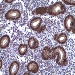
آنتی بادی PAX-2 (EP235)
اطلاعات بیشترname: PAX-2 Antibody clone EP235
Description and aplications: PAX2 is a member of the paired box family of transcription factors, which is required for development and proliferation of the kidney, brain, and müllerian organs. PAX2 genes contain a highly conserved DNA sequence within the paired box region, which encodes a DNA-binding domain, enabling PAX proteins to bind the promoters of specific genes to transcriptionally regulate their expression. Defects in PAX2 gene are related with the renal coloboma syndrome (RCS) (also known as papillorenal syndrome) which is a condition that primarily affects kidney (renal) and eye development. PAX2 is specifically expressed in the developing central nervous system, eye, ear, and urogenital tract, and is essential for the development of these organs. In normal adult tissues PAX2 was mainly detected in the urogenital system, including kidney, uretericepithelium, fallopian tube epithelium, ovary and uterus. In tumors, PAX2 has been detected in renal cell carcinomas, Wilms’ tumors, nephrogenic adenomas and papillary serous carcinoma of the ovary. For these reasons, PAX2 has been used as a marker for the identification of renal cell carcinoma and ovarian carcinoma by immunohistochemistry. It has been also suggested as a useful tool in diagnosis and classification of hyperplastic endometrial epithelial proliferations
Composition: anti-human PAX2 rabbit monoclonal antibody purified from ascites fluid by chromatography. Prepared in 10mM PBS, pH 7.4, with 0.2% BSA and 0.09% sodium azide
-
 آنتی بادیهای ایمونوهیستوشیمی
آنتی بادی Pax-5 (MX017)
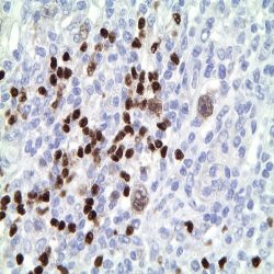
اطلاعات بیشترName: PAX-5 Antibody clone MX017
Description and applications: The PAX-5 gene is essential for B-cell differentiation. There are at least four isoforms, of which PAX-5a has been most studied. PAX-5 encodes the 50 kDa B-cell specific activator protein, BSAP. PAX-5 is expressed by pro-, pre-, and mature B-cells, but is downregulated during terminal differentiation of plasma cells. PAX-5 influences the expression of other B-cell specific genes, including CD19 and CD20 and CD79a, preceding the expression of CD20. PAX-5 is silenced at the plasma cell stage under the influence of Blymphocyte-induced maturation protein-1 (PRDM1). Pax-5 is consistently expressed in mature B cell lymphomas. It is useful to distinguish between classical Hodgkin lymphoma (+) and ALCL (-). In Hodgkin lymphoma, the neoplastic cells show a weaker staining compared with the accompanying B cells. Occasionally it expressed in acute myeloid leukemias especially those with t (8; 21). CD20 negative B-cell lymphomas of patients treated with anti-CD20 monoclonal antibodies, maintain PAX5 staining the antibody representing a good tool to confirm their cell of origin. Pax-5 expression was detected in hyperplastic mesonephric remains, epididymitis, neurons of the adult nervous tissue, small cell carcinomas, Merkel cell tumor and other neuroendocrine tumors.
Composition: anti-human PAX5 mouse monoclonal antibody purified from serum and prepared in 10mM PBS, pH 7.4, with 0.2% BSA and 0.09% sodium azide
-
 آنتی بادیهای ایمونوهیستوشیمی
آنتی بادی CA 19-9 (121SLE)
اطلاعات بیشترName : Antibody Ca19-9 clone 121SLE
Description and applications: CA19-9 antigen is highly expressed in gastrointestinal (gastric, pancreatic, and colonic) adenocarcinomas and salivary gland mucoepidermoid carcinomas. Anti- CA19-9 antibody is usually not reactive with breast, kidney, and prostate carcinomas. Anti-human CA19-9 mouse monoclonal antibody purified from serum and prepared in 10mM PBS, pH 7.4, with 0.2% BSA and 0.09% sodium azide
Composition:Anti-human CA19-9 mouse monoclonal antibody purified from serum and prepared in 10mM PBS, pH 7.4, with 0.2% BSA and 0.09% sodium azide.
-
 آنتی بادیهای ایمونوهیستوشیمی
آنتی بادی PAX-8 (MD-50 also known as MRQ-50)
اطلاعات بیشترName: Pax-8 Antibody clone MD-50 also known as MRQ-50
Description and applications:PAX8 antibody recognizes a protein of 62kDa, identified as PAX8. It is a member of the paired box (PAX) family of transcription factors. This nuclear protein is involved in thyroid follicular cell development and expression of thyroid-specific genes. Mutations in this gene have been associated with thyroid dysgenesis, thyroid follicular carcinomas, and atypical thyroid adenomas. PAX8 is expressed in the normal thyroid follicular cells and associated carcinomas. As a marker of mesonephric and müllerian organs, ovarian and renal normal and neoplasms are frequently positive. For that, non-ciliated mucosal cells of the fallopian tubes, and simple ovarian inclusion cysts and normal ovarian surface epithelial cells are positive. PAX8 is expressed in a high percentage of ovarian serous, endometrioid, and clear cell carcinomas, but only rarely in primary ovarian mucinous adenocarcinomas. PAX8 antibody may be used as an additional immunohistochemical marker for renal epithelial tumors as it is expressed by all the segments of the nephron and the great majority of renal tumours including sarcomatoid carcinomas. Thymic and parathyroid carcinomas and isolated cases of urothelial carcinomas, sex-cord stromal tumours of the ovary or squamous carcinomas of the lung have showed weak and focal positivity.
Composition:Anti-human PAX-8 mouse monoclonal antibody purified from serum and prepared in 10mM PBS, pH 7.4, with 0.2% BSA and 0.09% sodium azide
-
 آنتی بادیهای ایمونوهیستوشیمی
آنتی بادی Cadherin 17 (SP183)
اطلاعات بیشترName:Cadherin 17 Antibody clone SP183
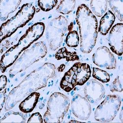
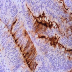
Description and applications: Cadherins represents a big family of Ca2+ dependent transmembrane cell adhesion glycoproteins that interact homophilically with each other and thus participate in the heterologous cell selection. They play a key role in the embryonic development and in the maintenance of the normal structure in adult tissues. Molecular defects in the structure of cadherins represent important pathogenic aspects of many diseases, including cancer. The cadherin 17 gene (CDH17) is located on the chromosome region 8q22.1 that encodes a protein of 832 amino acids and approximately 92kDa of molecular weight. Cadherin 17, also known as human peptide transporter 1 (HPT-1) or liver-intestine cadherin (LICadherin), is located in the lateral contact areas of the intestinal epithelial cells, being undetectable in the stomach, kidney, lung or liver, among other organs. It
features many structural similarities with the kidney specific cadherin, both belonging to the 7D-cadherin family. In tumors, the great specificity of cadherin 17 to identify the primary or metastatic colorectal adenocarcinomas has been demonstrated. Less frequently, cadNherin 17 has been described in gastroesophageal junction adenocarcinomas or adenocarcinomas with gastric origin. In the few available studies, cadherin 17 has shown staining in all well-differentiated neuroendocrine tumors (carcinoid tumors) originated in the small intestine and in the appendix. Up to 80% of the primary pancreatic adenocarcinomas and only half of their metastases are usually positive. Up to 50% of cholangiocarcinomas show membrane staining against cadherin 17, showing greater sensitivity than other markers such as villin, CDX2 or SATB2, which can be used to refine the diagnosis. Cadherin 17 shows along with SATB2 greater sensitivity in the detection of medullar carcinomas of the large intestine that are usually negative for CDX2 and cytokeratins 7 and 20.Composition: Anti-human Cadherin 17 rabbit monoclonal antibody purified from serum and prepared in 10mM PBS, pH 7.4, with 0.2% BSA and 0.09% sodium azide
-
 آنتی بادیهای ایمونوهیستوشیمی
آنتی بادی CD3 (EP41)
اطلاعات بیشترName: CD3 Antibody clone EP41
Description and applications: CD3 (Cluster of Differentiation 3) is a complex of proteins that associates directly with the T cell antigen receptor (TCR). CD3 is composed of five invariant polypeptide chains that associate to form three dimers. The five invariant chains of CD3 are labeled gamma, delta, epsilon, zeta, and eta. The CD3 is involved in T cell development and survival. It is expressed on T cells in thymus, peripheral lymphoid tissue, blood and bone marrow. CD3 is a commonly used marker for identification of T cell and T cell derived malignancies. This CD3 antibody has been validated by the 9th International Conference on Human Leukocyte Differentiation Antigens (HLDA9).
Composition: Anti-human CD3 rabbit monoclonal antibody purified from serum and prepared in 10mM PBS, pH 7.4, with 0.2% BSA and 0.09% sodium azide
Intended use: Immunohistochemistry (IHC) on paraffin embedded tissues. Not tested on frozen tissues or Western-Blotting
Species reactivity: In vitro diagnostics in humans. Not tested in other species
-
 آنتی بادیهای ایمونوهیستوشیمی
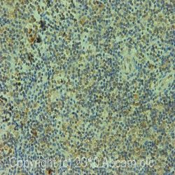
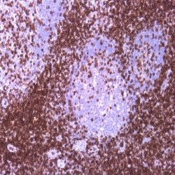
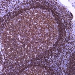
آنتی بادی CD45RA (SPM504)
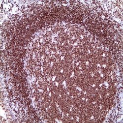
اطلاعات بیشترName: CD45RA Monoclonal Antibody clone SPM504
Description and aplications: This antibody reacts with the CD45RA isoform with a molecular mass of 220 kD. CD45RA is present in peripheral blood B cells, follicular mantle and centre, medullary thymocytes, monocytes, and mature T cells that have not been modulated by antigenic contact. In this regard, and after immune processing in the thymus, T cells are released into peripheral blood to colonize secondary lymphoid organs, including spleen, lymph nodes, and mucosa-associated lymphoid tissue (MALT). These lymphocytes, not yet activated in vivo by antigens (naive cells), are phenotypically characterized by the co-expression of the highest molecular mass CD45 isoforms, CD45RAC and CD45RA, and CD62L (peripheral lymph node homing receptor), a decisive molecule for the fixation of cells in thymus-dependent areas of secondary lymphoid organs. When these cells are stimulated by its specific antigen expressed by the corresponding antigen-presenting cells and are transformed into effector T cells, either CD4- or CD8-positive or memory T cells, CD45RA and CD62L expression disappears, the former being replaced by the lower molecular mass CD45R0 isoform, and the latter, by various type 1 and type 2 integrins and APO-1/Fas receptor (CD95), an apoptosis inducer. The CD45 molecule, also called leukocyte common antigen, is a membrane glycoprotein that occupies up to 10% of the surface of cells expressing it. In mammals, its homology varies markedly between the intracytoplasmic region (greater than 90%) and the extracellular region (only 35%, although with similar domain organization). Structurally, there are multiple CD45 isoforms determined by the different possible ways of joining exons 4, 5, and 6 during the synthesis of the mRNA encoding the extracellular domain of the protein. These isoforms are referred to as A, B, and C, using the RABC terminology for the bigger isoform, which includes all three exons, RA and RB when the exonic expression is restricted to one of these exons, and RO to designate the smaller molecule, which lacks all three exons. The sequence of these three exons includes multiple oxygen-linked glycosylation sites that can be variably modified by sialic acid. This, on the one hand, causes the molecular mass of the different isoforms to vary between 180 kD (RO) and 240 kD (RABC); on the other hand, it gives these molecules remarkable differences in terms of their shape and anionic charge. The rest of CD45’s extracellular domain shows marked nitrogen-linked glycosylation and contains a cysteine-rich region followed by three amino acid repeats analogous to fibronectin type III domain. CD45’s high degree of glycosylation, attributable to both sialic acid and oligosaccharides on the variable domains and N-glycoconjugates on the constant extracellular domain, plays an important role in the functional activity of the molecule, as demonstrated by the interaction between CD45RO of T cells and B cells expressing CD22 or the mannose hybrids on CD45 in the development of immature thymocytes. The rest of the CD45 molecule consists of a single transmembrane domain followed by a long intracytoplasmic tail containing two tandem-placed identical domains (D1 and D2) with protein-tyrosine phosphatase (PTPase) homology. Of these domains, only D1 possesses enzymatic activity capable of rescuing TCR signal activation in CD45-deficient cell lines. The function of D2 is hardly understood, although it seems to contribute to the molecule’s intracytoplasmic stability. Although in in vitro models the extracellular domain of CD45 is not essential for its intracytoplasmic function, the exonic transcription of this domain is strongly regulated depending on the cell type that expresses it, its development, and its degree of activation, thus playing an important functional role in vivo.The CD45RA epitope recognized by the MB1 antibody is useful for the study of B-cell lymphomas; however, as some of them are not positive against it, it is advisable to use it as part of a large panel of antibodies. Because the epitope recognized by MB1 may be altered by prolonged formol fixation, and in order to ensure a correct interpretation of results, it is appropriate to check that at least some normal B cells in the histological section are stained. The antibody shows no reactivity against cortical thymocytes, immature T cells, and activated B cells. This antibody reacts against human tissue. In other species, it has not been tested.
Composition: Anti-human CD45RA mouse monoclonal antibody purified from serum and prepared in 10mM PBS, pH 7.4, with 0.2% BSA and 0.09% sodium azide
-
 آنتی بادیهای ایمونوهیستوشیمی
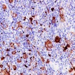
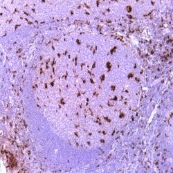
آنتی بادی CD68 (KP-1)
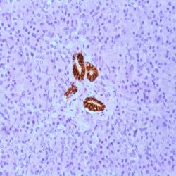
اطلاعات بیشترName: CD68 Monoclonal Antibody clone KP-1
Description and aplications: Anti-CD68 marks cells of monocyte/macrophage lineage. This antibody is capable of staining monocytes, Kupffer cells, osteoclasts, NK cells, granulocytes and their precursors; lymphomas are negative or show a few granules. This antibody may be useful for the identification of myelomonocytic and histiocytic tumors. Anti-CD68 may help to distinguish malignant fibrous histiocytoma from other pleomorphic sarcomas. However, since this detects a formalinresistant epitope that may be associated with lysosomal granules, other lysosome-rich cells may also stain.
Composition: Anti-human CD68 mouse monoclonal antibody purified from serum and prepared in 10mM PBS, pH 7.4, with 0.2% BSA and 0.09% sodium azide