مونوکلونال
-
 آنتی بادیهای ایمونوهیستوشیمی
آنتی بادی Glucagón (EP74)
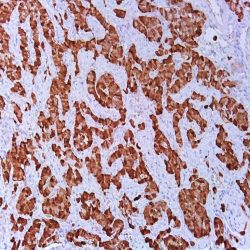
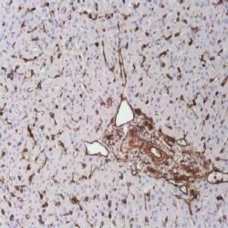
اطلاعات بیشتر.(Name: Glucagon Monoclonal Antibody (Clone EP74
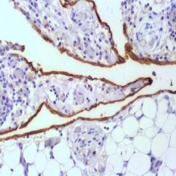
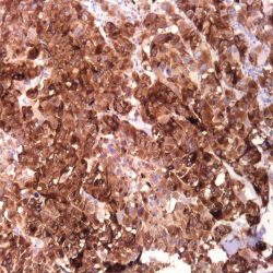
Description and application: Glucagon is synthesized and released by the alpha-cells of the islets of Langerhans’ in pancreas. It regulates blood glucose by increasing gluconeogenesis and decreasing glycolysis, stimulates fluid secretions from the intestine and suppresses the release of gastrin. Antiglucagon is useful in identification of glucagonoma.
Composition: Anti-human Glucagon rabbit monoclonal antibody purified from serum and prepared in 10mM PBS, pH 7.4, with 0.2% BSA and 0.09% sodium azide.
Immunogen: A synthetic peptide corresponding to residues in human Glucagon protein.
-
 آنتی بادیهای ایمونوهیستوشیمی
آنتی بادی Glycophorin A (JC159)
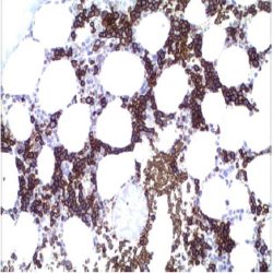
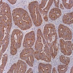
اطلاعات بیشتر.(Name: Glycophorin A Antibody (Clone JC159
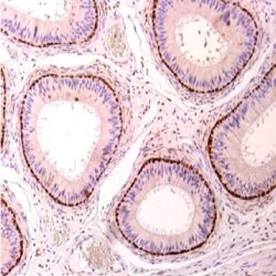
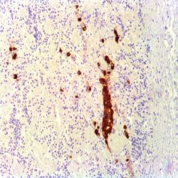
Description and aplications: This antibody reacts with a resistant paraffin-embedded and formol fixed epitope located in the extracellular domain of the protein, probably between amino acids 27 and 40. Therefore, this antibody recognizes normal erythroid cells in all stage of differentiation from erythroblasts to mature red blood cells. Once glycophorin A is at its maximum expression, the quantity in each erythroid cell remains constant and shows no change during maturation despite the decreasing size of the cell. The neoplastic erythroblast in most erythroleukemias are identified by Glycophorin A.
Composition: anti-Glycophorin A mouse monoclonal antibody obtained from supernatant culture and prediluted in a tris buffered solution pH 7.4 containing 0.375mM sodium azide solution as bacteriostatic and bactericidal.
Immunogen: membrane preparation from splenic hairy cell leukemia cells.
-
 آنتی بادیهای ایمونوهیستوشیمی
آنتی بادی Glypican 3 (1G12)
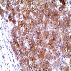
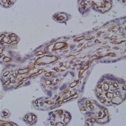
اطلاعات بیشتر.(Name: Glypican 3 Antibody (Clone 1G12
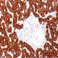
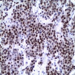
Description and applications: Glypican‐3 (GPC3) is a member of the glypican family of glycosyl phosphatidylinositol‐anchored cell‐surface heparin sulfate proteoglycans. The 1G12 monoclonal antibody has been used to assess GPC3 expression in malignant and nonmalignant liver tissue samples and for enzyme‐linked immunosorbent assay (ELISA) for detection of GPC3 in the serum. Studies have shown that GPC3 is expressed at the protein level in most HCCs, but it is undetectable in normal liver and benign hepatic lesions, including dysplastic and cirrhotic nodules. In addition, GPC3 is significantly elevated in the serum of a large proportion of patients with HCC. Based on these results, it has been proposed that GPC3 could be a useful marker to differentiate between benign and malignant liver diseases. The GPC3 is expressed in the great majority of the gonadal yolk sac tumors. The endodermal component of endometrial or ovarian endometrioid carcinoma cases with also show intense cytoplasmic and / or membrane positivity. It is positive in sporadic cases of choriocarcinoma and presents focal positivity in embryonal carcinomas and immature teratomas; is not expressed in seminomas an intratubular germ cell neoplasia. Isolated cases spermatocytic seminomas show weak positivity. Other tumors of gonadal origin that can express GPC3 are endometrioid, clear cell, mucinous or serous carcinomas. 80% of placental site trophoblastic tumors and 100% of uterine placental site nodules are also positive. In other no gynecological tumors, GPC3 is expressed to 23% of lung carcinomas (55% squamous cell carcinomas and only 8% of adenocarcinomas) and 3% of renal cell carcinomas (chromophobe carcinomas). Along with other markers such as E-cadherin (negative) and β-catenin (positive nuclear and cytoplasmic), GPC3 is a useful marker in the diagnosis of pseudopapillary solid tumors of the pancreas and helps the differential diagnosis of neuroendocrine tumors. 34.7% of rhabdomyosarcomas, more frequent embryonal type, show positivity for this antibody while other types of sarcomas arenegative. In a recent study, 52% of liposarcomas and 29% of melanomas have been positive. Varying percentages of other tumors of epithelial and mesenchymal have shown variable positivity, demonstrating the lack of specificity of this antibody and the need to analyze the results in context with other antibodies staining.
Composition: anti-human Glypican 3 mouse monoclonal antibody purified from serum and prepared in 10mM PBS, pH 7.4, with 0.2% BSA and 0.09% sodium azide.
Immunogen: Balb/C mice immunized with a single i.p. injection of 50 μg of a fragment containing the last 70 amino acids of the core protein
-
 آنتی بادیهای ایمونوهیستوشیمی
آنتی بادی Granzyme B (GrB-7)
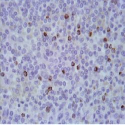
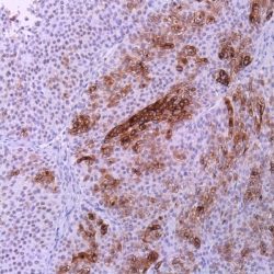
اطلاعات بیشتر.(Name: Granzyme B Antibody (Clone GrB-7
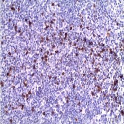
Description and aplications: This antibody reacts with human Granzyme B. Granzyme B is a neutral serine protease which is stored in granules of specialized cytotoxic T lymphocytes (CD8) and NK cells. From mouse CD8 and NK cells vaious types of granzymes have been identifies and named alphabetically (A-G). They are involved in the apoptosis mediated by the cytotoxic lymphocytes. The exocytosis of granules containing granzyme in the cytoplasm of the target cells leads to DNA fragmentation and subsequent apoptosis. Granzyme B has also been located in EBV positive Reed Sternberg cells of Hodgkin’s disease.
Composition: anti-Granzyme B mouse monoclonal antibody obtained from supernatant culture and prediluted in a tris buffered solution pH 7.4 containing 0.375mM sodium azide solution as bacteriostatic and bactericidal.
Immunogen: Recombinant protein encoding the N-terminus of the mature granzyme B.
-
 آنتی بادیهای ایمونوهیستوشیمی
آنتی بادی HBME-1 (HBME-1)
اطلاعات بیشتر.(Name: Mesothelial Cell Antibody (Clone HBME-1
Description and aplications: Antibody against HBME-1 stains the cytoplasmic membrane of normal pleural and peritoneal mesothelial cells and mesothelioma. Although both the membrane and the cytoplasm of mesothelioma cells stain positive with HBME-1, a thick membrane pattern turns out to be more useful for the diagnosis of malignant mesothelioma and it correlates with the presence of abundant “microvilli” in the surface membrane of these cells. There are other tissues (both malignant and normal) which are also positive with HBME-1, although immunostaining can be much weaker. Although HBME-1 positivity has been proven in 19 of 50 adenocarcinomas of different origin, the pattern of staining observed was cytoplasmic, although occasionally weak membrane staining also appeared. This antibody shows useful to distinguish malignant lesions of the thyroid (papillary and follicular) of benign lesions (goiter, adenomas, etc..). No cross reaction was observed with tissues such as kidney, liver, lung, ovary and pancreas. Storage and stability: up to 18 months; stored at 2- 8ºC. Do not freeze.
Composition: anti-HBME-1 mouse monoclonal antibody obtained from supernatant culture and prediluted in a tris buffered solution pH 7.4 containing 0.375mM sodium azide solution as bacteriostatic and bactericidal.
-
 آنتی بادیهای ایمونوهیستوشیمی
آنتی بادی Heat Shock Protein 70 (HSP70) (W27)
اطلاعات بیشترName: Heat Shock Protein 70 (HSP70) Monoclonal Antibody clone W27
Description and applications: Heat shock proteins (HSP’s) or stress response proteins (SRP’s) are synthesized in variety of environmental and pathophysiological stressful conditions. Many HSP’s are involved in processes such as protein denaturation-renaturation, folding-unfolding, transporttranslocation, activation-inactivation, and secretion.HSP70 is found to be associated with steroid receptors, actin, p53, polyoma T antigen, nucleotides, and other unknown proteins. Also, HSP70 has been shown to be involved in protective roles against thermal stress, cytotoxic drugs, and other damaging conditions. This antibody recognizes both the constitutive (HSP73), and inducible (HSP72) forms of HSP70. Most normal cells show moderate cytoplasmic and nuclear immunostaining. Pancreatic islets, cardiac muscle cells and most CNS cells are weakly stained or negative. HSP70 in tumors is significantly increased in patients with cholangiocarcinoma. HSP70 is positive in the majority of the hepatocellular carcinoma. In oral squamous cell carcinomas HSP70 expression has been associated with increased survival compared with HSP70 negative tumors, suggesting that low HSP70 expression in epidermoid carcinomas require more aggressive treatment.
Composition: anti-human HSP70 rabbit monoclonal antibody purified from serum and prepared in 10mM PBS, pH 7.4, with 0.2% BSA and 0.09% sodium azide
Immunogen: HSP70 protein from HeLa cells
Species reactivity: In vitro diagnostics in humans. Not tested in other species
-
 آنتی بادیهای ایمونوهیستوشیمی
آنتی بادی Hepatocyte Specific Antigen (Hep Par 1) (OCH1E5)
اطلاعات بیشترName: Hepatocyte Specific Antigen (Hep Par 1) Antibody (Clone OCH1E5)
Description and applications: Anti-hepatocyte specific antigen, also known as anti-Hep-Par1, recognizes both benign and malignant liver-derived tissues including such tumors as hepatoblastoma, hepatocellular carcinoma (including its fibrolamellar variant), and hepatic adenoma. It recognizes both normal adult and fetal liver tissue. The typical pattern is a granular cytoplasmic staining. This antibody is useful in differentiating hepatocellular carcinomas with adenoid features from adenocarcinomas, either primary in the liver or metastatic lesions to the liver. In labeling hepatoblastoma, it is useful in differentiating this entity from other small round cell tumors. No reaction is usually seen with other human tumors with the exception of some gastrointestinal tumors. Occasionally it might be expressed by some tumors which include adrenal carcinomas, yolk sac tumors, adenocarcinomas of the colon, lung, ovarian carcinomas and adenocarcinomas of the endocervix.
Composition: anti-human HepPar 1 mouse monoclonal antibody purified from ascites. Prepared in 10mM PBS, pH 7.4, with 0.2% BSA and 0.09% sodium azide.
-
 آنتی بادیهای ایمونوهیستوشیمی
آنتی بادی TCR B (G-11)
اطلاعات بیشتر.(Name: TCR beta Antibody (Clone G-11
Description and applications: The T cell antigen receptor (TCR) recognizes foreign antigens and translates such recognition events into intracellular signals that elicit a change in the cell from a dormant to an activated state. TCR is a heterodimer composed of either α and β or γ and δ chains. The vast majority of circulating T cells (95%) express the α/β heterodimer while roughly 2-5% express the γ/δ heterodimer.
Composition: anti-human TCR beta mouse monoclonal antibody purified from serum and prepared in 10mM PBS, pH 7.4, with 0.2% BSA and 0.09% sodium azide.
Immunogen: human constant region of T cell receptor beta.
-
 آنتی بادیهای ایمونوهیستوشیمی
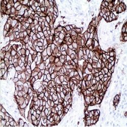
آنتی بادی C-erbB-2/HER2/NEU (SP3)
اطلاعات بیشترName: Rabbit Anti-c-erbB2 Monoclonal Antibody clone SP3
Description and applications: C-erbB-2 is a receptor tyrosine kinase of the c-erbB family. The C-erbB-2 is a proto-oncogene located on human chromosome 17, band 21. It has structural similarities to the EGFR (Epidermal Growth Factor Receptor). Immunohistochemical staining of the oncoprotein is related to gene amplification. Different studies have demonstrated immunohistochemical detectable levels in more than 20% of adenocarcinomas of different locations, including ovarian, gastro-intestinal tract and breast. In the case of breast cancer, the overexpression has been shown to be associated with poor prognosis. This antibody is recommended to determine the expression levels of the oncoprotein C-erb B-2 in breast adenocarcinomas and other locations.
Composition: anti-cerbB2 rabbit monoclonal antibody obtained from supernatant culture and prediluted in a tris buffered solution pH 7.4 containing 0.375mM sodium azide solution as bacteriostatic and bactericidal. Intended use : Immunohistochemistry (IHC) on paraffin embedded tissues. Not tested on frozen tissues or Western-Blotting
Immunogen: Recombinant protein encoding extracellular domain of human c-erbB2
Species reactivity: In vitro diagnostics in humans. Not tested in other species
-
 آنتی بادیهای ایمونوهیستوشیمی
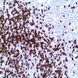
آنتی بادی ZAP-70 (2F3.2)
اطلاعات بیشتر(Name: ZAP-70 Antibody (Clone 2F3.2
Description and applications:ZAP-70 is a 70kDa protein tyrosine kinase found in T-cells and natural killer cells. Control of this protein translation is via the IgVH gene. In Western blotting of whole cell lysates of normal peripheral blood mononuclear cells, the antibody labels a band corresponding to ZAP-70. In Western blotting of whole cell lysates of CD19- positive purified leukemia cells from patients with lgunmutated and lg-mutated CLL, the antibody labels a band corresponding to ZAP-70 in the Ig-unmutated CLL samples, whereas no band is observed in the Igmutated CLL samples. In Western blotting of cell lysates of Jurkat cells (T-lymphoblastic cell line), the antibody labels a band of 70kDa protein. In Western blotting of cell lysates of A431 cells (carcinoma cell line), no band is observed. ZAP-70 protein is expressed in leukemic cells of approximately 25% ofchronic lymphocytic leukemia (CLL) cases as well. Anti-ZAP-70 expression is an excellent surrogate marker for the distinction between the g-mutated (anti-ZAP-70 negative) and Ig-unmutated (anti-ZAP-70 positive) CLL subtypes and can identify patient groups with divergent clinical courses. The anti-ZAP-70 positive Ig-unmutated CLL cases have been shown to have a poorer prognosis.
Composition: Anti-human ZAP-70 mouse monoclonal antibody purified from serum and prepared in 10mM PBS, pH 7.4, with 0.2% BSA and 0.09% sodium azide.
Immunogen: Recombinant ZAP-70 protein including residues 1-254 and encompassing SH2 domains of human ZAP-70.
-
 آنتی بادیهای ایمونوهیستوشیمی
آنتی بادی WT1 (Wilms Tumor) (6F-H2)
اطلاعات بیشتر(Name: WT-1 (Wilm´s Tumour Protein 1)Antibody (Clone 6F-H2
Description and aplications: WT-1 gene is located on chromosome 11p13 and is involved in the development of Wilm´s tumor (WT). WT is associated with mutations of WT1, a zinc-finger transcription factor that is essential for the development of the metanephric kidney and the urogenital system. The WT1 gene is normally expressed in fetal kidney and mesothelium, and its expression has been suggested as a marker for Wilms tumor and mesothelioma. Anti-WT1 is useful in the diagnosis of malignant mesotheliomas (nuclei) while the nuclei of lung adenocarcinomas are not labeled with this product. However, cytoplasmic labeling of lung adenocarcinomas
may be observed. Anti-WT1 labels 93% of serousovarian carcinomas and 0% (nuclei) of mucinous carcinomas of the ovary and pancreato-biliary carcinomas. Anti-WT1 can also be utilized in the differential diagnosis of small round cell tumors as 100% of desmoplastic small round cell tumors and 70% of nephroblastomas (Wilm’s tumor) are positive (nuclei) for this marker. Tumors such as Ewings sarcoma/PNET, neuroblastomas, rhabdomyosarcomas, and rhabdoid tumors are negative. Occasionally cytoplasmic labeling may be seen in these tumors, however.Composition: anti-human WT-1 mouse monoclonal antibody obtained from tissue culture supernatant prepared in 10mM PBS, pH 7.4, with 0.2% BSA and 0.09% sodium azide.
Immunogen: Truncated human WT1 protein corresponding to aa 1-181.
-
 آنتی بادیهای ایمونوهیستوشیمی
آنتی بادی VIP (H-6)
اطلاعات بیشتر.(Name: VIPAntibody(Clone H-6
Description and applications: Glucagon is a pancreatic hormone that functions as an antagonist to Insulin, stimulating the conversion of glycogen to glucose and increasing blood sugar levels. Glucagonlike peptide-1 (GLP-1), glucagon-like peptide-2 (GLP- 2), VIP (vasoactive intestinal peptide) and PACAP (pituitary adenylate cyclase activating polypeptide) are members of the glucagon family of hormones. GLP-1 functions as a transmitter in the central nervous system, inhibiting feeding and drinking behavior, whereas GLP-2 is a stimulator of intestinal epithelial growth. VIP causes vasodilation resulting in the lowering of blood pressure. PACAP is abundant in the hypothalamus and has been shown to increasethe synthesis of several hormones, including growth hormone.
Composition: Anti-human VIP mouse monoclonal antibody purified from serum and prepared in PBS with < 0.1% sodium azide and 0.1% gelatin.
Immunogen: Antibody raised against amino acids 1-95 of vasoactive intestinal peptide VIP of human origin.
-
 آنتی بادیهای ایمونوهیستوشیمی
آنتی بادی Vimentin (SP20)
اطلاعات بیشتر.(Name: Vimentin Antibody (Clone SP20
Description and applications: Vimentin is the main intermediate filament protein in mesenchymal cells and is therefore of value in the differential diagnosis of undifferentiated neoplasms. In mesenchymal tumors, vimentin expression is practically the rule, though, it must not forget that there are epithelial tumors that coexpressed Vimentin and cytokeratins, such as thyroid gland carcinomas, pleomorphic adenomas salivary of the salivary glands and some renal carcinomas. Furthermore, by the universal staining the anti-vimentin antibody in the blood vessels and lymphoid tissue, they are considered the best internal control to assess the results of immunostaining.
Composition: anti-human Vimentin, rabbit monoclonal antibody purified from culture supernatant, prepared in 10mM PBS, pH 7.4, with 0.2% BSA and 0.09% sodium azide.
Immunogen: Recombinant protein encoding human vimentin.
-
 آنتی بادیهای ایمونوهیستوشیمی
آنتی بادی Villin-1 (CWWB1)
اطلاعات بیشتر.(Name: Villin-1 Antibody (Clone CWWB1
Description and applications:Villin is a tissue-specific protein with a molecular massAnti-villin monoclonal antibody (CWWB1 clone) against the human antigen has a sensitivity of almost 100% and high specificity; thus, immunostaining is fully reproducible using standard amplification and development procedures performed as part of automated immunostaining systems. Villin is expressed in the brush border of enterocytes and kidney proximal tubule cells. In addition to allowing immunohistochemical identification of villin in normal human tissues, this antibody is a highly specific marker of gastrointestinal tumours and pancreatic cancer. Although with less sensitivity, renal, hepatic and pulmonary neoplasms, especially carcinoid tumours, neuroendocrine tumours and signet-ring cell carcinomas or carcinomas with microlumens filled with microvilli, as well as some breast and ovarian tumours, are also stained. of 95.5 kDa. Of all proteins belonging to the gelsolin family, it is the only one capable of assembling and disassembling actin filaments through three specific binding domains, two of them Ca2+-dependent. Villin is essential for the assembling of the cell brush border citoskeleton and the organization of the actin filament bundle forming the core of microvilli. In addition, it has been proposed that this protein, due to its dependence on cell activation via phosphatidylinositol, is essential for the control of cell polarity during embryonic development of tissues containing it, as well as to facilitate epithelial plasticity in response to cell injury and immune system cell elements migration through the intestinal barrier.
Composition:Anti-human Villin-1 mouse monoclonal antibody purified from serum and prepared in 10mM PBS, pH 7.4, with 0.2% BSA and 0.09% sodium azide.
Immunogen:uman villin protein.
-
 آنتی بادیهای ایمونوهیستوشیمی
آنتی بادی VEGF (Vascular Endothelial Growth Factor)(EP1176Y)
اطلاعات بیشتر.(Name: VEGF (Vascular Endothelial Growth Factor)Antibody (Clone EP1176Y
Description and applications: Growth factor active in angiogenesis, vasculogenesis and endothelial cell growth. Induces endothelial cell proliferation, promotes cell migration, inhibits apoptosis and induces permeabilization of blood vessels. Binds to the FLT1/VEGFR1 and KDR/VEGFR2 receptors, heparan sulfate and heparin. NRP1/Neuropilin-1 binds isoforms VEGF-165 and VEGF-145. Isoform VEGF165B binds to KDR but does not activate downstream signaling pathways, does not activate angiogenesis and inhibits tumor growth.
Composition: anti-human VEGF Rabbit monoclonal antibody purified from ascites fluid by Protein A chromatography prepared in 10mM PBS, pH 7.4, with 0.2% BSA and 0.09% sodium azide.
.Immunogen: Synthetic peptide corresponding to residues on the C-terminus of human VEGF
-
 آنتی بادیهای ایمونوهیستوشیمی
آنتی بادی Uroplakin III (BC17)
اطلاعات بیشتر.(Name: Uroplakin III Antibody (Clone BC17
Description and aplications: This antibody detects the Uroplakin III, a glycoprotein of 47 kDa that forms the asymmetric part of the membrane plates unit constituting the apical surface of the urothelial cell surface or “umbrella cells”. In non-neoplastic cells the uroplakin III is detected exclusively in the normal urothelium of the bladder, ureter and renal pelvis. The antibody stains the luminal surface of the plasma membrane of the “umbrella cells” and weakly in the cytoplasm of some of these cells and some deeper isolated urothelial cells. In tumors, Uroplakin III is detected in 60% of primary urothelial carcinomas and up to 53% of metastases (sensitivity 0.57). Brenner tumors of the ovary also show immunoreactivity for Uroplakin III, the remaining non-urothelial neoplasms being negative (specificity 1.00). Markers such as Uroplakin III, p63, thrombomodulin and GATA3 are expressed in a high percentage of urothelial carcinomas and are usually negative in renal carcinomas.
Composition: anti-Uroplakin III rabbit monoclonal antibody obtained from supernatant culture and prediluted in a tris buffered solution pH 7.4 containing 0.375mM sodium azide solution as bacteriostatic and bactericidal.
.Immunogen: Synthetic peptide derived from the Cterminus of human Uroplakin III
-
 آنتی بادیهای ایمونوهیستوشیمی
آنتی بادیTyrosinase -کلون T311
اطلاعات بیشتر.(Name: Tyrosinase Antibody (Clone T311
Description and applications:Melanin synthesis by melanocytes comprises a cascade of reactions involving a family of enzymes. One of the key enzymes is tyrosinase. L-tyrosine is the initial substrate for melanin synthesis and its conversion to dopaquinone is catalysed by tyrosinase, the expression of which is therefore a marker of melanocytes and melanoma. Tyrosinase deficiency is associated with various forms of albinism, specially oculocutaneous albinism. This antibody is useful for the identification of melanocytic tumours and it may be used in combination with other markers such as Melan A. Both markers are expressed in 80-100% of melanoma cases.
Composition: Anti-human Tyrosinase mouse monoclonal antibody purified from serum and prepared in 10mM PBS, pH 7.4, with 0.2% BSA and 0.09% sodium azide.
Immunogen: Recombinant protein corresponding to the tyrosinase molecule.
-
![[wpdm_package id=2137 template="link-template-calltoaction3.php"]l](https://samatashkhis.com/wp-content/uploads/2018/07/TTF1-e1577866769820-250x250.jpg) آنتی بادیهای ایمونوهیستوشیمی
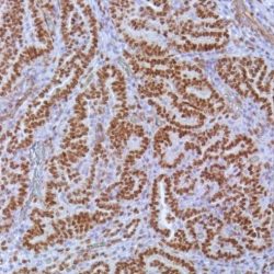
آنتی بادیهای ایمونوهیستوشیمیآنتی بادی TTF-1 (SPT24)
اطلاعات بیشتر.(Name: Thyroid Transcription Factor 1 (TTF-1)(clone SPT24
Description and aplications:This antibody recognizes a protein identified as thyroid transcription factor 1 (TTF-1), a member of the Nkx2 family homeodomain transcription factors. This antibody was designed against a short recombinant peptide and shows great sensitivity and provides a stronger and less cytoplasmic unspecific staining or cross-reaction with tissues other than thyroid and lung. The clone SPT24 identifies the presence of TTF-1 in thyroid follicular epithelium, as well as type II pneumocytes and Clara cells of the lung and their corresponding tumors. It can also be observed in certain brain reactive glial cells and some cases of colon adenocarcinomas. It shows no nuclear staining in a variety of normal tissues and no cytoplasmic reactivity was described in hepatocarcinomas.
Composition: anti- TTF1 mouse monoclonal antibody obtained from supernatant culture and prediluted in a tris buffered solution pH 7.4 containing 0.375mM sodium azide solution as bacteriostatic and bactericidal. The quantity of the active antibody was not determined.
.Immunogen: recombinant protein molecule corresponding to a fragment of 123 AA of the N-terminal region of human TTF-1
-
 آنتی بادیهای ایمونوهیستوشیمی
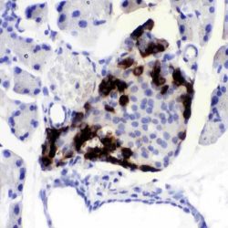
آنتی بادی TSH (Thyroid Stimulating Hormone) (EP254)
اطلاعات بیشتر.(Name: TSH (Thyroid Stimulating Hormone) Antibody(Clone EP254
Description and applications:TSH is a member of the glycoprotein hormone family, constituting a subset of the cystine-knot growth factor superfamily. TSH is produced by the pituitary thyrotrophs and released into circulation in a pulsatile manner. It stimulates thyroid functions using a specific membrane TSH receptor (TSHR) that belongs to the superfamily of G protein-coupled receptors (GPCRs). TSH beta is the beta subunit of thyroid stimulating hormone. This TSH antibody labels normal and neoplastic thyrotropic cells. It may be useful in classification of pituitary tumors.
Composition:Anti-human TSH (Thyroid Stimulating Hormone) rabbit monoclonal antibody purified from serum and prepared in 10mM PBS, pH 7.4, with 0.2% BSA and 0.09% sodium azide.
.Immunogen:A synthetic peptide corresponding to residues of human TSH (subunit beta) protein
-
 آنتی بادیهای ایمونوهیستوشیمی
آنتی بادی Transcriptional Regulator ERG (9FY)
اطلاعات بیشتر.(Name: Transcriptional Regulator ERG (Clone 9FY
Description and applications: TMPRSS2:ERG is the most prevalent gene rearrangement in prostate cancers leading to overexpression of a truncated ERG protein in a subset of prostate cancers. ERG expression is also reported in high-grade prostatic intraepithelial neoplasia (PIN). Publications suggest that the prevalence of the TMPRSS2:ERG rearrangement in prostate cancer cases ranges from ~25% to 70%1,3. ERG is expressed in many tissues including vascular tumors.
Composition: anti-human ERG mouse monoclonal antibody purified from ascites. Prepared in 10mM PBS, pH 7.4, with 0.2% BSA and 0.09% sodium azide.