Name: Proto-Oncogene Tyrosine-Protein Kinase Antibody clone D4D6
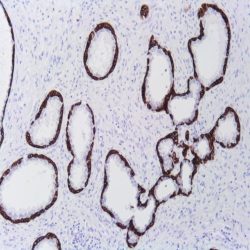
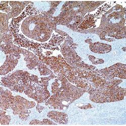
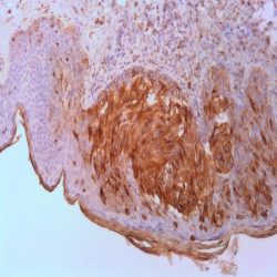
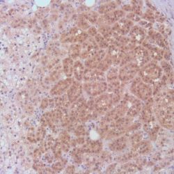
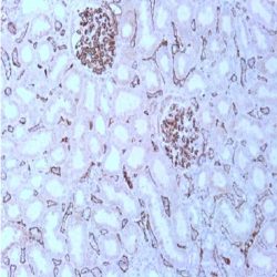
Description and applications: The transmembrane proto-oncogene tyrosine- kinase protein ROS, better known as ROS1, is a protein belonging to the insulin receptors tyrosine-kinase subfamily whose production is encoded by the ROS (MCF3) gene, located in the 6q22.1 chromosome region. After its activation, this gene intervenes in numerous molecular pathways related to the cell differentiation, proliferation, growth and survival including the activation of the PI3K-mTOR pathway. Chromosome aberrations affecting the ROS gene have been described in the glioblastoma multiforme through fusions of the C-terminal portion of ROS1 with the N-terminal domain of the FIG (Fused in Glioblastoma) encoded in the GOPC gene. The chimeric protein GOPC-ROS1 is located at the level of the Golgi apparatus and presents activity as tyrosine kinase receptor. Other fusions of ROS1 with different genes such as SLC34A2, CD74, EZR, LRIG3, SDC4, TPM3, CCDC6 or KDELR2 among others, have as a result the appearance and expression of several chimeric proteins in 1-3 % of cases of lung adenocarcinoma and represents the therapeutic target for Crizotinib and analog molecules. Non-specific staining on macrophages and reactive type II pneumocytes has also been described in isolated cases, whereas mucinous adenocarcinomas in general show low diffuse cytoplasmic staining in the absence of the translocation of the ROS1 gene. In order to identify the cases of lung carcinomas with translocations of ROS1, complex techniques of RT-PCR and FISH can be used, although the rabbit monoclonal antibody D4D6 has been recently validated as an useful screening tool for positive cases of immunostaining against ROS1, which in comparative studies through in situ hybridization techniques with break-apart probes has proven a sensitivity and specificity of over 95%. In order to consider a case as positive for ROS1, the immunostaining has to be strong or moderately intense on the membrane and/or the cytoplasm in more than 75% of tumor cells. Depending on the type of fusion of the ROS1 gene, other immunostaining patterns can be expressed as cytoplasmic punctiform in the CD74-ROS1 cases or cytoplasmic with linear accentuation in the lateral or apical membrane in the EZR-ROS1 cases. Although the morphology of the tumor cannot be considered as a criterion to select the positive ROS1 cases, the solid, micro-papillary, cribriform and signet ring cell growth patterns have been observed more frequently among the positive ROS1 cases. Focal and low-intensity staining can also be observed in up to 30% of the non-translocated cases. Therefore, the result must be confirmed with other analytical methods. 50% of the inflammatory myofibroblastic tumors also present translocation of the ALK gene, whereas the remaining ones show a little known genetic profile. It has been recently proven in a relatively low number of cases that up to 10% of these tumors show diffuse cytoplasmic or punctiform staining against the D4D6 clone (with translocation confirmed through FISH and RT-PCR techniques). In this same study, the antibody proved weak, focak nuclear staining in cases of gastrointestinal stromal tumors, myofibroblastic sarcomas, leiomyosarcomas and follicular dendritic cell sarcomas.Less than 1% of colorectal adenocarcinomas and up to 5% of stomach adenocarcinomas can show translocations of the ROS1 gene and consequential cytoplasmic staining. The low percentage of colorectal adenocarcinomas makes the gene as a potential therapeutic target, but in the case of the stomach adenocarcinomas, it can represent a therapeutic alternative, since, in general, the positive ROS1 cases show a non-amplified phenotype of HER2 and MET.
Composition: Anti-human ROS1 rabbit monoclonal antibody purified from serum and prepared in 10mM PBS, pH 7.4, with 0.2% BSA and 0.09% sodium azide
Immunogen: Human colon carcinoma extract.
Species reactivity: In vitro diagnostics in humans. Not tested in other species

Reviews
There are no reviews yet.