Name: S100-A1 Protein Antibody clone EP184
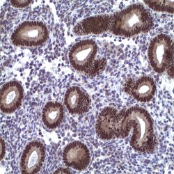
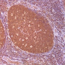
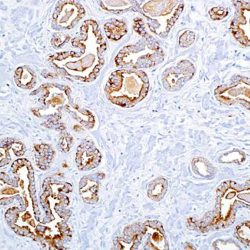
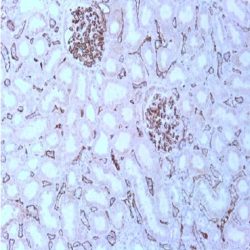
Description and applications: The S100-A1 protein belongs to the S100 protein family, a set of 25 small acidic molecules with a molecular mass of 10-12 kDa that are functionally grouped into homo or heterodimers composed of two subunits, alpha and beta, with extensive sequence homology and capable of combining with each other. There exist up to 14 variants of the alpha chain gene, which is located on chromosome 1q21; on the contrary, there is only one sequence of the beta chain gene, located on chromosome 21q22.3, showing its greater functional specificity. Depending on the combination between the alpha and beta single chains and the homology of their sequences, there exist three forms of S-100 protein: A, with at least 15 subtypes between A1 and A15, B, and G. In their dimeric form, S100 proteins, together with calmodulin and troponin C, belong to the calcium binding protein family; their affinity for this ion, as well as for other metals such as zinc, is remarkable. It is for this reason that the S100 protein is involved in numerous basic cellular activities such as cation diffusion across lipid membranes, microtubule assembly, and regulation of RNA polymerase activity. In neurons, the S100 protein also regulates the interaction between chromosomes and synaptosomes. As for the S100-A1 protein, its most important role is to regulate myocardial contractility. In normal tissues, S100-A1 protein expression is observed at the sarcolemma of the cardiac and the skeletal muscle, in a smaller proportion in the latter; likewise, this protein is found in renal tubule and brain cells. It is not expressed in other tissues. In neoplasms, S100-A1 protein expression has been investigated predominantly in renal tumour pathology, where it is important for the diagnosis of oncocytic tumours, both oncocytomas and oncocytic papillary carcinomas, and allows the differential diagnosis with chromophobe carcinomas, especially its eosinophilic variant, which does not express this protein. For this reason, the S100-A1 protein, together with cytokeratin 7, vimentin, and c-kit (CD117) has been included in the optimal panel for the diagnosis of renal oncocytomas. Additionally, and compared to normal urothelium, which may exhibit very slight staining, nephrogenic adenomas show intense cytoplasmic and/or nuclear/cytoplasmic staining. For all of the above reasons, the S100-A1 protein, together with PAX8, p63, PSA, and CEA, is a highly sensitive and specific marker for the diagnosis of nephrogenic adenomas, making it a useful marker in the differential diagnosis of male urinary and genital system tumours, including prostatic adenocarcinomas, which are regularly negative for this protein. The S100-A1 protein is also useful in the differential diagnosis between dysplastic melanocytic nevi and malignant melanoma, with slight staining in the former and more intense staining in the latter, in contrast to the S100-B protein, which tends to
present greater expression in nevus lesions, except in areas of melanoma with high proliferation. Finally, in both endometrial and ovarian endometrioid carcinomas, S100-A1 protein overexpression is an
indicator of a poorer prognosi .
Composition: Anti-human S100-A1 Protein rabbit monoclonal antibody purified from serum and prepared in 10mM PBS, pH 7.4, with 0.2% BSA and 0.09% sodium azide
Intended use: Immunohistochemistry (IHC) on paraffin embedded tissues. Not tested on frozen tissues or Western-Blotting
Clone: EP184
Ig isotype: IgG
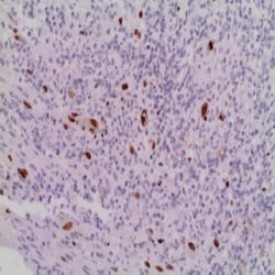
IHC positive control: Myocardium, skeletal muscle, or renal cell carcinoma tissue section.
Visualization: Cytoplasm.

Reviews
There are no reviews yet.