-
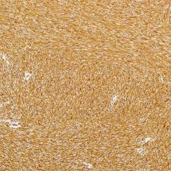
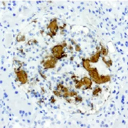
 آنتی بادیهای ایمونوهیستوشیمی
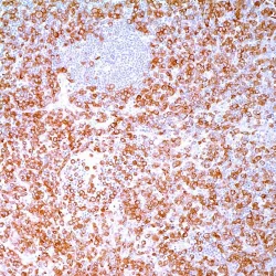
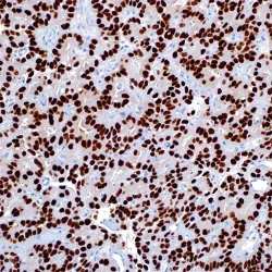
آنتی بادی Cdk4 کلون CDK4/7987R برند Vitro
Name: Cdk4 Monoclonal Antibody clone CDK4/7987R
DESCRIPTION AND APPLICATIONS: Cell cycle progression is controlled in part by a family of cyclin proteins and cyclin dependent kinases (Cdks). Cdk proteins work in concert with the cyclins to phosphorylate key substrates involved in each phase of cell cycle progression. Another family of proteins, Cdk inhibitors, also plays a role in regulating the cell cycle by binding to cyclin Cdk complexes and modulating their activity. Several Cdk proteins have been identified, including Cdk2 Cdk8, PCTAIRE-1-PCTAIRE-3, PITALRE and PITSLRE. Cdk4, in complex with D-type cyclins, is thought to regulate cell growth during the G1 phase of the cell cycle. This association with a D-type cyclin upregulates Cdk4 activity, whereas binding to the Cdk inhibitor p16 downregulates Cdk4 activity. Activation of the Cdk4-cyclin complexes requires phosphorylation on a single threonyl residue of Cdk4, catalyzed by a Cdk-activating protein (CAK).
COMPOSITION: Anti-human CDK4 rabbit monoclonal antibody diluted in a pH 7.6 buffer containing stabilizing protein and sodium azide as bacteriostatic and bactericidal agent.
INTENDED USE: Immunohistochemistry (IHC) on paraffin embedded tissues. Not tested on frozen tissues or Western-Blotting
IMMUNOGEN: Recombinant fragment corresponding to N-terminal of human CDK4 protein
SPECIES REACTIVITY: In vitro diagnostics in humans. Not tested in other species
-
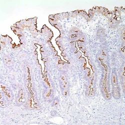
 آنتی بادیهای ایمونوهیستوشیمی
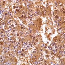
آنتی بادی WT1 (Wilms Tumor) کلون 6F-H2 برند PathoSage
Intended use:
This antibody is intended for in vitro diagnostic (IVD) use. Primary Antibody is intended for professional laboratory use in formalin-fixed, paraffin-embedded (FFPE) tissue stained in manual qualitative immunohistochemistry (IHC) testing. A qualified pathologist must interpret the results using this product to aid diagnosis in conjunction with the patient’s relevant clinical history, other diagnostic tests, and proper controls.
Presentation:
supernatant prepared in 10mM PBS, pH 7.4, with %0.2 BSA and %0.09 sodium azide Anti-human WT-1 mouse monoclonal antibody obtained from tissue culture
-
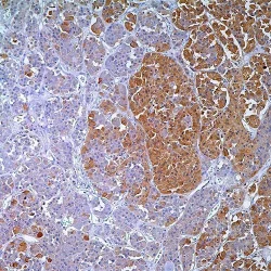
 آنتی بادیهای ایمونوهیستوشیمی
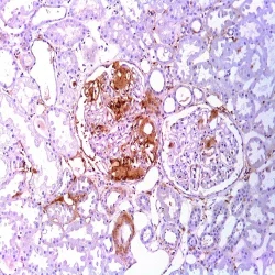
آنتی بادی VIP کلون H-6 برند PathoSage
Intended use:
This antibody is intended for in vitro diagnostic (IVD) use. Primary Antibody is intended for professional laboratory use in formalin-fixed, paraffin-embedded (FFPE) tissue stained in manual qualitative immunohistochemistry (IHC) testing. A qualified pathologist must interpret the results using this product to aid diagnosis in conjunction with the patient’s relevant clinical history, other diagnostic tests, and proper controls.
Presentation:
Anti-human VIP mouse monoclonal antibody purified from serum and prepared in PBS with < %0.1 sodium azide and %0.1 gelatin
-
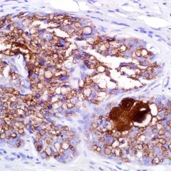
 آنتی بادیهای ایمونوهیستوشیمی
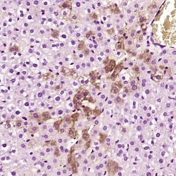
آنتی بادی Vimentin کلون SP20 برند PathoSage
Intended use:
This antibody is intended for in vitro diagnostic (IVD) use. Primary Antibody is intended for professional laboratory use in formalin-fixed, paraffin-embedded (FFPE) tissue stained in manual qualitative immunohistochemistry (IHC) testing. A qualified pathologist must interpret the results using this product to aid diagnosis in conjunction with the patient’s relevant clinical history, other diagnostic tests and proper controls.
Presentation:
Anti-human Vimentin rabbit monoclonal antibody purified from serum and prepare din 10mM PBS, pH 7.4, with %0.2 BSA and %0.09 sodium azide
-
 آنتی بادیهای ایمونوهیستوشیمی
آنتی بادی Villin 1 کلون CWWB1 برند PathoSage
Intended use:
This antibody is intended for in vitro diagnostic (IVD) use. Primary Antibody is intended for professional laboratory use in formalin-fixed, paraffin-embedded (FFPE) tissue stained in manual qualitative immunohistochemistry (IHC) testing. A qualified pathologist must interpret the results using this product to aid diagnosis in conjunction with the patient’s relevant clinical history, other diagnostic tests, and proper controls.
Presentation:
Anti-human Villin-1 mouse monoclonal antibody purified from serum and prepared in 10mM PBS, pH 7.4, with %0.2 BSA and %0.09 sodium azide
-
 آنتی بادیهای ایمونوهیستوشیمی
آنتی بادی VEGF (Vascular Endothelial Growth Factor) کلون EP1176Y برند PathoSage
Intended use:
This antibody is intended for in vitro diagnostic (IVD) use. Primary Antibody is intended for professional laboratory use in formalin fixed, paraffin-embedded (FFPE) tissue stained in manual qualitative immunohistochemistry (IHC) testing. A qualified pathologist must interpret the results using this product to aid diagnosis in conjunction with the patient’s relevant clinical history, other diagnostic tests, and proper controls.
Presentation:
Anti-human VEGF Rabbit monoclonal antibody purified from ascites fluid by Protein A chromatography prepared in 10mM PBS, pH 7.4, with %0.2 BSA and %0.09 sodium azide
-
آنتی بادیهای ایمونوهیستوشیمی
آنتی بادی Uroplakin 3 کلون BC17 برند PathoSage
Intended use:
This antibody is intended for in vitro diagnostic (IVD) use. Primary Antibody is intended for professional laboratory use in formalin-fixed, paraffin-embedded (FFPE) tissue stained in manual qualitative immunohistochemistry (IHC) testing. A qualified pathologist must interpret the results using this product to aid diagnosis in conjunction with the patient’s relevant clinical history, other diagnostic tests, and proper controls.
Presentation:
Anti-human Uroplakin III mouse monoclonal antibody purified from serum and prepared in 10mM PBS, pH 7.4, with %0.2 BSA and %0.09 sodium azide
-
 آنتی بادیهای ایمونوهیستوشیمی
آنتی بادی Uroplakin 2 کلون BC21 برند PathoSage
Intended use:
This antibody is intended for in vitro diagnostic (IVD) use. Primary Antibody is intended for professional laboratory use in formalin-fixed, paraffin-embedded (FFPE) tissue stained in manual qualitative immunohistochemistry (IHC) testing. A qualified pathologist must interpret the results using this product to aid diagnosis in conjunction with the patient’s relevant clinical history, other diagnostic tests, and proper controls.
Presentation:
Anti-Uroplakin 3 mouse monoclonal antibody obtained from supernatant culture and prediluted in a tris buffered solution pH 7.4 containing 0.375mM sodium azide solution as bacteriostatic and bactericidal
-
 آنتی بادیهای ایمونوهیستوشیمی
آنتی بادی TSH (Thyroid Stimulating Hormone) کلون EP254 برند PathoSage
Intended use:
This antibody is intended for in vitro diagnostic (IVD) use. Primary Antibody is intended for professional laboratory use in formalin-fixed, paraffin-embedded (FFPE) tissue stained in manual qualitative immunohistochemistry (IHC) testing. A qualified pathologist must interpret the results using this product to aid diagnosis in conjunction with the patient’s relevant clinical history, other diagnostic tests, and proper controls.
Presentation:
Anti-human TSH (Thyroid Stimulating Hormone) rabbit monoclonal antibody purified from serum and prepared in 10mM PBS, pH 7.4, with %0.2 BSA and %0.09 sodium azide
-
 PSV
PSVآنتی بادی ACTH(Adrenocorticotropic Hormone) کلون Polyclonal برند PathoSage
Intended use:
This antibody is intended for in vitro diagnostic (IVD) use. Primary Antibody is intended for professional laboratory use in formalin-fixed, paraffin-embedded (FFPE) tissue stained in manual qualitative immunohistochemistry (IHC) testing. A qualified pathologist must interpret the results using this product to aid diagnosis in conjunction with the patient’s relevant clinical history, other diagnostic tests, and proper controls.Presentation:
Anti-human ACTH rabbit polyclonal antibody purified from serum and prepared in 10mM PBS, pH 7.4, with 0.2% BSA and 0.09% sodium azide -
 PSV
PSVآنتی بادی Adipophilin کلون Polyclonal برند PathoSage
Intended use:
This antibody is intended for in vitro diagnostic (IVD) use. Primary Antibody is intended for professional laboratory use in formalin-fixed, paraffin-embedded (FFPE) tissue stained in manual qualitative immunohistochemistry (IHC) testing. A qualified pathologist must interpret the results using this product to aid diagnosis in conjunction with the patient’s relevant clinical history, other diagnostic tests, and proper controls.Presentation:
Anti-human Adipophilin rabbit polyclonal antibody purified from serum and prepared in 10mM PBS, pH 7.4, with 0.2% BSA and 0.09% sodium azide -
 PSV
PSVآنتی بادی ALK/p80 کلون 5A4 برند PathoSage
Intended use:
This antibody is intended for in vitro diagnostic (IVD) use. Primary Antibody is intended for professional laboratory use in formalin-fixed, paraffin-embedded (FFPE) tissue stained in manual qualitative immunohistochemistry (IHC) testing. A qualified pathologist must interpret the results using this product to aid diagnosis in conjunction with the patient’s relevant clinical history, other diagnostic tests, and proper controls.Presentation:
Anti-human ALK/p80 mouse monoclonal antibody purified from serum and prepared in 10mM PBS, pH 7.4, with 0.2% BSA and 0.09% sodium azide -
 PSV
PSVآنتی بادی Alpha Fetoprotein کلون EP209 برند PathoSage
Intended use:
This antibody is intended for in vitro diagnostic (IVD) use. Primary Antibody is intended for professional laboratory use in formalin-fixed, paraffin-embedded (FFPE) tissue stained in manual qualitative immunohistochemistry (IHC) testing. A qualified pathologist must interpret the results using this product to aid diagnosis in conjunction with the patient’s relevant clinical history, other diagnostic tests, and proper controls.Presentation:
Anti-human AFP rabbit monoclonal antibody purified from serum and prepared in 10mM PBS, pH 7.4, with 0.2% BSA and 0.09% sodium azide -
 PSV
PSVآنتی بادی Amyloid P کلون EP1018Y برند PathoSage
Intended use:
This antibody is intended for in vitro diagnostic (IVD) use. Primary Antibody is intended for professional laboratory use in formalin-fixed, paraffin-embedded (FFPE) tissue stained in manual qualitative immunohistochemistry (IHC) testing. A qualified pathologist must interpret the results using this product to aid diagnosis in conjunction with the patient’s relevant clinical history, other diagnostic tests, and proper controls.Presentation:
Anti-human Amyloid P rabbit monoclonal antibody purified from serum and prepared in 10mM PBS, pH 7.4, with 0.2% BSA and 0.09% sodium azide -
 PSV
PSVآنتی بادی Alpha1 Antichymotrypsin کلون Polyclonal برند PathoSage
Intended use:
This antibody is intended for in vitro diagnostic (IVD) use. Primary Antibody is intended for professional laboratory use in formalin-fixed, paraffin-embedded (FFPE) tissue stained in manual qualitative immunohistochemistry (IHC) testing. A qualified pathologist must interpret the results using this product to aid diagnosis in conjunction with the patient’s relevant clinical history, other diagnostic tests, and proper controls.Presentation:
Anti-human Alpha-1-Antichymotrypsin rabbit polyclonal antibody purified from serum and prepared in 10mM PBS, pH 7.4, with 0.2% BSA and 0.09% sodium azide -
 PSV
PSVآنتی بادی Amyloid A کلون MC1 برند PathoSage
Intended use:
This antibody is intended for in vitro diagnostic (IVD) use. Primary Antibody is intended for professional laboratory use in formalin-fixed, paraffin-embedded (FFPE) tissue stained in manual qualitative immunohistochemistry (IHC) testing. A qualified pathologist must interpret the results using this product to aid diagnosis in conjunction with the patient’s relevant clinical history, other diagnostic tests, and proper controls.Presentation:
Anti-human Amyloid-A rabbit monoclonal antibody purified from Bioreactor Concentrate by Protein A/G and diluted in a pH7.6 buffer containing stabilizing protein and sodium azide as bacteriostatic and bactericidal agent -
 PSV
PSVآنتی بادی Anexin A1 کلون 29 برند PathoSage
Intended use:
This antibody is intended for in vitro diagnostic (IVD) use. Primary Antibody is intended for professional laboratory use in formalin-fixed, paraffin-embedded (FFPE) tissue stained in manual qualitative immunohistochemistry (IHC) testing. A qualified pathologist must interpret the results using this product to aid diagnosis in conjunction with the patient’s relevant clinical history, other diagnostic tests, and proper controls.Presentation:
Anti-human Annexin 1 mouse monoclonal antibody purified from serum and prepared in 10mM PBS, pH 7.4, with 0.2% BSA and 0.09% sodium azide -
 PSV
PSVآنتی بادی ARG1 کلون EP261 برند PathoSage
Intended use:
This antibody is intended for in vitro diagnostic (IVD) use. Primary Antibody is intended for professional laboratory use in formalin-fixed, paraffin-embedded (FFPE) tissue stained in manual qualitative immunohistochemistry (IHC) testing. A qualified pathologist must interpret the results using this product to aid diagnosis in conjunction with the patient’s relevant clinical history, other diagnostic tests, and proper controls.Presentation:
Anti-human ARG-1 rabbit monoclonal antibody purified from serum and prepared in 10mM PBS, pH 7.4, with 0.2% BSA and 0.09% sodium azide -
 PSV
PSVآنتی بادی BCL2 کلون EP36 برند PathoSage
Intended use:
This antibody is intended for in vitro diagnostic (IVD) use. Primary Antibody is intended for professional laboratory use in formalin-fixed, paraffin-embedded (FFPE) tissue stained in manual qualitative immunohistochemistry (IHC) testing. A qualified pathologist must interpret the results using this product to aid diagnosis in conjunction with the patient’s relevant clinical history, other diagnostic tests, and proper controls.Presentation:
Anti-human Bcl-2 rabbit monoclonal antibody purified from serum and prepared in 10mM PBS, pH 7.4, with 0.2% BSA and 0.09% sodium azide -
 PSV
PSVآنتی بادی AR(Androgen Receptor) کلون SP107 برند PathoSage
Intended use:
This antibody is intended for in vitro diagnostic (IVD) use. Primary Antibody is intended for professional laboratory use in formalin-fixed, paraffin-embedded (FFPE) tissue stained in manual qualitative immunohistochemistry (IHC) testing. A qualified pathologist must interpret the results using this product to aid diagnosis in conjunction with the patient’s relevant clinical history, other diagnostic tests, and proper controls.Presentation:
Anti-human Androgen Receptor (AR) rabbit monoclonal antibody purified from serum and prepared in 10mM PBS, pH 7.4, with 0.2% BSA and 0.09% sodium azide
برای تماس کلیک کنید
سما تشخیص آریا ، عرضه کننده متنوع ترین و کامل ترین محصولات کیت های آزمایشگاهی