-
 PSV
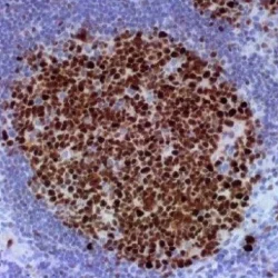
PSVآنتی بادی BCL6 کلون LN22 برند PathoSage
Intended use:
This antibody is intended for in vitro diagnostic (IVD) use. Primary Antibody is intended for professional laboratory use in formalin-fixed, paraffin-embedded (FFPE) tissue stained in manual qualitative immunohistochemistry (IHC) testing. A qualified pathologist must interpret the results using this product to aid diagnosis in conjunction with the patient’s relevant clinical history, other diagnostic tests, and proper controls.Presentation:
Anti-human Bcl-6 mouse monoclonal antibody purified from serum and prepared in 10mM PBS, pH 7.4, with 0.2% BSA and 0.09% sodium azide -
 PSV
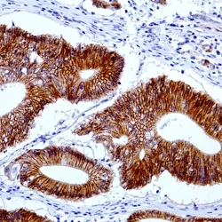
PSVآنتی بادی Beta Catenin 1 کلون EP35 برند PathoSage
Intended use:
This antibody is intended for in vitro diagnostic (IVD) use. Primary Antibody is intended for professional laboratory use in formalin-fixed, paraffin-embedded (FFPE) tissue stained in manual qualitative immunohistochemistry (IHC) testing. A qualified pathologist must interpret the results using this product to aid diagnosis in conjunction with the patient’s relevant clinical history, other diagnostic tests, and proper controls.Presentation:
Anti-human Beta-catenin rabbit monoclonal antibody purified and prepared in 10mM PBS, pH 7.4, with 0.2% BSA and 0.09% sodium azide -
 PSV
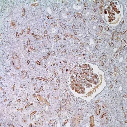
PSVآنتی بادی C3d کلون Polyclonal برند PathoSage
Intended use:
This antibody is intended for in vitro diagnostic (IVD) use. Primary Antibody is intended for professional laboratory use in formalin-fixed, paraffin-embedded (FFPE) tissue stained in manual qualitative immunohistochemistry (IHC) testing. A qualified pathologist must interpret the results using this product to aid diagnosis in conjunction with the patient’s relevant clinical history, other diagnostic tests, and proper controls.Presentation:
Anti-human C3d Complement Component rabbit polyclonal antibody purified from serum and prepared in 10mM PBS, pH 7.4, with 0.2% BSA and 0.09% sodium azide -
 PSV
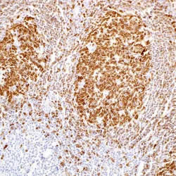
PSVآنتی بادی BOB1 کلون SP92 برند PathoSage
Intended use:
This antibody is intended for in vitro diagnostic (IVD) use. Primary Antibody is intended for professional laboratory use in formalin-fixed, paraffin-embedded (FFPE) tissue stained in manual qualitative immunohistochemistry (IHC) testing. A qualified pathologist must interpret the results using this product to aid diagnosis in conjunction with the patient’s relevant clinical history, other diagnostic tests, and proper controls.Presentation:
Anti-human BOB-1 rabbit monoclonal antibody purified from serum and prepared in 10mM PBS, pH 7.4, with 0.2% BSA and 0.09% sodium azide -
 PSV
PSVآنتی بادی BRST-2(GCDFP-15) کلون EP95 برند PathoSage
Intended use:
This antibody is intended for in vitro diagnostic (IVD) use. Primary Antibody is intended for professional laboratory use in formalin-fixed, paraffin-embedded (FFPE) tissue stained in manual qualitative immunohistochemistry (IHC) testing. A qualified pathologist must interpret the results using this product to aid diagnosis in conjunction with the patient’s relevant clinical history, other diagnostic tests, and proper controls.Presentation:
Anti-human BRST-2/GCDFP-15 rabbit monoclonal antibody purified from serum and prepared in 10mM PBS, pH 7.4, with 0.2% BSA and 0.09% sodium azide -
 PSV
PSVآنتی بادی Cadherin 17 کلون SP183 برند PathoSage
Intended use:
This antibody is intended for in vitro diagnostic (IVD) use. Primary Antibody is intended for professional laboratory use in formalin-fixed, paraffin-embedded (FFPE) tissue stained in manual qualitative immunohistochemistry (IHC) testing. A qualified pathologist must interpret the results using this product to aid diagnosis in conjunction with the patient’s relevant clinical history, other diagnostic tests, and proper controls.Presentation:
Anti-human Cadherin 17 rabbit monoclonal antibody purified from serum and prepared in 10mM PBS, pH 7.4, with 0.2% BSA and 0.09% sodium azide -
 PSV
PSVآنتی بادی C4d کلون Polyclonal برند PathoSage
Intended use:
This antibody is intended for in vitro diagnostic (IVD) use. Primary Antibody is intended for professional laboratory use in formalin-fixed, paraffin-embedded (FFPE) tissue stained in manual qualitative immunohistochemistry (IHC) testing. A qualified pathologist must interpret the results using this product to aid diagnosis in conjunction with the patient’s relevant clinical history, other diagnostic tests, and proper controls.Presentation:
Anti-human C4d rabbit polyclonal antibody purified from serum and prepared in 10mM PBS, pH 7.4, with 0.2% BSA and 0.09% sodium azide -
 PSV
PSVآنتی بادی BRG1 کلون EPNCIR111A برند PathoSage
Intended use:
This antibody is intended for in vitro diagnostic (IVD) use. Primary Antibody is intended for professional laboratory use in formalin-fixed, paraffin-embedded (FFPE) tissue stained in manual qualitative immunohistochemistry (IHC) testing. A qualified pathologist must interpret the results using this product to aid diagnosis in conjunction with the patient’s relevant clinical history, other diagnostic tests, and proper controls.Presentation:
Anti-human BRG1 rabbit monoclonal antibody
purified from serum and prepared in 10mM PBS, pH
7.4, with 0.2% BSA and 0.09% sodium azide -
 PSV
PSVآنتی بادی Carbonic Anhydrase 9 کلون EP161 برند PathoSage
Intended use:
This antibody is intended for in vitro diagnostic (IVD) use. Primary Antibody is intended for professional laboratory use in formalin-fixed, paraffin-embedded (FFPE) tissue stained in manual qualitative immunohistochemistry (IHC) testing. A qualified pathologist must interpret the results using this product to aid diagnosis in conjunction with the patient’s relevant clinical history, other diagnostic tests, and proper controls.Presentation:
Anti-human Carbonic Anhydrase 9 rabbit monoclonal antibody purified from serum and prepared in 10mM PBS, pH 7.4, with 0.2% BSA and 0.09% sodium azide -
 PSV
PSVآنتی بادی Cadherin E کلون HECD-1 برند PathoSage
Intended use:
This antibody is intended for in vitro diagnostic (IVD) use. Primary Antibody is intended for professional laboratory use in formalin-fixed, paraffin-embedded (FFPE) tissue stained in manual qualitative immunohistochemistry (IHC) testing. A qualified pathologist must interpret the results using this product to aid diagnosis in conjunction with the patient’s relevant clinical history, other diagnostic tests, and proper controls.Presentation:
Anti-human Cadherin E mouse monoclonal antibody purified from serum and prepared in 10mM PBS, pH 7.4, with 0.2% BSA and 0.09% sodium azide -
 PSV
PSVآنتی بادی Calponin کلون EP63 برند PathoSage
Intended use:
This antibody is intended for in vitro diagnostic (IVD) use. Primary Antibody is intended for professional laboratory use in formalin-fixed, paraffin-embedded (FFPE) tissue stained in manual qualitative immunohistochemistry (IHC) testing. A qualified pathologist must interpret the results using this product to aid diagnosis in conjunction with the patient’s relevant clinical history, other diagnostic tests, and proper controls.Presentation:
Anti-human Calponin rabbit monoclonal antibody purified from serum and prepared in 10mM PBS, pH 7.4, with 0.2% BSA and 0.09% sodium azide -
 PSV
PSVآنتی بادی CD117(c-kit) کلون EP10 برند PathoSage
Intended use:
This antibody is intended for in vitro diagnostic (IVD) use. Primary Antibody is intended for professional laboratory use in formalin-fixed, paraffin-embedded (FFPE) tissue stained in manual qualitative immunohistochemistry (IHC) testing. A qualified pathologist must interpret the results using this product to aid diagnosis in conjunction with the patient’s relevant clinical history, other diagnostic tests, and proper controls.Presentation:
Anti-human CD117/c-kit rabbit monoclonal antibody purified from serum and prepared in 10mM PBS, pH 7.4, with 0.2% BSA and 0.09% sodium azide -
 PSV
PSVآنتی بادی Cathepsin K کلون 3F9 برند PathoSage
Intended use:
This antibody is intended for in vitro diagnostic (IVD) use. Primary Antibody is intended for professional laboratory use in formalin-fixed, paraffin-embedded (FFPE) tissue stained in manual qualitative immunohistochemistry (IHC) testing. A qualified pathologist must interpret the results using this product to aid diagnosis in conjunction with the patient’s relevant clinical history, other diagnostic tests, and proper controls.Presentation:
Anti-human Cathepsin K mouse monoclonal antibody purified from serum and prepared in 10mM PBS, pH 7.4, with 0.2% BSA and 0.09% sodium azide -
 PSV
PSVآنتی بادی Calcitonin کلون SP17 برند PathoSage
Intended use:
This antibody is intended for in vitro diagnostic (IVD) use. Primary Antibody is intended for professional laboratory use in formalin-fixed, paraffin-embedded (FFPE) tissue stained in manual qualitative immunohistochemistry (IHC) testing. A qualified pathologist must interpret the results using this product to aid diagnosis in conjunction with the patient’s relevant clinical history, other diagnostic tests, and proper controls.Presentation:
Anti-human Calcitonin rabbit monoclonal antibody purified from serum and prepared in 10mM PBS, pH 7.4, with 0.2% BSA and 0.09% sodium azide -
 PSV
PSVآنتی بادی Calretinin کلون BSR235 برند PathoSage
Intended use:
This antibody is intended for in vitro diagnostic (IVD) use. Primary Antibody is intended for professional laboratory use in formalin-fixed, paraffin-embedded (FFPE) tissue stained in manual qualitative immunohistochemistry (IHC) testing. A qualified pathologist must interpret the results using this product to aid diagnosis in conjunction with the patient’s relevant clinical history, other diagnostic tests, and proper controls.Presentation:
Anti-Calretinin monoclonal antibody obtained from purified ascitic fluid and prepared in 10mM PBS, pH 7.4, with 0.2% BSA and 0.09% sodium azide -
 PSV
PSVآنتی بادی CD103 کلون EP206 برند PathoSage
Intended use:
This antibody is intended for in vitro diagnostic (IVD) use. Primary Antibody is intended for professional laboratory use in formalin-fixed, paraffin-embedded (FFPE) tissue stained in manual qualitative immunohistochemistry (IHC) testing. A qualified pathologist must interpret the results using this product to aid diagnosis in conjunction with the patient’s relevant clinical history, other diagnostic tests, and proper controls.Presentation:
Anti-human CD103 rabbit monoclonal antibody purified from serum and prepared in 10mM PBS, pH 7.4, with 0.2% BSA and 0.09% sodium azide -
 PSV
PSVآنتی بادی CD11c(Integrin Alpha-X) کلون EP157 برند PathoSage
Intended use:
This antibody is intended for in vitro diagnostic (IVD) use. Primary Antibody is intended for professional laboratory use in formalin-fixed, paraffin-embedded (FFPE) tissue stained in manual qualitative immunohistochemistry (IHC) testing. A qualified pathologist must interpret the results using this product to aid diagnosis in conjunction with the patient’s relevant clinical history, other diagnostic tests, and proper controls.Presentation:
Anti-human CD11c rabbit monoclonal antibody purified from serum and prepared in 10mM PBS, pH 7.4, with 0.2% BSA and 0.09% sodium azide -
 PSV
PSVآنتی بادی CD123(Interleukin-3 Recepctor Alpha Chain) کلون 7G3 برند PathoSage
Intended use:
This antibody is intended for in vitro diagnostic (IVD) use. Primary Antibody is intended for professional laboratory use in formalin-fixed, paraffin-embedded (FFPE) tissue stained in manual qualitative immunohistochemistry (IHC) testing. A qualified pathologist must interpret the results using this product to aid diagnosis in conjunction with the patient’s relevant clinical history, other diagnostic tests, and proper controls.Presentation:
Anti-human CD123 mouse monoclonal antibody purified from serum and prepared in 10mM PBS, pH 7.4, with 0.2% BSA and 0.09% sodium azide -
 PSV
PSVآنتی بادی CD11b(Integrin Alpha-M) کلون EP45 برند PathoSage
Intended use:
This antibody is intended for in vitro diagnostic (IVD) use. Primary Antibody is intended for professional laboratory use in formalin-fixed, paraffin-embedded (FFPE) tissue stained in manual qualitative immunohistochemistry (IHC) testing. A qualified pathologist must interpret the results using this product to aid diagnosis in conjunction with the patient’s relevant clinical history, other diagnostic tests, and proper controls.Presentation:
Anti-human CD11b (Integrin Alpha-M) rabbit monoclonal antibody purified from serum and prepared in 10mM PBS, pH 7.4, with 0.2% BSA and 0.09% sodium azide -
 PSV
PSVآنتی بادی CD138 کلون EP201 برند PathoSage
Intended use:
This antibody is intended for in vitro diagnostic (IVD) use. Primary Antibody is intended for professional laboratory use in formalin-fixed, paraffin-embedded (FFPE) tissue stained in manual qualitative immunohistochemistry (IHC) testing. A qualified pathologist must interpret the results using this product to aid diagnosis in conjunction with the patient’s relevant clinical history, other diagnostic tests, and proper controls.Presentation:
Anti-human CD138 rabbit monoclonal antibody purified from serum and prepared in 10mM PBS, pH 7.4, with 0.2% BSA and 0.09% sodium azide
برای تماس کلیک کنید
سما تشخیص آریا ، عرضه کننده متنوع ترین و کامل ترین محصولات کیت های آزمایشگاهی