دسته: مونوکلونال
نمایش 381–400 از 412 نتیجه
فیلتر ها-
Quartett
آنتی بادی PMS2 کلون QR009 برند Quartett
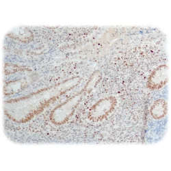
نمره 0 از 5DNA mismatch repair (MMR) system consists of four major proteins called MLH1, MSH2, MSH6, and PMS2. These proteins work two by two, MLH1 with PMS2 and MSH2 with MSH6. Loss of function of one of the four proteins leads to inactivation of the MMR system, resulting in a loss of fidelity of the replication and an accumulation of mutations thereby leading to microsatellite instability (MSI). MSI is associated with hereditary nonpolyposis colorectal cancer (HNPCC, Lynch syndrome), which is characterized by the development of colorectal cancer, endometrial cancer and various other tumors at early age.
Loss of PMS2 function due to gene mutation or epigenetic changes is characterized by absence of nuclear expression in neoplastic cells, whereas intact nuclear PMS2 expression indicates normal PMS2 function and no gene mutations. PMS2 is normally expressed in most cases of sporadic colorectal cancer (CRC). Isolated loss of PMS2 staining is an uncommon immunophenotype among CRCs, accounting for approximately 4%. Anti-PMS2 is useful in detection of MSI, especially in a panel with MSH2 (QR010), MSH6 (QR011) and MLH1 (QM003). -
Quartett
آنتی بادی PR(Progesterone Receptor) کلون QR014 برند Quartett
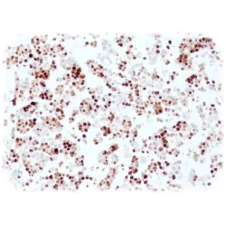
نمره 0 از 5Progesterone receptor (PR) is a nuclear hormone receptor that activates transcription of various genes, especially in reproductive tissues, regulated by its ligand progesterone. PR is predominantly expressed in female sex steroid responsive tissues such as the mammary gland, uterus and ovary. Two isoforms exist: PR-A (94 kDa) and PR-B (114 kDa). Ratio of these isoforms varies in reproductive tissues and during carcinogenesis. PR is expressed in about 65-75% of breast carcinomas. The expression of PR is usually closely linked to the expression of the estrogen receptor (ER), as both receptors are hormonally regulated. PR-positive status is often used as an indicator for a better prognosis and a better response to hormone therapies.
-
Quartett
آنتی بادی PAX8 کلون QR016 برند Quartett
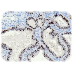
نمره 0 از 5PAX8 is a member of the PAX family (Paired Box) of transcription factors. It is vital for organogenesis during the embryonic development of the kidneys, Müller organs and thyroid. Due to the restrictive expression in normal tissues, PAX8 is a sensitive and specific marker for primary tumors as well as for metastatic tumors from the above-mentioned organs and tissues. PAX8 is expressed in thyroid carcinoma (~90%), endometrial carcinoma (84-98%), ovarian carcinoma (71 99%) and renal cell carcinoma (~90%).
-
Quartett
آنتی بادی P53 کلون QR025 برند Quartett
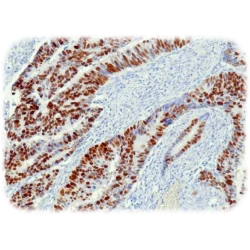
نمره 0 از 5Anti-human antibody for in vitro diagnostic use. The primary antibody is intended for the qualitative detection of associated antigens as listed in the section ‘Summary and explanation’. It is intended to be used within an immunohistochemistry (IHC) procedure on formalin-fixed, paraffin-embedded (FFPE) tissue sections followed by light microscopy visualization to aid tumor diagnosis. The antibody may be used manually or with any automated staining platform. Authorized and skilled personnel may only use the product. The clinical interpretation of any test results should be evaluated within the context of the patient’s medical history and other diagnostic laboratory test results. A qualified pathologist must perform evaluation.
-
Quartett
آنتی بادی P16-INK4 کلون QR019 برند Quartett
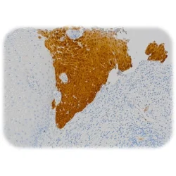
نمره 0 از 5p16, also designated p16(INK4a), cyclin-dependent kinase inhibitor 2A or CDKN2A, is a tumor suppressor protein that slows cell division by slowing the progression of the cell cycle from the G1 phase to the S phase. Mutations in the p16 gene – associated with loss or overexpression of the protein – are associated with increased risk of a wide range of cancers and cancer precursor lesions. When overexpressed, the protein is detected both in the nucleus and cytoplasm. p16 is expressed in almost 100% of cervical carcinomas, especially in squamous cell carcinomas of the cervix. The vast majority of these carcinomas are associated with human papilloma virus (HPV) infection, especially high-risk HPV types such as HPV 16 and 18. The overexpression of p16 is caused by the inactivation of the retinoblastoma protein (Rb) by the viral E7 oncogene, which leads to the upregulation of p16. Strong and full thickness staining of p16 in the cervix epithelium is highly supportive of high grade squamous epithelial lesion (HSIL), while weak and intermediate staining favors low grade squamous epithelial lesion (LSIL).
-
Quartett
آنتی بادی P63 کلون QR007 برند Quartett
نمره 0 از 5p63 is a transcription factor with significant structural similarity to p53 and p73. p63 plays a critical role in the growth and development of many epithelial organs.
p63 is highest expressed in squamous cell carcinoma irrespective of their origin (96-100%). Anti-p63 may be useful in the detection of squamous cell carcinoma of the head and neck (HNSCC), of the lung and skin. Prostate adenocarcinoma is almost always p63 negative, so p63 is used for the differentiation between prostate adenocarcinoma and benign prostate tissue.
-
Quartett
آنتی بادی PIN Cocktail(AMACR/p504s+p63) کلون QR108-QM006 برند Quartett
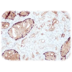
نمره 0 از 5AMACR is an important enzyme for the b-oxidation of branched-chain fatty acids. High expression of the AMACR protein is found in prostate cancer (88-94%), but not in normal prostate and benign prostate diseases. The expression of AMACR is also detected in premalignant lesions of the prostate, in prostatic intraepithelial neoplasia (PIN) the positive rate of AMACR ranges from 13-72%. The p63 protein, a homolog of the tumor suppressor p53, is highly expressed in the basal or precursor layer of many epithelial tissues. p63 is detected in the basal cells of the prostate in normal prostate glands and PIN. Prostate adenocarcinoma is almost always negative for p63. The combination of AMACR and p63 can be extremely useful in the diagnosis of PIN and small focus adenocarcinomas, especially in difficult cases and cases with limited tissue. AMACR stains the cytoplasm of prostate adenocarcinomas and PIN, while p63 stains the basal cell nuclei of PIN and benign prostate glands.
-
Quartett
آنتی بادی PSA(Prostate-Specific Antigen) کلون QR038 برند Quartett
نمره 0 از 5PSA is a glycoprotein enzyme that is secreted by the epithelial cells of the prostate gland. PSA is produced for the ejaculate, where it liquefies semen in the seminal coagulum and allows sperm to swim freely. It is also believed to be instrumental in dissolving cervical mucus, allowing the entry of sperm into the uterus. PSA is present in small quantities in the serum of men with healthy prostates but is often elevated in the presence of prostate cancer or other prostate disorders. It is not uniquely an indicator of prostate cancer but may also detect prostatitis or benign prostatic hyperplasia.
-
Quartett
آنتی بادی PTEN کلون QR042 برند Quartett
نمره 0 از 5PTEN is a phosphoinositide3-phosphatase and is identified as a tumor suppressor that is mutated in a large number of cancers at high frequency. PTEN contains a tensin-like domain as well as a catalytic domain similar to that of the dual specificity protein tyrosine phosphatases. Expression is found at a relatively high levels in all adult tissues including heart, brain, placenta, lung, liver, muscle, kidney, and pancreas.
-
Quartett
آنتی بادی PSMA کلون QR079 برند Quartett
نمره 0 از 5PSMA (Prostate specific membrane antigen) is a zinc metalloenzyme/class II membrane glycoprotein that resides in membranes. It is mainly expressed in four tissues of the body, including prostate epithelium, the proximal tubules of the kidney, the jejunal brush border of the small intestine and ganglia of the nervous system. Human PSMA is highly expressed in the prostate, roughly a hundred times greater than in most other tissues. In some prostate cancers, PSMA is the second-most upregulated gene product, with an 8- to 12-fold increase over levels in noncancerous prostate cells. Because of this high expression, PSMA is being developed as potential biomarker for therapy and imaging of some cancers. In human prostate cancer, the higher expressing tumors are associated with quicker time to progression and a greater percentage of patients suffering relapse.
-
Quartett
آنتی بادی Plap(Placental Alkaline Phosphatase) کلون QR084 برند Quartett
نمره 0 از 5Alkaline phosphatase, placental type is a membrane bound glycosylated dimeric enzyme that is expressed primarily in the placenta. This antibody detects germ cell tumors; it discriminates between these and other neoplasms. It also reacts with somatic neoplasms, such as breast, gastrointestinal, prostatic and urinary cancers. PLAP antibody has been useful in the diagnosis of gestational trophoblastic disease.
-
Quartett
آنتی بادی Podoplanin کلون QR048 برند Quartett
نمره 0 از 5Podoplanin is a type-I integral membrane glycoprotein that is selectively expressed in lymphatic endothelium. The exact molecular function of podoplanin in the lymphatic endothelium has remained unclear. This antibody detects lymphangiomas, Kaposi sarcomas and a subset of angiosarcomas with probable lymphatic differentiation.
-
Quartett
آنتی بادی PAX2 کلون QR060 برند Quartett
نمره 0 از 5PAX2 is a transcription factor that is strongly expressed during kidney development, in the metanephric mesenchyma after ureter bud induction and is a key factor for the mesenchyma-epithelium conversion, in renal carcinoma cell lines and, in papillary renal cell carcinoma, and chromophobe renal cell carcinomas.
-
Quartett
آنتی بادی PAX5 کلون QR056 برند Quartett
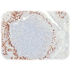
نمره 0 از 5PAX5 is a transcription factor and important regulator in early development. Alterations in the expression of PAX5 gene are thought to contribute to neoplastic transformation. The PAX5 gene encodes the B-cell lineage specific activator protein (BSAP) that is expressed at early, but not late stages of B-cell differentiation. Its expression has also been detected in developing CNS and testis, therefore, PAX5 gene product may not only play an important role in B-cell differentiation, but also in neural development and spermatogenesis.
-
Quartett
آنتی بادی P21(Waf1) کلون QR085 برند Quartett
نمره 0 از 5p21/WAF1 is a potent cyclin-dependent kinase inhibitor that binds and inhibits the activity of cyclin-CDK2 or -CDK1 complexes, and thus functions as a regulator of cell cycle progression at G1. It is expressed in all adult human tissues, with lower levels in the brain.
-
Quartett
آنتی بادی SATB2(DNA-Binding Protein SATB2) کلون QR023 برند Quartett
نمره 0 از 5SATB2 is a nuclear matrix protein that influences craniofacial formation mechanisms, such as jaw and palate development, and is part of a transcriptional network regulating skeletal development and osteoblast differentiation. It is highly expressed in adult and fetal brain, and weak expressed in liver, kidney, and spinal cord as well as some brain regions. Defects in the gene encoding SATB2 are the cause of cleft palate manifested in conjunction with severe mental retardation.
-
Quartett
آنتی بادی S100 Protein کلون QR031 برند Quartett
نمره 0 از 5Anti-human antibody for in vitro diagnostic use. The primary antibody is intended for the qualitative detection of associated antigens as listed in the section ‘Summary and explanation’. It is intended to be used within an immunohistochemistry (IHC) procedure on formalin-fixed, paraffin-embedded (FFPE) tissue sections followed by light microscopy visualization to aid tumor diagnosis. The antibody may be used manually or with any automated staining platform.
Authorized and skilled personnel may only use the product. The clinical interpretation of any test results should be evaluated within the context of the patient’s medical history and other diagnostic laboratory test results. A qualified pathologist must perform evaluation. -
Quartett
آنتی بادی Synaptophysin کلون QR054 برند Quartett
نمره 0 از 5Synaptophysin is an integral transmembrane protein with four transmembrane domains. The Protein plays a role in endocytosis of synaptic vesicles in mammalian central neurons. It is present in all neurons, all types of pancreastic islet cells and paraganglia cells. Additional, it is expressed in almost all neuroendocrine neoplasms. Synaptophysin is helpful for the detection of tumors with neuronal and neuroendocrine differentiation.
-
Quartett
آنتی بادی STAT6 کلون QR041 برند Quartett
نمره 0 از 5STAT family consists of seven genes, STAT1 to STAT4, STAT5A, STAT5B and STAT6, that are involved in regulating cellular proliferation, apoptosis, angiogenesis and the immune system response. STAT6 is a transcription factor that carries out a dual function: signal transduction and activation of transcription. It is involved in interleukin-4 signaling and seems to play an important role in cancer. STAT6 has been found to form recurrent fusions with NAB2 on chromosome 12q13 in the majority of solitary fibrous tumors (SFTs) excluding meningiomas, hemangioblastomas, schwannomas, and hemangiomas. The fusion enables cytosolic STAT6 to migrate to the nucleus. The detection of nuclear relocation of STAT6 with immunohistochemistry is a characteristic of SFTs and may serve as a diagnostic marker that indicates NAB2 STAT6 fusion.
-
Quartett
آنتی بادی SOX10 کلون QR006 برند Quartett
نمره 0 از 5SOX10 is a nuclear transcription factor that acts as a shuttle protein between the cytoplasm and the nucleus. It is important for the development of the neural crest and the peripheral nervous system as well as for the differentiation, maturation and maintenance of Schwann cells and melanocytes. SOX10 is expressed in Schwann and glial cells of the nervous system and in melanocytes and epithelial cells of the salivary and mammary glands.
Studies show that SOX10 is expressed in 100% of benign nevi and in 95-100% of malignant melanomas, both in primary and metastatic tumors.

