-
 آنتی بادیهای ایمونوهیستوشیمی
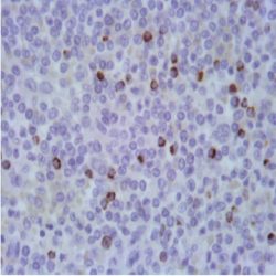
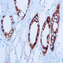
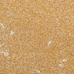
آنتی بادی – H3K27me3 (Lys27) (clone C36B11)
Name: Rabbit Anti-Human H3K27me3 (Lys27) Monoclonal Antibody (Clone C36B11)
Description and aplications: The nucleosome, which consists of four core histone proteins (H2A, H2B, H3 and H4), is the main component of chromatin. Originally thought to function as a static scaffold for DNA packaging, histones have subsequently been shown to be dynamic proteins, undergoing multiple types of posttranslational modifications, including acetylation, phosphorylation, methylation, and ubiquitination. Histone methylation is a fundamental determinant for
the formation of active and inactive regions of the genome and is crucial for their proper programming during embryonic development. Arginine methylation of histones H3 (Arg2, 17, 26) and H4 (Arg3) promotes transcriptional activation and is mediated by a family of arginine methyltransferase (PRMT)-like proteins, including the coactivators PRMT1 and CARM1(PRMT4). In contrast, a more diverse set of lysine methyltransferase-type histones has been identified, among which all but one contains a conserved catalytic SET domain, and that was originally identified in the Zeste and Trithorax regulatory proteins of the fly Drosophila Su (var) 3-9. Lysine methylation occurs primarily at histones H3 (Lys4, 9, 27, 36, 79) and H4 (Lys20) and has been implicated in both transcriptional activation and gene silencing. Additionally, methylation of these lysine residues coordinates the recruitment of chromatinmodifyinenzymes containing methyl lysine binding modules, either as chromodomains (HP1, PRC1), PHD fingers (BPTF, ING2), Tudor domains (53BP1) and WD-40 domains (WDR5). Finally, the discovery of histone demethylases such as PADI4, LSD1, JMJD1, JMJD2 and JHDM1 has demonstrated that methylation is a reversible epigenetic marker. NF1 mutations and inactivation of CDKN2A are found
in most malignant peripheral nerve sheath tumors (TMVNP), where inactivation of CDKN2A is an early event during their development, occurring in the course of progression from conventional to atypical neurofibroma. Furthermore, inactivation of the polycomb repressive complex 2 (PRC2) resulting from mutually exclusive mutations of its SUZ12 or
EED1 portions has recently been identified in 70-90% of TMVNPs. In this regard, inactivation of PRC2 leads to loss of trimethylation at histone H3 lysine 27 (H3K27me3).Composition: Anti-human H3K27me3 (Lys27) rabbit polyclonal antibody obtained from ascitic fluid and prepared in 10mM PBS, pH 7.4, with 0.2% BSA and 0.09% sodium azide.
Immunogen: Synthetic peptide corresponding to the terminal amino acids of human histone 3 in which Lys27 is trimethylated.
-
 آنتی بادیهای ایمونوهیستوشیمی
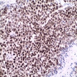
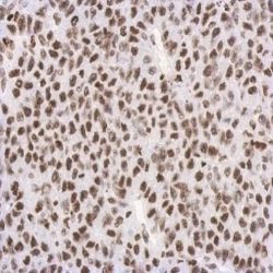
آنتی بادی PRAME 1 (EPR20330)
Name: Rabbit Anti-Human PRAME 1 Monoclonal Antibody (Clone EPR20330)
Description and aplications: The PReferentially expressed Antigen in MElanoma (PRAME), or LB33-E antigen, is part of the family of melanoma nuclear proteins recognized by cytolytic T lymphocytes and is encoded by a gene of the same
name (PRAME) located on chromosomal region 22q11.22. The PRAME protein has been characterized as a dominant inhibitor of the retinoic acid receptor, thus participating in the blockade of cell proliferation, differentiation or apoptosis induced by retinoic acid through the RARA, RARB and RARG receptors. Thus, overexpression of PRAME in tumor cells confers a survival advantage over normal cells. In melanomas, PRAME inhibition restores retinoic acid signaling an reinstates the sensitivity of tumor cells to the antiproliferative effects of this molecule.Composition: Anti-human PRAME 1 rabbit monoclonal antibody obtained from ascitic and prepared in 10mM PBS, pH 7.4, with 0.2% BSA and 0.09% sodium azide.
Immunogen: N/A
-
 آنتی بادیهای ایمونوهیستوشیمی
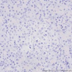
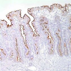
آنتی بادی Renal cell carcinoma marker (PN-15)
Name: Mouse anti- Human Renal cell carcinoma marker (PN-15) Monoclonal Antibody
Description and aplications:This antibody detects the membrane glycoprotein gp200. In a normal kidney, gp200 is located along the brush border of the contoured and straight segments of the proximal tubule, as well as focally along the luminal surface of Bowman’s capsule, contiguous with the exit of the proximal tubule. Gp200 is also located on the luminal surface of the mammary ducts and lobes and the tubular epithelium of the epididymis, in the cytoplasm of parathyroid and parenchymal cells, and focally within the colloid of thyroid follicles.
Composition: Anti-human renal cell carcinoma marker (PN-15) mouse monoclonal antibody purified and prepared in 10mM PBS, pH 7.4, with 0.2% BSA and 0.09% sodium azide.
Immunogen: Microsomal fraction of human renal cortical tissue homogenate.
-
 آنتی بادیهای ایمونوهیستوشیمی
آنتی بادی SMAD4/Dpc4 (B8)
Name: Mouse Anti-Human SMAD4/Dpc4 Monoclonal Antibody (Clone B8)
Description and applications: The Mothers Against Decapentaplegic homolog 4(SMAD4) protein, also known as MADH4, MANrelatedprotein 4, MAD, pancreatic carcinoma deletion factor 4 or DPC4, is linked to the SMAD4 suppressor gene, which consists of 11 exons, and is located in the genomic region 18q21.1. This gene shows allelic or biallelic loss in 50% of pancreatic carcinomas
Composition: Anti-human SMAD4/Dpc4 mouse monoclonal antibody obtained from ascitic fluid and prepared in 10mM PBS, pH 7.4, with 0.2% BSA and 0.09% sodium azide
Immunogen: N/A -
 آنتی بادیهای ایمونوهیستوشیمی
آنتی بادی Parvalbumin (EP300)
NAME: Rabitt anti-Parvalbumin Antibody (Clon EP300)
Description and applications: Parvalbumin is a protein with wide calcium-ionbinding affinity and thus intervenes in its cytosolic homeostasis. The PVALB gene, which encodes the production of this protein, is located in the chromosome region 22q12.3 and shows structural and functional similarities with the one of the Calmodulin and Troponin C, whose coding genes constitute a superfamily.
Composition: anti-human Parvalbumin rabbit monoclonal antibody purified from serum and prepared in 10mM PBS, pH 7.4, with 0.2% BSA and 0.09% sodium azide
-
 آنتی بادیهای ایمونوهیستوشیمی
آنتی بادی CD38 (CD38/4328)
Name: CD38 Antibody Clone CD38/4328
Description and applications: CD38 is a single chain type II integral transmembrane protein, which is
highly expressed on thymocytes. It is also present on activated T cells and terminally differentiated B cells
(plasma cells). Other reactive cells include NK cells, monocytes, macrophages, and dendritic cells. CD38
may be detected on cells from multiple myeloma, ALL (B and T) and some AML. It is, however, not found on most mature resting peripheral lymphocytes. CD38 functions as a multicatalytic ectoenzyme serving as
ADP-ribosyl cyclase, cyclic ADP-ribose hydrolase and possibly NAD+ glycohydrolase or as a cell surface receptor.Composition: Anti-human CD38 mouse monoclonal antibody purified from serum and prepared in 10mM PBS, pH 7.4, with 0.2% BSA and 0.09% sodium azide.
Immunogen: Recombinant full-length human CD38 protein.
-
 آنتی بادیهای ایمونوهیستوشیمی
آنتی بادی CD38 کلون 38C03 برند Vitro
Name: CD38 Monoclonal Antibody clone 38C03
DESCRIPTION AND APPLICATIONS: CD38 is a single chain type II integral transmembrane protein, which is highly expressed on thymocytes. It is also present on activated T cells and terminally differentiated B cells (plasma cells). Other reactive cells include NK cells, monocytes, macrophages, and dendritic cells. CD38 may be detected on cells from multiple myeloma, ALL (B and T) and some AML. It is, however, not found on most mature resting peripheral lymphocytes. CD38 functions as a multicatalytic ectoenzyme serving as ADP-ribosyl cyclase, cyclic ADP-ribose hydrolase and possibly NAD+ glycohydrolase or as a cell surface receptor. Antibodies to CD38 are useful in subtyping of lymphomas and leukemias, detection of plasma cells (i.e. identification of myelomas), and as a marker for activated B and T cells.
COMPOSITION: Anti-human CD38 mouse monoclonal antibody purified from serum and prepared in 10mM PBS, pH 7.4, with 0.2% BSA and 0.09% sodium azide
INTENDED USE: Immunohistochemistry (IHC) on paraffin embedded tissues. Not tested on frozen tissues or Western-Blotting
IMMUNOGEN: Recombinant protein encoding the extracellular domain of human CD38.
SPECIES REACTIVITY: In vitro diagnostics in humans. Not tested in other species
-
آنتی بادیهای ایمونوهیستوشیمی
آنتی بادی CD45 کلون 2B11 & PD7/26 برند Vitro
Name: CD38 Monoclonal Antibody clone 2B11 & PD7/26
DESCRIPTION AND APPLICATIONS: Anti-CD45 (anti leukocyte common antigen) is routinely used to aid the differential diagnosis of undifferentiated neoplasms, whenever malignant lymphoma is suspected by the morphological or clinical data. It is a highly specific antibody; therefore a positive result is highly indicative of hematolymphoid origin. Certain types of hematolymphoid neoplasms may lack CD45(Hodgkin lymphoma, some T-cell lymphomas, and some leukemias) so its absence does not rule out a hematolymphoid tumor. This antibody is expressed almost exclusively by cells of hematopoietic lineage and is present in most benign and malignant lymphocytes as well as plasma cell precursors.
COMPOSITION: Anti-human CD45, also known as leukocyte common antigen, is a mouse monoclonal antibody purified from serum and prepared in 10mM PBS, pH 7.4, with 0.2% BSA and 0.09% sodium azide
INTENDED USE: Immunohistochemistry (IHC) on paraffin embedded tissues. Not tested on frozen tissues or Western-Blotting
SPECIES REACTIVITY: In vitro diagnostics in humans. Not tested in other species
-
آنتی بادیهای ایمونوهیستوشیمی
آنتی بادی CD42b کلون EPR19204 برند Vitro
Name: CD42b Monoclonal Antibody clone EPR19204
DESCRIPTION AND APPLICATIONS: The CD42b glycoprotein, also known as GPIb, is a co-factor of ristocetin-induced aggregation and is involved in the binding of platelets to blood vessel walls. The CD42b antigen is expressed on platelets and on megakaryocytes in bone marrow. The absence of CD42b antigen on platelets may indicate Bernard Soulier disease. The antibody is of value in theimmunophenotyping of megakaryoblastic leukaemias that express one or more markers associated with platelets (CD41, CD61 and CD42b). In chronic myeloproliferative processes, like chronic idiopathic myelofibrosis in prefibrotic stage besides appearing in greater numbers the megakaryocytes are markedly abnormal while megakaryocytes in unclassifiable myelodysplastic / myeloproliferative diseases there is an increased of normal or small megakaryocytes (micromegakaryocytes).
COMPOSITION: Anti-human CD42b rabbit monoclonal antibody purified from hybridoma and, diluted in a pH7.6 buffer containing stabilizing protein and sodium azide as bacteriostatic and bactericidal agent.
INTENDED USE: Immunohistochemistry (IHC) on paraffin embedded tissues. Not tested on frozen tissues or Western-Blotting
IMMUNOGEN: Recombinant CD42b.
SPECIES REACTIVITY: In vitro diagnostics in humans. Not tested in other species
-
آنتی بادیهای ایمونوهیستوشیمی
آنتی بادی Cdk4 کلون CDK4/7987R برند Vitro
Name: Cdk4 Monoclonal Antibody clone CDK4/7987R
DESCRIPTION AND APPLICATIONS: Cell cycle progression is controlled in part by a family of cyclin proteins and cyclin dependent kinases (Cdks). Cdk proteins work in concert with the cyclins to phosphorylate key substrates involved in each phase of cell cycle progression. Another family of proteins, Cdk inhibitors, also plays a role in regulating the cell cycle by binding to cyclin Cdk complexes and modulating their activity. Several Cdk proteins have been identified, including Cdk2 Cdk8, PCTAIRE-1-PCTAIRE-3, PITALRE and PITSLRE. Cdk4, in complex with D-type cyclins, is thought to regulate cell growth during the G1 phase of the cell cycle. This association with a D-type cyclin upregulates Cdk4 activity, whereas binding to the Cdk inhibitor p16 downregulates Cdk4 activity. Activation of the Cdk4-cyclin complexes requires phosphorylation on a single threonyl residue of Cdk4, catalyzed by a Cdk-activating protein (CAK).
COMPOSITION: Anti-human CDK4 rabbit monoclonal antibody diluted in a pH 7.6 buffer containing stabilizing protein and sodium azide as bacteriostatic and bactericidal agent.
INTENDED USE: Immunohistochemistry (IHC) on paraffin embedded tissues. Not tested on frozen tissues or Western-Blotting
IMMUNOGEN: Recombinant fragment corresponding to N-terminal of human CDK4 protein
SPECIES REACTIVITY: In vitro diagnostics in humans. Not tested in other species
-
 آنتی بادیهای ایمونوهیستوشیمی
آنتی بادی WT1 (Wilms Tumor) کلون 6F-H2 برند PathoSage
Intended use:
This antibody is intended for in vitro diagnostic (IVD) use. Primary Antibody is intended for professional laboratory use in formalin-fixed, paraffin-embedded (FFPE) tissue stained in manual qualitative immunohistochemistry (IHC) testing. A qualified pathologist must interpret the results using this product to aid diagnosis in conjunction with the patient’s relevant clinical history, other diagnostic tests, and proper controls.
Presentation:
supernatant prepared in 10mM PBS, pH 7.4, with %0.2 BSA and %0.09 sodium azide Anti-human WT-1 mouse monoclonal antibody obtained from tissue culture
-
 آنتی بادیهای ایمونوهیستوشیمی
آنتی بادی VIP کلون H-6 برند PathoSage
Intended use:
This antibody is intended for in vitro diagnostic (IVD) use. Primary Antibody is intended for professional laboratory use in formalin-fixed, paraffin-embedded (FFPE) tissue stained in manual qualitative immunohistochemistry (IHC) testing. A qualified pathologist must interpret the results using this product to aid diagnosis in conjunction with the patient’s relevant clinical history, other diagnostic tests, and proper controls.
Presentation:
Anti-human VIP mouse monoclonal antibody purified from serum and prepared in PBS with < %0.1 sodium azide and %0.1 gelatin
-
 آنتی بادیهای ایمونوهیستوشیمی
آنتی بادی Vimentin کلون SP20 برند PathoSage
Intended use:
This antibody is intended for in vitro diagnostic (IVD) use. Primary Antibody is intended for professional laboratory use in formalin-fixed, paraffin-embedded (FFPE) tissue stained in manual qualitative immunohistochemistry (IHC) testing. A qualified pathologist must interpret the results using this product to aid diagnosis in conjunction with the patient’s relevant clinical history, other diagnostic tests and proper controls.
Presentation:
Anti-human Vimentin rabbit monoclonal antibody purified from serum and prepare din 10mM PBS, pH 7.4, with %0.2 BSA and %0.09 sodium azide
-
 آنتی بادیهای ایمونوهیستوشیمی
آنتی بادی Villin 1 کلون CWWB1 برند PathoSage
Intended use:
This antibody is intended for in vitro diagnostic (IVD) use. Primary Antibody is intended for professional laboratory use in formalin-fixed, paraffin-embedded (FFPE) tissue stained in manual qualitative immunohistochemistry (IHC) testing. A qualified pathologist must interpret the results using this product to aid diagnosis in conjunction with the patient’s relevant clinical history, other diagnostic tests, and proper controls.
Presentation:
Anti-human Villin-1 mouse monoclonal antibody purified from serum and prepared in 10mM PBS, pH 7.4, with %0.2 BSA and %0.09 sodium azide
-
 آنتی بادیهای ایمونوهیستوشیمی
آنتی بادی VEGF (Vascular Endothelial Growth Factor) کلون EP1176Y برند PathoSage
Intended use:
This antibody is intended for in vitro diagnostic (IVD) use. Primary Antibody is intended for professional laboratory use in formalin fixed, paraffin-embedded (FFPE) tissue stained in manual qualitative immunohistochemistry (IHC) testing. A qualified pathologist must interpret the results using this product to aid diagnosis in conjunction with the patient’s relevant clinical history, other diagnostic tests, and proper controls.
Presentation:
Anti-human VEGF Rabbit monoclonal antibody purified from ascites fluid by Protein A chromatography prepared in 10mM PBS, pH 7.4, with %0.2 BSA and %0.09 sodium azide
-
آنتی بادیهای ایمونوهیستوشیمی
آنتی بادی Uroplakin 3 کلون BC17 برند PathoSage
Intended use:
This antibody is intended for in vitro diagnostic (IVD) use. Primary Antibody is intended for professional laboratory use in formalin-fixed, paraffin-embedded (FFPE) tissue stained in manual qualitative immunohistochemistry (IHC) testing. A qualified pathologist must interpret the results using this product to aid diagnosis in conjunction with the patient’s relevant clinical history, other diagnostic tests, and proper controls.
Presentation:
Anti-human Uroplakin III mouse monoclonal antibody purified from serum and prepared in 10mM PBS, pH 7.4, with %0.2 BSA and %0.09 sodium azide
-
 آنتی بادیهای ایمونوهیستوشیمی
آنتی بادی Uroplakin 2 کلون BC21 برند PathoSage
Intended use:
This antibody is intended for in vitro diagnostic (IVD) use. Primary Antibody is intended for professional laboratory use in formalin-fixed, paraffin-embedded (FFPE) tissue stained in manual qualitative immunohistochemistry (IHC) testing. A qualified pathologist must interpret the results using this product to aid diagnosis in conjunction with the patient’s relevant clinical history, other diagnostic tests, and proper controls.
Presentation:
Anti-Uroplakin 3 mouse monoclonal antibody obtained from supernatant culture and prediluted in a tris buffered solution pH 7.4 containing 0.375mM sodium azide solution as bacteriostatic and bactericidal
-
 آنتی بادیهای ایمونوهیستوشیمی
آنتی بادی TSH (Thyroid Stimulating Hormone) کلون EP254 برند PathoSage
Intended use:
This antibody is intended for in vitro diagnostic (IVD) use. Primary Antibody is intended for professional laboratory use in formalin-fixed, paraffin-embedded (FFPE) tissue stained in manual qualitative immunohistochemistry (IHC) testing. A qualified pathologist must interpret the results using this product to aid diagnosis in conjunction with the patient’s relevant clinical history, other diagnostic tests, and proper controls.
Presentation:
Anti-human TSH (Thyroid Stimulating Hormone) rabbit monoclonal antibody purified from serum and prepared in 10mM PBS, pH 7.4, with %0.2 BSA and %0.09 sodium azide
-
 Quartett
آنتی بادی ALK/p80(CD246) کلون QR017 برند Quartett
Anaplastic lymphoma kinase (ALK) belongs to the insulin receptor superfamily acting as a transmembrane receptorprotein-tyrosine kinase. In normal tissues, ALK protein is expressed only few cells within the developing and mature nervous system. ALK can be active in cancer through multiple mechanisms. The most common mechanism is through the formation of a fusion protein from chromosomal translocations. The protein expression by tumor cells is an independent prognostic factor that predicts a favorable outcome. ALK staining is positive in 50-60% of anaplastic large cell lymphoma (ALCL). NSCLC is found to express ALK 3-7%.
-
 Quartett
آنتی بادی BCL2 کلون QR062 برند Quartett
Bcl2 (B-cell lymphoma 2) plays an important role in regulation of apoptosis. It is expressed in different cells and tissues. Overexpression due to chromosomal translocation, gene amplification, increased gene transcription and/or altered post-translational processing is found in many cancer types. These include B-cell malignancies, such as B-cell lymphoma, follicular lymphoma (FL) and chronic lymphocytic leukemia (CLL), as well as some T-cell lymphomas, breast cancer, lung cancer, ovarian cancer, and prostat a cancer.
برای تماس کلیک کنید
سما تشخیص آریا ، عرضه کننده متنوع ترین و کامل ترین محصولات کیت های آزمایشگاهی