-
 آنتی بادیهای ایمونوهیستوشیمی
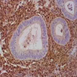
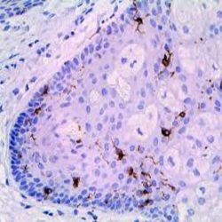
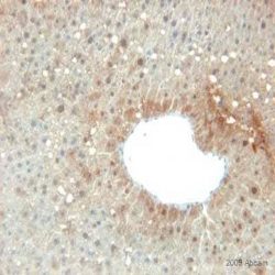
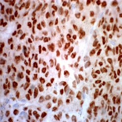
آنتی بادی HuC/HuD Neuronal Protein (16A11)
Name: Mouse anti-human HuC/HuD Neuronal Protein Monoclonal Antibody (Clone 16A11)
Description and aplications: The Hu antigen is a protein of the Elav (embryonic lethal abnormal visual) family. This antibody recognises only some protein members of the Elav family (HuC, HuD, and Hel-N1), all of them neuronal proteins. It does not recognise HuR, another Elav family member that is present in all proliferating cells. This antibody can be considered as a neuron marker. This antibody identifies neuronal cells and neoplasms derived from them.
Composition:Anti-human HuC/HuD Neuronal Protein mouse monoclonal antibody purified from serum and prepared in 10mM PBS, pH 7.4, with 0.2% BSA and 0.09% sodium azide
-
 آنتی بادیهای ایمونوهیستوشیمی
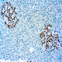
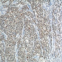
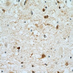
آنتی بادی INSM1 (A-8)
Name: Mouse anti-human INSM1 (Insulinoma-associated protein 1) Monoclonal Antibody (Clone A-8)
Description and aplications: INSM1 protein, also known as IA1, is a nuclear transcription factor encoded by INSM1 gene, located in the chromosome region 20p11.23 and composed of 510 amino acids with a molecular mass of approximately 52.8kDa. The protein, which shows a wide homology in different species, contains a Cterminal portion with 5 finger-binding domains and an N-terminal domain with four pro-hormone conversion sites as well as an amidation signal sequence. INSM1 factor plays an important role in neurogenesis and differentiation during the embryonic and fetal development of the pancreatic neuroendocrine, anterior pituitary, intestine cells and synapto-adrenal cells of the peripheral nervous system. INSM1 gene, which does not express in normal endocrine and non-endocrine tissues in adults, acts as a suppressor gene of the transmission pathways NEUROD1 and INS after interaction with Cyclin D1, inhibits the specific genes of the skeletal muscle in neuroendocrine cells of the pituitary and suppress neural factors such as HDAC1, HDAC2, HDAC3 and KDM1A (involved in the incorporation of chromatin-modifying factors). In addition to this, it suppresses the activity of the histone deacetylase RCOR1.
Composition: Anti-human INSM1 mouse monoclonal antibody purified from serum and prepared in 10mM PBS, pH
7.4, with 0.2% BSA and 0.09% sodium azide -
 آنتی بادیهای ایمونوهیستوشیمی
آنتی بادی Langerin/CD207 (EPR15863)
Name: Rabbit anti-human Langerin/CD207 Monoclonal Antibody (Clone EPR15863)
Description and aplications: Calciumdependent lectin displaying mannose-binding specificity. Induces the formation of Birbeck granules (BGs); is a potent regulator of membrane superimposition and zippering. Binds to sulfated as well as mannosylated glycans, keratan sulfate (KS) and beta-glucans. Facilitates uptake of antigens and is involved in the routing and/or processing of antigen for presentation to T cells. Major receptor on primary Langerhans cells for Candida species, Saccharomyces species, and Malassezia furfur. Protects against human immunodeficiency virus-1 (HIV-1) infection. Binds to high-mannose structures present on the envelope glycoprotein which is followed by subsequent targeting of the virus to the Birbeck granules leading to its rapid degradation.
Composition: Anti-human Langerin/CD207 rabbit monoclonal antibody purified from serum and prepared in 10mM
PBS, pH 7.4, with 0.2% BSA and 0.09% sodium azide
Immunogen: Recombinant fragment within Human Langerin aa 200 to the C-terminus.
-
 آنتی بادیهای ایمونوهیستوشیمی
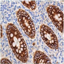
آنتی بادی Mucin 4 (8G7)
Name: Mouse anti-human Mucin 4 (MUC4) Monoclonal Antibody (Clone 8G7)
Description and aplications: Mucins are a group of high molecular weight glycoproteins consisting of a mucin core protein and O-linked carbohydrates. Mucin 4, a membrane-bound mucin, is the human homolog of the rat sialomucin complex (SMC). Mucin 4 protein consists of Mucin 4α, a large amino mucin type subunit and Mucin 4β, a transmembrane subunit containing three EGF-like domains. The Mucin 4 gene is the predominant mucin gene expressed in the normal urothelium and is also expressed in several normal tissues such as trachea, lung and testis.
Composition: Anti-human MUC4 mouse monoclonal antibody purified from serum and prepared in 10mM PBS, pH
7.4, with 0.2% BSA and 0.09% sodium azide
Immunogen: A synthetic peptide directed against the Mucin 4 tandem repeats of human origin.
-
 آنتی بادیهای ایمونوهیستوشیمی
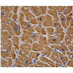
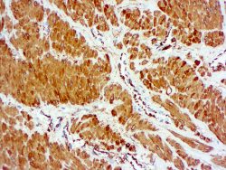
آنتی بادی Myosin – Skeletal Muscle (MY-32)
Name: Mouse anti-human Myosin – Skeletal Muscle Monoclonal Antibody (Clone MY-32)
Description and aplications: This antibody reacts against the skeletal muscle myosin heavy chain. Myosin is a 480-kDa protein present in muscle and non-muscle cells. This antibody may be used as a tool for the diagnosis of tumours of skeletal muscle origin. It may also be useful in studies of the architecture of thick filament cytoskeleton. It does not react with human or animal cardiac myosin or smooth muscle myosin. It reacts with myosin of human, rabbit, rat, murine, bovine, and guinea pig origin.
Composition: Anti-human Myosin – Skeletal Muscle mouse monoclonal antibody purified from serum and prepared in 10mM PBS, pH 7.4, with 0.2% BSA and 0.09% sodium azide
Immunogen: Rabbit myosin.
-
 آنتی بادیهای ایمونوهیستوشیمی
آنتی بادی NKX2.2 (EP336)
Name: Rabbit anti-NKX2.2 Monoclonal Antibody (clone EP336)
Description and aplications: The NKX2.2 antibody, also known as NK2 or NKX2B, is a nuclear transcription factor that belongs to the NK2 family of homeobox genes and is encoded in the chromosome region 20p11.22. This gene is involved in the development of beta cells of the pancreas that produce insulin, as well as in the neuroendocrine, glial, neuronal and oligodendroglial differentiation. Likewise, it is also involved in several process of diencephalic development and organization as well as in the control of the genes involved in the axonal orientation.
Composition: Rabbit anti-NKX2.2 monoclonal antibody obtained from purified ascitic fluid and prepared in 10mM PBS,
pH 7.4, with 0.2% BSA and 0.09% sodium azide.
Immunogen: synthetic peptide corresponding to the human NKX2.2 protein
-
 آنتی بادیهای ایمونوهیستوشیمی
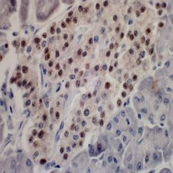
آنتی بادی NKX3.1 (EP356)
Name: Rabbit Anti-Human NKX3.1 Monoclonal Antibody (EP356)
Description and aplications: NKX3.1 is a prostatespecific tumor suppressor protein that is encoded bythe homeobox gene NKX3.1 located in chromosomeregion 8p21 and whose expression is regulated byandrogens. The protein acts as an importanttranscription factor in the normNKX3.1 is a prostatespecifical development of theprostate since it regulates the proliferation of theglandular epithelium and the formation of excretoryducts. For this reason, the protein, which is located in the luminal cells (considered the stem of the prostate epithelium), intervenes in prostate regeneration and issusceptible to oncogenic transformation.
Composition: anti-NKX3.1 rabbit monoclonal antibody obtained from supernatant culture and prediluted in a tris buffered solution pH 7.4 containing 0.375mM sodium azide solution as bacteriostatic and bactericidal.
Immunogen: Synthetic peptide corresponding to residues in the human protein of the NKX3.1.
-
 آنتی بادیهای ایمونوهیستوشیمی
آنتی بادی Pan-Cytokeratin (BS5)
Name: Mouse anti-human Pan Cytokeratin Monoclonal Antibody (Clone BS5)
Description and aplications: Cytokeratins are structural intermediate filaments encoded in more than 49 different genes in the chromosomes 17 (I) and 12 (II). The nomenclature adopted in 1982 by Moll and Franke assigns the ranks 1 to 8 for the type II cytokeratins (neutral or basic) and between 9 and 21 for the type I (acid). Nowadays, an analogue nomenclature has been defined to name the hair keratin with the addition of the Ha and Hb letters to separate the ones of the group I from group II. Structurally speaking, cytokeratins share with the rest of intermediate filaments a central axis of 310 amino acids made up of four α-helices domains (1A, 1B, 2A and 2B) highly preserved that define the type of
intermediate filament that they will make up after its packing, separated by three non-helix binding sites (L1, L12 and L2) and two extreme domains highly different in size and sequence (head-1- and tail-2-), each of them with constant (E1/E2), variable (V1/V2) and homology regions (H1/H2), the last ones are characteristic of the type II keratins and absent of the type I. Usually, keratins assemble in heterodimers I/II and they are co-expressed in pairs specifically in each tissue.Composition:Anti-human Pan Cytokeratin mouse monoclonal antibody purified from serum and prepared in 10mM PBS, pH 7.4, with 0.2% BSA and 0.09% sodium azide
-
 آنتی بادیهای ایمونوهیستوشیمی
آنتی بادی Parvovirus B19 (R92F6)
Name: Mouse anti-Parvovirus B19 Monoclonal Antibody (Clone R92F6)
Description and aplications: Parvovirus B19 is one of the smallest viruses (20 kDa), whose nucleic acid is linear single-stranded DNA of negative polarity and without lipoprotein envelope. The viral genome encodes three proteins: the nonstructural protein NS1 and two viral capsid proteins, VP1 and VP2. The minor capsid protein VP1 has the same sequence of amino acids as VP2 plus 227 amino acids in the amino-terminus region, the VP1 unique region (VP1u). Recently, the phospholipase A2 (sPLA2) motif has been identified in VP1u in members of the Parvoviridae family, including B19, classified as an erythrovirus because of its ability to invade erythrocyte precursors in the bone marrow.
Composition: Anti-Parvovirus B19 mouse monoclonal antibody purified from serum and prepared in 10mM PBS, pH 7.4, with 0.2% BSA and 0.09% sodium azide
Immunogen: Amino acids 328-344 of the VP2 protein of the viral capsid
-
 آنتی بادیهای ایمونوهیستوشیمی
آنتی بادی Prealbumin (EPR3219)
Name: Rabbit anti-human Prealbumin Monoclonal Antibody (Clone EPR3219)
Description and aplications: This antibody recognizes a protein of 16 kDa, a tetramer of 127 amino acids, identified as prealbumin whose secretion is controlled by a gen located in the chromosome region 18q12.1. The prealbumin, also known as transthyretin (TTR), TBPA or PALB is a transport protein of hormones such as the thyroxine (T4) and retinol (vitamin A). Different forms of amyloidosis have been described: systemic amyloidosis which encompass secondary
(AA), primary (AL), senile and those associated with hemodialysis or hereditary cases. Localized amyloidosis is limited to an organ or tissue and includes cerebral amyloidosis, dystrophic (age-related or senile); endocrine, located or tumoral
Composition:Anti-human Prealbumin rabbit monoclonal antibody purified from serum and prepared in 10mM PBS, pH
7.4, with 0.2% BSA and 0.09% sodium azide
Immunogen: Synthetic peptide of the human prealbumin.
-
 آنتی بادیهای ایمونوهیستوشیمی
آنتی بادی Prohibitina (Mitochondrial Marker) (MTC02)
Name: Mouse anti- human Prohibitina (Mitochondrial Marker) Monoclonal Antibody (Clone MTC02)
Description and aplications: Prohibitina (Mitochondrial Marker) recognizes a 60kDa nonglycosylated protein component of mitochondria found in human cells. It can be used to stain the mitochondria in cell or tissue preparations
and can be used as a marker of the mitochondria in subcellular fractions. This monoclonal antibody is part of a new panel of reagents which recognizes subcellular organelles or compartments of human cells. These markers may be useful in identification of these organelles in cells, tissues, and biochemical preparations. Prohibitina (Mitochondrial Marker)
produces”spaghetti”-like staining pattern in the cytoplasm of normal and malignant human cells.Composition: anti-human Prohibitina (Mitochondrial Marker) Monoclonal Antibody purified from the ascites fluid by Protein G chromatography. Prepared in 10mM PBS, pH 7.4, with 0.2% BSA and 0.09% sodium azide
Immunogen: Semi purified mitocondrial preparation.
-
 آنتی بادیهای ایمونوهیستوشیمی
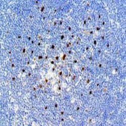
آنتی بادی PHOX2B (Paired Mesoderm Homeobox Protein 2B) (EP312)
Name: Rabbit anti-human PHOX2B Monoclonal Antibody (Clone EP312)
Description and aplications: The PHOX2B gene (also known as PMX2B or NBPHOX) is located in the chromosome region 4p13 and encodes a nuclear transcription factor that, during the embryonic development, predominantly expresses in the autonomic nervous system, being essential in the differentiation and survival of vegetative neurons and chromaffin cells. Mutations of the PHOX2B gene are responsible for the central hypoventilation syndrome, a syndrome with autosomal dominant transmission in which the hypoventilation is secondary to the reduction or absence of ventilatory response to hypercapnia or progressive hypoxemia and, as a consequence, new born die during sleep. By definition, there must not be specific neuromuscular diseases of the central nervous system; metabolic, lung, heart diseases or other lesions that explain hypercapnia. This syndrome is associated with tumors of neural crest origin, including neuroblastoma, as well as Hirschsprung’s disease because of the lack of development of the vegetative neurons of the myenteric plexuses.
Composition: Anti-human PHOX2B rabbit monoclonal antibody purified from culture supernatant, filtered, sterilized
and prepared in 10mM PBS, pH 7.4, with 0.2% BSA and 0.09% sodium azide
Immunogen: Synthetic peptide corresponding to the human PHOX2B.
-
 آنتی بادیهای ایمونوهیستوشیمی
آنتی بادی Smooth Muscle Actin (1A4)
Name: Mouse anti-human Smooth Muscle Actin (SMA) Monoclonal Antibody (Clone 1A4)
Description and aplications: Actin is a major component of the cytoskeleton and is present in every cell type. Actin can be resolved on the basis of its isoelectric points into three distinctive components: alpha, beta, and gamma in order of increasing isoelectric point. Anti-Smooth Muscle Actin antibody does not stain cardiac or skeletal muscle;
however, it will stain myofibroblasts and myoepithelial cells. This antibody could be used together with Muscle Specific Actin to distinguish leiomyosarcoma from rhabdomyosarcoma. In most cases of rhabdomyosarcoma, this antibody gives negative results whereas Muscle Specific Actin is positive in the rhabdomyoblasts. Leiomyosarcomas are positive with both Muscle Specific Actin and Smooth Muscle Actin antibodies.Composition: Anti-human SMA mouse monoclonal antibody purified from serum and prepared in 10mM PBS, pH
7.4, with 0.2% BSA and 0.09% sodium azide
Immunogen: N-Terminal decapeptide of alpha smooth muscle isoform of actin; acetylated at the Nterminus.
-
 آنتی بادیهای ایمونوهیستوشیمی
آنتی بادی SOX 9 (EP317)
Name: Rabitt anti-human SOX9 Antibody (Clon EP317)
Description and aplications: SOX9 is a nuclear transcription factor involved in the differentiation of the chondrocytes and the skeleton as well as of sexual development. The factor is encoded by the gene with the same name located in the chromosome region 17q24.3. This gene encodes a peptide consisting of 509 amino acids containing a
domain homologous to SRY. The co-expression of the genes SOX9 and COL2A1 as well as the overexpression of the first, which is induced by the fibroblast growth factor of primordial chondrocytes, intervenes in the differentiation into mature chondrocytes. About the latter ones, SOX9 interacts additionally with the parathyroid hormone related protein (PTHRP) and helps in keeping its phenotype, inhibiting maturation of hypertrophic chondrocytes. In the determination of the sex of the product of fertilization, SOX9 intervenes together with the steroidogenic factor 1 (SF-1), most probably through SRY gene, functioning as an essential factor in the differentiation of Sertoli cells. The transient activation of the SRY gene of the Y chromosome initializes a set of interactions leading to the formation of the testicle from the undifferentiated gonad; so SOX9 plays a crucial role in this pathway. In fact, the SOX9 gene is overexpressed during the activation of the SRY gene with transfer of its protein from a cytoplasmic to a nuclear location. In contrast, in the female gonad, the SOX9 gene is regulated downwards.Composition: anti-human SOX9 rabbit monoclonal antibody purified from serum and prepared in 10mM PBS, pH 7.4, with 0.2% BSA and 0.09% sodium azide
-
 آنتی بادیهای ایمونوهیستوشیمی
آنتی بادی SOX-10 (EP268)
Name: Rabbit anti-human SOX-10 Monoclonal Antibody (Clone EP268)
Description and aplications: SOX10 is a member of the SOX (SRY-related HMG-box) family of transcription factors involved in the regulation of embryonic development and in the determination of cell fate. During development, SOX10 first appears in the forming neural crest and continues to be expressed in Schwann cells. It is important for differentiation, maturation and maintenance of Schwann cells and melanocytes. In normal tissues, SOX10 is expressed in Schwann cells and glial cells in the nervous system. It is also detected in melanocytes and epithelial cells of salivary gland and mammary gland. In tumor tissues, SOX10 labels melanoma and the tumor of neural crest origin. A recent study reported the expression of SOX10 in basal-like, unclassified triple-negative breast carcinoma. Thus, breast carcinoma must be considered in the differential diagnosis of melanoma for a SOX10-positive metastatic malignant neoplasm.
Composition: Anti-human SOX-10 rabbit monoclonal antibody purified from serum and prepared in 10mM PBS, pH 7.4, with 0.2% BSA and 0.09% sodium azide
Immunogen: A recombinant fragment corresponding to residues in human SOX10 protein.
-
 آنتی بادیهای ایمونوهیستوشیمی
آنتی بادی Stathmin 1 (STMN1) (SP49)
Name: Rabbit Anti-Human Stathmin 1 (STMN1) Monoclonal Antibody (Clone SP49)
Description and aplications: Stathmin 1 [also known as leukemia-associated phosphoprotein p18 (LAP18), metablastin or OP18] is a cytosolic protein of 18kDa involved in the regulation of the filament system of micotubules (MTs), destabilizing them after the sequestration of molecules of the tubulin. Its production is coded by the STM1 gene located in the chromosome region 1p36.11. The protein prevents the assembly and supports the disassembly of microtubules. Ser-16 phosphorylation might be necessary to form axons during neurogenesis. Furthermore, many different phosphorylated forms can be observed depending on specific combinations among the locations that can be phosphorylated. MAPK is responsible for the phosphorylation of the stathmin in response to NGF. Ser-16 phosphorylation seems to be necessary for the polarization of neurons (by similarity). Ser-63 phosphorylation reduces 10-fold the binding of tubulin and suppresses the inhibition of the polymerization of the MTs. It is involved in the control of the learned and innate fear.
Composition: anti-Stathmin 1 rabbit monoclonal antibody obtained from supernatant culture and prediluted in a tris buffered solution pH 7.4 containing 0.375mM sodium azide solution as bacteriostatic and bactericidal.
Immunogen: Synthetic peptide among the amino acids 1 to 100 of the C-terminal subunit of the human Stathmin 1
-
 آنتی بادیهای ایمونوهیستوشیمی
آنتی بادی STAT6 (EP325)
Name: Rabbit anti-human STAT6 Monoclonal Antibody (Clone EP325)
Description and aplications: Signal transducer and activator of transcription 6 (STAT6) is a member of the STAT family, which encompasses several cytoplasmic transcription factors that are responsible for the transmission of the activation signals induced by several cytokines and cell growth factors to the nucleus. Specifically, the STAT6 molecule is essential in the Jak/STAT transduction pathway, where it is responsible for regulating the signal that controls embryonic
development, the regulation of cell differentiation and apoptosis and certain aspects of the immune adaptive response, essentially the secretion of interleukins 3 and 4. The nuclear or cytoplasmic expression of STAT6 is a useful marker to distinguish the solitary fibrous tumor from other soft tissue neoplasms.Composition: Anti-human STAT6 rabbit monoclonal antibody purified from culture supernatant, filtered, sterilized and prepared in 10mM PBS, pH 7.4, with 0.2% BSA and 0.09% sodium azide
Immunogen: Synthetic peptide corresponding to the human STAT6.
-
 آنتی بادیهای ایمونوهیستوشیمی
آنتی بادی SF1 – FACTOR 1 ESTEROIDOGÉNICO (N1665)
Name: Mouse anti-Steroidogenic Factor 1 Monoclonal Antibody (clone N1665)
Description and aplications: Steroidogenic factor 1 (SF-1), also known as Ad4BT (adrenal 4 binding protein) or NR5A1 (nuclear receptor subfamily, group 5, member 1) is a transcription factor that belongs to the nuclear receptor superfamily and functions as a key regulator in the development and function of the adrenal gland and the male reproductive system after the enzymatic control of the biosynthesis of corticosteroids and aldosterone. It also intervenes at pituitary level for the development of the gonads. SF-1 is encoded by the NR5A1 gene, located in the region q33 of chromosome 9. It is a gene of 22kb containing 7 exons. Mutations of the gene are responsible for the development of dysgenetic gonads and ovarian insufficiency in women or female gonads or ambiguous gonads in men. It is also responsible for the absence of puberty and infertility, especially in men.
Composition: Mouse anti-Steroidogenic Factor 1 (SF1) monoclonal antibody obtained from purified ascitic fluid and prepared in 10mM PBS, pH 7.4, with 0.2% BSA and 0.09% sodium azide.
-
 آنتی بادیهای ایمونوهیستوشیمی
آنتی بادی Thyrosinase (T311)
Name: Mouse anti-human Tyrosinase Monoclonal Antibody (Clone T311)
Description and aplications: Melanin synthesis by melanocytes comprises a cascade of reactions involving a family of enzymes. One of the key enzymes is tyrosinase. L-tyrosine is the initial substrate for melanin synthesis and its conversion to dopaquinone is catalysed by tyrosinase, the expression of which is therefore a marker of melanocytes and melanoma.
Tyrosinase deficiency is associated with various forms of albinism, specially oculocutaneous albinism. This antibody is useful for the identification of melanocytic tumours and it may be used in combination with other markers such as Melan A. Both markers are expressed in 80-100% of melanoma cases.Composition: Anti-human Tyrosinase mouse monoclonal antibody purified from serum and prepared in 10mM PBS, pH
7.4, with 0.2% BSA and 0.09% sodium azide
Immunogen: Recombinant protein corresponding to the tyrosinase molecule.
-
 آنتی بادیهای ایمونوهیستوشیمی
آنتی بادی Uroplakin II (BC21)
Name: Mouse Anti-Human Uroplakin II Monoclonal Antibody (Clone BC21)
Description and aplications: Uroplakin II (UP2) is a membrane-specialized protein of 15kDa that, along with the Uroplakins 1A, 1B and 3, compose the asymmetric membrane unit located in the apical cell plates of the human urothelium. UP2 is synthesized by urothelial cells in the final stage of differentiation. The synthesis of the protein is coded by the UPK2 gene located in the chromosome region 11q23.3. In normal tissues, the antibody specifically recognizes the UP2 in the surface layers of the normal urothelium, being negative in the rest of the human epithelia.
Composition: anti-Uroplakin 3 mouse monoclonal antibody obtained from supernatant culture and prediluted in a tris buffered solution pH 7.4 containing 0.375mM sodium azide solution as bacteriostatic and bactericidal.
Immunogen: Residues corresponding to amino acids 3650 of the human Uroplakin II
برای تماس کلیک کنید
سما تشخیص آریا ، عرضه کننده متنوع ترین و کامل ترین محصولات کیت های آزمایشگاهی