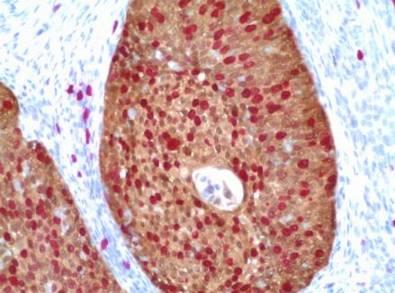
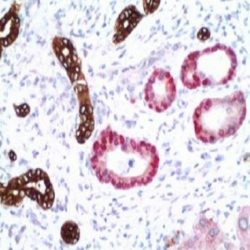
ANALYTICAL PRINCIPLE OF THE METHOD
The purpose of immunohistochemically or immunocytochemical staining is to convert the antibody-antigen specific
reaction of molecules intended to be studied in cells or tissues into a visible stain.
This way, the p16-INK4/Ki67 Dual Staining Kit presents a set of highly specific and sensitive reagents that allows the
double visualization of the binding of two antibodies to their specific antigens in the same sample.
The functioning of this kit is based on the use of a mix of two antibodies and a mix of two micropolymers, each of them
marked with different enzymes (Peroxidase and Alkaline Phosphatase), which recognize each one the immunoglobulins
(antibodies) developed in rabbit and mouse respectively.
In the event of a reaction between the primary antibodies and their antigens, the micropolymers are specifically bound
to each one of them and due to the enzyme marking, adding the chromogens and the corresponding substrates,
precipitates of different colors are obtained that allow the presence of specific antigens to be detected under a
microscope.
COMPONENTS AND REAGENTS INCLUDED IN THE KIT:
MAD-021540Q-10 – Peroxidase Blocking Reagent : 10 mL
MAD-000788QD-10 – p16-INK4/Ki67 (MX007/SP6)* : 10 mL
MAD-001882QK-C – POLYMER MIX : 10 mL
MAD-001812QK-A – DAB Substrate Buffer : 15 mL
MAD-001812QK-B – DAB Chromogen Concentrate : 0.5 mL
MAD-001818QK-A – AP Substrate Buffer ; 2×15 mL
MAD-001818QK-B – AP Chromogen Concentrate : 0.5 mL
MAD-001560Q-10 – DAB Enhancer : 10 mL
1. Dewaxing and Heat-induced epitope retrieval
a. Incubate the slides with the paraffin-embedded tissue sections in the oven at 60ºC all night long.
b. Place the slides in the PT module using the following conditions:
– Pre-heat at 65ºC
– Dewax and retrieve 20 minutes at 95ºC
In case you do not have a PT module, you can use other instruments such as a pressure cook or microwave for antigen
retrieval under previous validation of the functioning conditions in each laboratory, using the following
recommendations:
c. Dewax with xylene.
d.Hydrate the samples with decreasing alcoholic solutions.
e. Hydrate with bidistilled or deionized water.
f. Heat-induced antigen retrieval
2. Detection and development
a. Wash in TBS Buffer – Tween 20 at RT
b. Apply 100 µl of Peroxidase Blocking Reagent and incubate for 10 min at RT
c. Wash 3 time in TBS buffer – Tween 20.
d. Apply 100 µl of p16-INK/Ki67 (MX007/SP6) and incubate for 10 min at RT.
e. Wash 3 times in TBS buffer – Tween 20.
f. Apply 100 µl of POLYMER MIX and incubate for 30 min at RT.
g. Wash 3 times in Distilled Water
h. Mix a drop of DAB Chromogen Concentrate in 1ml of DAB Substrate Buffer and apply the resulting solution on the
tissue; incubate for 5 min at RT
i. Wash 3 times in TBS buffer – Tween 20.
j. Mix a drop of AP Chromogen Concentrate in 2,5ml of AP Substrate Buffer and apply the resulting solution on the
tissue; incubate for 15 min at RT
k. Wash 3 times in Distilled water.
l. Apply 100 µl of DAB Enhancer and incubate for 2 min at RT.
m. Wash 3 times in TBS buffer – Tween 20.
3. Counterstaining and mounting
a. Stain with Contrast Hematoxylin for 30 seconds.
b. Dye blue in running water.
c. After washing with water, dry the slides at 50-60ºC for at least 30 minutes or 1 hour at Room Temperature, rinse
in xylene and mount with permanent mounting medium.


دیدگاهها
هیچ دیدگاهی برای این محصول نوشته نشده است.