-
 آنتی بادیهای ایمونوهیستوشیمی
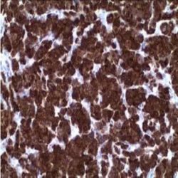
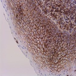
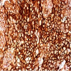
آنتی بادی ALK/p80 (5A4)
Name: ALK (Anaplastic Lymphoma Kinase) / p80 Antibody
Description and aplications: This Antibody recognizes the hybrid p80NPM/ALK chimeric protein od 80kDa, a product of the t (2; 5) (p23; q35) translocation between the anaplastic lymphoma kinase gene and the nucleophosmin gene which is specifically expressed in 40% of Ki-1 lymphoma (anaplastic large cell lymphoma). In addition to the anaplastic large cell lymphoma, the antibody can be used for the identification of lung adenocarcinomas harbouring rearrangements of the ALK gene and which have shown dramatic improvement Other neoplasm and reactive conditions like some diffuse large B cell lymphomas (DLBCL), neuroblastomas, rare anaplastic carcinomas of the thyroid, kidney, oesophagus, colon and breast, various sarcomas (rare rhabdomyosarcomas, lipogenic tumors, Ewing/PNET sarcomas and leyomiosarcomas) or the inflammatory myofibroblastic tumour might also express the protein not necessary related with the specific translocation.
Composition: anti- ALK mouse monoclonal antibody obtained from supernatant culture and prediluted in a Tris buffered solution pH 7.4 containing 0.375mM sodium azide solution as bacteriostatic and bactericidal. The quantity of the active antibody was not determined.
Immunogen: Recombinant protein corresponding to a region which spans the tyrosine kinase catalytic domain and part of the C-terminus of the NPM-ALK transcript (419-520aa)
-
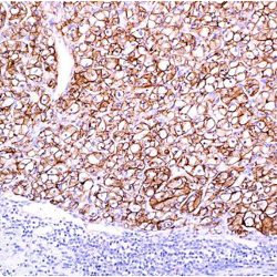
 آنتی بادیهای ایمونوهیستوشیمی
آنتی بادی Alpha-Fetoprotein (EP209)
Name: Antibody AFP(alpha-fetoprotein) clone EP209
Description and applications: Alpha-fetoprotein (AFP) is the most abundant plasma protein found in the human fetus. It is thought to be the fetal form of serum albumin. AFP binds to copper, nickel, fatty acids and bilirubin and is found in monomeric, dimeric and trimeric forms. Alpha-Fetoprotein (AFP) is synthesized by the cells of the embryonic yolk sac, fetal liver and fetal intestinal tract. AFP levels decrease soon after birth. In abnormal tissues, expression of AFP has been demonstrated in hepatocellular carcinoma, hepatoid adenocarcinoma, germ cell tumors, particularly yolk sac tumor. The anti-AFP antibody may be useful for the identification of neoplastic liver diseases or yolk sac tumors.
Composition: anti-human AFP rabbit monoclonal antibody purified by protein affinity and prepared in 10mM PBS, pH 7.4, with 0.2% BSA and 0.09% sodium azide
Immunogen: A synthetic peptide corresponding to residues of human AFP protein
-
 آنتی بادیهای ایمونوهیستوشیمی
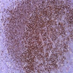
آنتی بادی Amyloid A (MC1)
Name: Amyloid-A Monoclonal Antibody
Description and applications: Este anticuerpo reacciona con depósitos de amiloide de tipo AA, tanto la forma nativa como desnaturalizada de la proteína fibrilar. Este anticuerpo reacciona con depósitos de amiloide en todos los órganos y tejidos, así como con el precursor sérico de la proteína AA.
Composition: anti-Amyloid-A mouse monoclonal antibody obtained from supernatant culture and prediluted in a tris buffered solution pH 7.4 containing 0.375mM sodium azide solution as bacteriostatic and bactericidal.
-
 آنتی بادیهای ایمونوهیستوشیمی
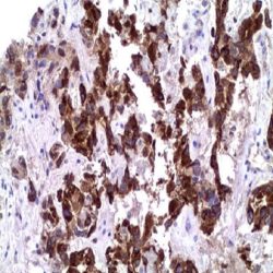
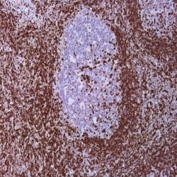
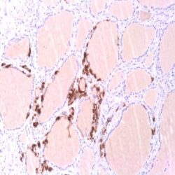
آنتی بادی CD23 (SP23)
Name: CD23 Monoclonal Antibody clone SP23
Description and applications: The role of IgE immunoglobulins in the initiation and development of acute allergic processes is well known. These processes are generally initiated through its interaction with high affinity receptors for Fc portion (FceRI) located mainly on the surface of mast cells and basophil granulocytes where it regulates exocytosis phenomena and subsequent degranulation of mediators of hypersensitivity reactions type I. Furthermore, the immunoglobulin IgE also binds specifically to an integral membrane glycoprotein of 45- 55 kDa type 2 (321 aa) that does not belong to the immunoglobulin superfamily but to the selectins and are included under the name of low affinity receptor of IgE (FceRIIa+b / CD23). CD23 is expressed in a wide range of cell types, including B lymphocytes, eosinophils, granulocytes, platelets, T and NK cells, dendritic reticulum cells and Langerhans cells. CD23 expression has been detected in neoplastic lymphoid cells of chronic lymphocytic leukemia, in a variable percentage of follicular lymphomas and in a small proportion of large B-cell lymphomas. Furthermore and although the CD23 molecule is produced by B cells, is adsorbed in a large amount on follicular dendritic reticulum cells thus is also used for the identification of its neoplasms (dendritic reticulum cell sarcoma).
Composition: anti-CD23 mouse monoclonal antibody obtained from supernatant culture and prediluted in a tris buffered solution pH 7.4 containing 0.375mM sodium azide solution as bacteriostatic and bactericidal.
Immunogen: Recombinant external domain of CD23 protein.
-
 آنتی بادیهای ایمونوهیستوشیمی
آنتی بادی CD44 (SP37)
Name:Mouse anti-human anti-CD44 Std./HCAM Monoclonal Antibody Clone SP37.
Description and aopplications: CD44 is a transmembrane type I glycoprotein involved in adhesion between cells and different components of the extracellular matrix, expr many human tissues. CD44 is also known as homing cell adhesion molecule (H-CAM), Phagocytic glycoprotein-1 (PgP-1), ECM-III, HUTCH-1, or Hermes-1. This antibody recognize CD44 standard isoform (CD44std), which comprises exons 1-5 and 16-20, present in most cells as a glycoprotein of approximately 90 kDa that acts as the main ligand for hyaluronic acid. For its behavior as a receptor for hyaluronic acid, CD44s molecule which is expressed on leukocytes including many T and B lymphocytes, medullary thymocytes, monocytes and granulocytes, plays an important role in intercellular adhesion of cells to the basement membrane and in maintaining its polarity. In tumors, the loss of CD44s expression is generally considered a factor of poor prognostic and in most cases is associated with the presence of distant metastases. Besides determining CD44s may be useful in the diagnosis of malignant urothelial lesions in their early stages, which would be characterized by a membrane staining restricted to the basal layers of the transformed epithelium compared with diffuse staining normally present in the normal urothelium.
Composition: anti-human CD44std mouse monoclonal antibody purified from ascites fluid by Protein A chromatography. Prepared in 10mM PBS, pH 7.4, with 0.2% BSA and 0.09% sodium azide.
.Immunogen: Stimulated human leukocytes
-
 آنتی بادیهای ایمونوهیستوشیمی
آنتی بادی p27 (EP104)
(Name:Mouse Anti-p27 Monoclonal Antibody (Clone EP104
Description and aplications: This antibody recognizes a protein of 27 kDa identified as p27kip1, a mitotic inhibitor cell cycle regulator. The epitope extends between amino acids 83 to 197 of p27. This antibody is highly specific and does not cross react with other mitotic inhibitors. The p27 functions as a negative regulator of G1 progression and has been proposed as a possible mediator of cell G1 arrest induced by TGF-Beta. Is structurally related with Kip1 Cip1 / WAF1, with a similar sequence of 60 amino acids at the N-terminal region. In vitro, p27Kip1 strongly binds to cyclin D-Cdk4, cyclin E-Cdk2 and cyclin A-Cdk2 and inhibits cdk activity. In normal cells the level of Kip1 activity gradually decreases as the cells reach the stage S. The addition of TGF-b in early G1 prevents this decrease of activity, apparently by preventing Kip1 sequestration that occurs in cells that have not been treated with this factor. The treatment with TGF-b also reduces the levels of Cdk4. Kip1 preferentially binds to cyclin D-Cdk4, but the lower levels of Cdk4 in cells treated by TGF-b, presumably allow for some of the Kip1 to bind to the cyclin E-Cdk2 and cyclin A-Cdk2.
Composition: anti-p27 mouse monoclonal antibody obtained from supernatant culture and prediluted in atris buffered solution pH 7.4 containing 0.375mM sodium azide solution as bacteriostatic and bactericidal.
.Immunogen: mouse p27 recombinant protein Ig
-
 آنتی بادیهای ایمونوهیستوشیمی
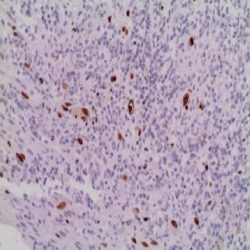
آنتی بادی Cytomegalovirus (DDG9/CCH2)
(Name: Mouse anti-CMV (Cytomegalovirus) cocktail of Monoclonal Antibodies (Clone DDG9/CCH2
Description and aplications: Cytomegalovirus is a member of the herpes virus group, which includes herpes simplex virus types 1 and 2, varicella zoster virus (which causes chicken pox), and Epstein Barr virus (which causes infectious mononucleosis). These viruses share a characteristic ability to remain dormant within the body over a long period.This antibody cocktail reacts with an immediate early antigen and an early CMV antigen. DDG9 is isotype IgG2a, kappa whilst CCH2 is isotype IgG1, kappa. The antibodies stain infected cells giving a nuclear staining pattern with an immediate early and an early antigen.
Composition: anti-CMV is a cocktail of two mouse monoclonal antibodies from tissue culture supernatant diluted in tris buffered saline, pH 7.3-7.7, with protein base, and preserved with sodium azide.
.Immunogen: Cytomegalovirus-infected cell lysate
-
 آنتی بادیهای ایمونوهیستوشیمی
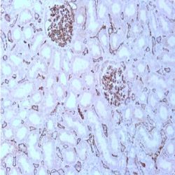
آنتی بادی C4d (Polyclonal)
Name: Rabbit anti-human C4d Polyclonal Antibody
Description and applications: This antibody recognizes the C4d fraction of complement. C4d fraction of complement represents the alpha-2 (42 kDa) portion of the C4 fraction of complemnto. Among the different complement proteins, the opsonins C3 and C4 have a protector thioester radical. This molecule allows the C4 (and C3) complement fraction to form covalent bonds when activated with the target molecule, thereby generating the C4b (C3b) molecule. When the proteolytic cleavage of C4b occurs, C4d fragment remains covalently attached to the target structure, while the C4c fragment is free.
Consecutively, C4d is a stable split product remnant of classical complement activation and an established marker of antibody-mediated acute renal allograft rejection. Due to its proclivity for endothelium, this component can be detected in peritubular capillaries in both chronic renal allograft rejection as well as hyperacute rejection, acute vascular rejection, acute cellular rejection, and borderline rejection. It has been shown to be a significant predictor of transplantkidney graft survival and is an aid in treating acute rejection.Composition: anti-human C4d rabbit polyclonal antibody purified from ascites. Prepared in 10mM PBS, pH 7.4, with 0.2% BSA and 0.09% sodium azide
Intended use: Immunohistochemistry (IHC) on paraffin embedded tissues. Not tested on frozen tissues or Western-Blotting
-
 آنتی بادیهای ایمونوهیستوشیمی
آنتی بادی CA125 (Mucin-16) (OC125)
(Name: Mouse anti-human CA125 (Mucin-16) Monoclonal Antibody (Clone OC125
Description and applications: CA 125 (Cancer Antigen 125) is a mucin-like glycoprotein with a molecular weight greater than 200 kD. OC 125 recognizes an epitope that is considered to be a peptide in nature. Anti-CA-125 antibody reacts with epitheloid malignancies of the ovary, papillary serous carcinoma of the cervix, adenocarcinoma of the endometrium, clear cell adenocarcinoma of the bladder, and epitheloid mesothelioma. The antigen is formalin resistant, permitting the detection of ovarian cancer by immunohistochemistry, although serum assays for this protein are widely used to monitor ovarian cancer. CA 125 is located on the surface of ovarian tumor cells with essentially no expression in normal adult ovarian tissue. CA125 is also found in sera of patients with pancreatic, liver, colon, and other adenocarcinomas.1 These references are for presentation in vials of Low Density Polyethylene (LDPE) dropper. In case the products are used in automated stainers, a special reference is assigned as follows: – / L: Cylindrical screw-cap vials (QD-3 / L, QD-7 / L, QD-12 / L). – / N: Polygonal screw-cap vials (QD-3 / N, QD-7 / N, QD-12 / N). For different presentations (references / volumes) please contact the supplier. CA 125 is found in the epithelial cells of normal colon, gall bladder, mammary gland, and stomach. Normal breast, liver, skin, and spleen are usually negative. When used with other antibodies, CA 125 antibodies are useful for the separation of colonic carcinoma from ovarian endometroid carcinoma in the pelvis and distinguishing renal clear cell carcinoma from clear cell carcinoma of the ovary and other Mullerian tumors.
Composition: anti-human CA125 mouse monoclonal antibody purified from ascites. Prepared in 10mM PBS, pH 7.4, with 0.2% BSA and 0.09% sodium azide.
Immunogen: Ovarian cancer cell line OVCA433 derived from patient with serous papillary cystadenocarcinoma.
-
 آنتی بادیهای ایمونوهیستوشیمی
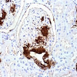
آنتی بادی Calcitonin (SP17)
.(Name: Mouse anti-human Calcitonin Monoclonal Antibody (Clone SP17
Description and applications: Calcitonin is a 32 amino acid peptide which can be demonstrated in C cells of the normal and hyperplastic thyroid. Staining for calcitonin may be used for the identification of a spectrum of C cell proliferative abnormalities ranging from C cell hyperplasia to invasive tumors. Staining for calcitonin in medullary carcinoma of the thyroid produces a fine granular pattern in the cytoplasm. Amyloid deposits within the tumor may also exhibit varying degrees of calcitonin activity.
Composition: anti-human Calcitonin rabbit monoclonal antibody purified from ascites fluid by Protein A chromatography. Prepared in 10mM PBS, pH 7.4, with 0.2% BSA and 0.09% sodium azide.
Immunogen: Synthetic human calcitonin 1-32 amino acid peptide.
-
 آنتی بادیهای ایمونوهیستوشیمی
آنتی بادی Caldesmón (h-CALD)
.(Name: Mouse anti-human Caldesmon Monoclonal Antibody clone (h-CALD
Description and applications: The antibody recognizes a protein of 150kDa, which is identified as the high molecular weight variant of Caldesmon. Two closely related variants of human caldesmon have been identified which are different in their electrophoretic mobility and cellular distribution. The h-caldesmon variant (120–150kDa) is predominantly expressed in smooth muscle whereas l-caldesmon (70–80kDa) is found in non- muscle tissue and cells. Neither of the two variants has been detected in skeletal muscle. This MAb recognizes only the 150kDa variant (hcaldesmon) in Western blots of human aortic media extracts and is unreactive with fibroblast extracts from cultivated human foreskin. Caldesmon is a developmentally regulated protein involved in smooth muscle and non-muscle contraction.This antibody may be useful for the study of muscle contraction and as a useful marker of neoplasms of smooth muscle origin. It is not expressed in myofibroblastic cells or tumors derived from them. Nodular fasciitis, fibromatosis and myofibroblastic sarcomas are negative for h-caldesmón.
COMPOSITION: Anti-human Caldesmon mouse monoclonal antibody purified from serum and prepared in 10mM PBS, pH 7.4, with 0.2% BSA and 0.09% sodium azide.
Immunogen: Crude human uterus extract.
-
 آنتی بادیهای ایمونوهیستوشیمی
آنتی بادی Calretinin (BSR235)
.(Name:Rabbit anti-human Calretinin Monoclonal Antibody (Clone BSR235
Description and aplications: This antibody recognizes a protein of 31.5kDa, identified as Calretinin. Calretinin is an intracellular calcium-binding protein belonging to the troponin C superfamily characterized by a structural motif described as the EF-hand domain. The immunohistochemical detection of calretinin in developing cerebellum is restricted to the later stages indicated by weak staining from week 21 of gestation, in Purkinje and basket cells and in neurons of the dentate nucleus. The intensity of staining increases as the cerebellum matures. In tumors, calretinin has been detected in mesotheliomas and some pulmonary adenocarcinomas. However, occasional positivity for calretinin has been described in other tumors such as ameloblastomas, sex cord tumors of the ovary and testis.
Composition: anti-human Calretinin rabbit monoclonal antibody purified from ascites. Prepared in 10mM PBS, pH 7.4, with 0.2% BSA and 0.09% sodium azide.
Immunogen: Recombinant full length mouse calretinin protein.
-
 آنتی بادیهای ایمونوهیستوشیمی
آنتی بادی Carbonic Anhydrase 9 (EP161)
(Name: Rabbit anti-human Carbonic Anhydrase 9 Monoclonal Antibody (Clone EP161.
Description and applications: Carbonic anhydrases (CAs) are a large family of zinc metalloenzymes that catalyze the reversible hydration of carbon dioxide. They participate in a variety of biological processes, including respiration, calcification, acid-base balance, bone resorption, and the formation of aqueous humor, cerebrospinal fluid, saliva, and gastric acid. They show extensive diversity in tissue distribution and subcellular localization. Carbonic Anhydrase 9, a member of the carbonic anhydrase family, is thought to play a role in the regulation of cell proliferation in response to hypoxic conditions and may be involved in oncogenesis and tumor progression.Carbonic Anhydrase 9 (CA9) has a distinctive expression pattern in normal and cancer tissues. The most abundant expression of CA9 was found in normal mucosa of the stomach and gallbladder. Other normal tissues have lower or no expression. Relatively high levels of CA9 are expressed in carcinomas of the cervix, kidney, lung, breast and many other tumors. Most studies have shown that decreased CA9 levels are independently associated with poor survival. Low levels of CA9 maybe benefit more from adjuvant treatment than patients with high levels.
COMPOSITION: Anti-human Carbonic Anhydrase 9 rabbit monoclonal antibody purified from serum and prepared in 10mM PBS, pH 7.4, with 0.2% BSA and 0.09% sodium azide.
IMMUNOGEN: A synthetic peptide corresponding to residues in the extracellular domain in human Carbonic Anhydrase 9 was used as an immunogen.
-
 آنتی بادیهای ایمونوهیستوشیمی
آنتی بادی Carcinoembryonic Antigen (CEAp) (Polyclonal)
Name: Carcino embryonic Antigen (CEAp) Antibody Polyclonal
Description and applications:This antibody has a high affinity for CEA and shows no detectable reactivity to nonspecific cross-reacting antigen (NCA), biliary glycoprotein (BGP) and human polymorphonuclear leucocytes. Ab-3 shows no reaction with a variety of normal tissues .CEA is not found in benign glands, stroma, or malignant prostatic cells. Antibody to CEA is useful in detecting early foci of gastric carcinoma and in distinguishing pulmonary adenocarcinomas (60-70% are CEA+) from pleural mesotheliomas (rarely or weakly CEA+).
Composition: anti-human CEA rabbit polyclonal antibody purified from serum and prepared in 10mM PBS, pH 7.4, with 0.2% BSA and 0.09% sodium azide.
Immunogen: Human CEA isolated from hepatic metastasis of colon adenocarcinoma.
-
 آنتی بادیهای ایمونوهیستوشیمی
آنتی بادی Cathepsin K (3F9)
.(Name: Mouse anti-human Cathepsin K Monoclonal Antibody (Clone 3F9
Description and Applications: Cathepsin K is a lysosomal papain–like cystine proteinase and can be expressed in normal osteoblasts, fibroblasts, and skin. Recently, cathepsin K has been reported as useful in distinguishing translocation renal cell carcinoma (RCC) from other subtypes of RCC. Mortignoni et al. tested 17 cases of translocation RCC and found that cathepsin K stained 6 of 8 cases of translocation RCC with t(X;1), all seven cases of translocation RCC with t(6;11), but was negative for one case with t(X;3) and one with t(X;17).1 Additionally, the study compared IHC detection of TFE3 antibody in the 17 cases and demonstrated that all translocation RCC with t(6;11) were completely negative for TFE3, but positive for cathepsin K; all other translocation RCC with different chromosomal tranlocations other than t(X;1) expressed TFE3. Thus, it is recommended that antibodies against both TFE3 and cathepsin K should be used for evaluating translocation renal cell carcinomas. None of the 305 other renal cell neoplasms representing the most common renal cell neoplasm subtypes were positive for cathepsin K. Zheng et al. reported only 2.7% of 1,140 carcinomas from various organs exhibited cathepsin K labeling, indicating that among carcinomas cathepsin K labeling is highly specific for translocation RCC and can be used to help differentiate translocation RCC from other carcinomas.
COMPOSITION: Anti-human Cathepsin K mouse monoclonal antibody purified from serum and prepared in 10mM PBS, pH 7.4, with 0.2% BSA and 0.09% sodium azide.
-
 آنتی بادیهای ایمونوهیستوشیمی
آنتی بادی Anti-Glycophorin -کلون GA-R2
.(Name: Mouse Anti-Glycophorin A Monoclonal Antibody (Clone GA-R2
Description and aplications: This antibody reacts with a resistant paraffin-embedded and formol fixed epitope located in the extracellular domain of the protein, probably between amino acids 27 and 40. Therefore, this antibody recognizes normal erythroid cells in all stage of differentiation from erythroblasts to mature red blood cells. Once glycophorin A is at its maximum expression, the quantity in each erythroid cell remains constant and shows no change during maturation despite the decreasing size of the cell. The neoplastic erythroblast in most erythroleukemias are identified by Glycophorin A.
Composition: anti-Glycophorin A mouse monoclonal antibody obtained from supernatant culture and prediluted in a tris buffered solution pH 7.4 containing 0.375mM sodium azide solution as bacteriostatic and bactericidal.
Immunogen: membrane preparation from splenic hairy cell leukemia cells.
-
 آنتی بادیهای ایمونوهیستوشیمی
آنتی بادی Cytokeratin (AE1)
.(Name: Mouse anti-human Cytokeratin Monoclonal Antibody (Clone AE1
Description and applications: Due to the high homology among the different molecules, it is common for a monoclonal antibody to react with different types of cytokeratins; for example, anti-cytokeratin AE1 labels medium molecular weight acidic cytokeratins 10, 14, 15, 16, and 19, and low molecular weight acidic cytokeratin 19. Proteins generically called intermediate filaments, because they measure between 7 and 22 nm in diameter (i.e., with a size between actin, 5-7 nm, and tubulin, 22-25 nm), are part, along with actin and tubulin, of the vertebrate cytoskeleton. This superfamily is composed of six subfamilies of molecules with different tissue expression patterns. Cytokeratins constitute homology groups I and II, and in humans they are encoded by more than 49 different genes located in chromosomes 17 (I) and 12 (II). The nomenclature chosen in 1982 by Moll and Franke assigns numbers from 1 to 8 to type II cytokeratins (neutral or basic) and from 9 to 21 to type I cytokeratins (acidic). An analogous nomenclature for hair keratins has now been defined, with the addition of letters Ha and Hb to distinguish type I from type II. Structurally, cytokeratins share with the rest of intermediate filaments a central axisof 310 amino acid residues consisting of four highlypreserved α-helical domains (1A, 1B, 2A, and 2B) that define the type of intermediate filament that will be formed after assembly; these domains are separated by three non-helical linking regions (L1, L12, and L2) and two end domains which are highly different in, size and sequence (head, 1, and tail, 2), each with constant (E1/E2), variable (V1/V2), and homology (H1/H2) regions, the latter characteristic of type II keratins and absent in type I keratins. The major immunogenic properties and the main differences between each keratin class are found in the variable domains. Keratins are usually assembled into I/II heterodimers and are specifically co-expressed in pairs in each tissue. This antibody is widely reactive and labels almost all types of epithelia, thus being one of the best antibodies for the preliminary screening of neoplasms of an epithelial origin, and specially for the identification of malignant metastatic tumours of this origin. It does not react against all hepatocarcinomas, basal cell carcinomas, and adrenocortical carcinomas, and it partially reacts against renal cell carcinomas, small cell lung carcinomas, and large-cell carcinomasof the lung.
IMMUNOGEN: Epidermal cytokeratin.
-
 آنتی بادیهای ایمونوهیستوشیمی
آنتی بادی Cytokeratin (AE3)
.(Name: Mouse anti-human Cytokeratin Monoclonal Antibody (AE3
Description and applications: Due to the high homology among the different molecules, it is common for a monoclonal antibody to react with different types of cytokeratins; for example, anti-cytokeratin AE3 labels high molecular weight basic cytokeratins 1, 2, 3, 4, 5, and 6, as well as low molecular weight basic cytokeratins 7 and 8. Proteins generically called intermediate filaments, because they measure between 7 and 22 nm in diameter (i.e., with a size between actin, 5-7 nm, and tubulin, 22-25 nm), are part, along with actin and tubulin, of the vertebrate cytoskeleton. This superfamily is composed of six subfamilies of molecules with different tissue expression patterns. Cytokeratins constitute homology groups I and II, and in humans they are encoded by more than 49 different genes located in chromosomes 17 (I) and 12 (II). The nomenclature chosen in 1982 by Moll and Franke assigns numbers from 1 to 8 to type II cytokeratins (neutral or basic) and from 9 to 21 to type I cytokeratins (acidic). An analogous nomenclature for hair keratins has now been defined, with the addition of letters Ha and Hb to distinguish type I from type II. Structurally, cytokeratins share with the rest of intermediate filaments a central axis of 310 amino acid residues consisting of four highly preserved α- helical domains (1A, 1B, 2A, and 2B) that define the type of intermediate filament that will be formed after assembly; these domains are separated by three non-helical linking regions (L1, L12, and L2) and two end domains which are highly different in size and sequence (head, 1, and tail, 2), each with constant (E1/E2), variable (V1/V2), and homology (H1/H2) regions, the latter characteristic of type II keratins and absent in type I keratins. The major immunogenic properties and the main differences between each keratin class are found in the variable domains. Keratins are usually assembled into I/II heterodimers and are specifically co-expressed in pairs in each tissue. This antibody is highly reactive and positively stains almost all epithelial tissues and their neoplasms, including the skin, the esophagus, and the urinary bladder. Moreover, it labels pancreatic ducts and parenchymal cells and it does not stain mesenchymal cells. It does not react against all hepatocarcinomas. It does not cross-react with other types of filaments, including vimentin, desmin, and neurofilaments.
COMPOSITION: Anti-human Cytokeratin mouse monoclonal antibody purified from serum and prepared in 10mM PBS, pH 7.4, with 0.2% BSA and 0.09% sodium azide.
-
 آنتی بادیهای ایمونوهیستوشیمی
آنتی بادی Cytokeratin (AE1/AE3)
Name: Mouse Anti-Human Keratin AE1-AE3 Cocktail Antibody clone AE1-AE3
Description and applications: The nomenclature coined in 1982 by Moll and Franke assigned ranges from 1 to 8 for keratin type II (neutral or basic) and between 9 and 21 for type I (acidic). However and because of the high homology among the different molecules is common for a single monoclonal antibody to react with different types of keratins. This antibody, comprising a cocktail of AE1 and AE3 monoclonal antibodies, simultaneously recognizes keratins 10, 14, 15, 16 and 19 and 1, 2, 3, 4, 5, 6, 7 and 8, so that it is considered as one of the broadest spectrum reagents for the identification of epithelial origin in a given neoplasm. However, it should be borne in mind that besides hepatocarcinomas other tumors that often can be negative to the antibody AE1-AE3 are kidney, adrenal, prostate and neuroendocrine carcinomas. For these reasons the AE1-AE3 keratin antibody should be used in conjunction with other broad spectrum keratins.
Composition: anti-Keratin AE1-AE3 mouse antibody obtained obtained from mouse ascites by protein A chromatography in a tris buffered solution pH 7.4 containing 0.375mM sodium azide solution as bacteriostatic and bactericidal. The quantity of the active antibody in the sample is of approximately 4 μ/mL..
Intended use : Immunohistochemistry (IHC) on paraffin embedded tissues. Not tested on frozen tissues or Western-Blotting
Immunogen: Human epidermal cells
-
 آنتی بادیهای ایمونوهیستوشیمی
آنتی بادیCaspase 3-کلونPolyclonal
Name: Caspase 3 Polyclonal Antibody
Description and aplications: Caspase 3 is ubiquitously expressed and like other caspases is synthesized as an inactive (32kDa) proenzyme. Upon activation, Caspase 3 is cleaved at Asp28-Ser29 and Asp175-Ser176 thereby generating two subunits of 17kDa and 12kDa, respectively. Recent studies have implicated that Caspase 3 is associated with the induction of apoptosis. Activation of Caspase 3 occurs in response to variety of apoptotic inducers including Fas mediated apoptosis. This antibody detects both active and inactive forms of Caspase-3 protein. The antibody is useful for studying the phenomenon of cell death by apoptosis as a pathogenic mechanism of numerous pathologies: nerve damage, neurodegenerative diseases, cardiovascular diseases, autoimmune diseases, AIDS and malignancies.
Composition: anti-human Caspase 3 rabbit polyclonal antibody purified from rabbit anti-serum by Protein A chromatography. Prepared in 10mM PBS, pH 7.4, with 0.2% BSA and 0.09% sodium azide
Immunogen: Recombinant full length human.
برای تماس کلیک کنید
سما تشخیص آریا ، عرضه کننده متنوع ترین و کامل ترین محصولات کیت های آزمایشگاهی