برای تماس کلیک کنید

به وب سایت سما تشخیص آریا خوش آمدید.

جستجوی پیشرفته
جستجوی عینی کلمه
جستجو در محتوا
جستجو در عنوان
دنبال محصول یا مطلبی میگردید؟ در این کادر جستجو کنید

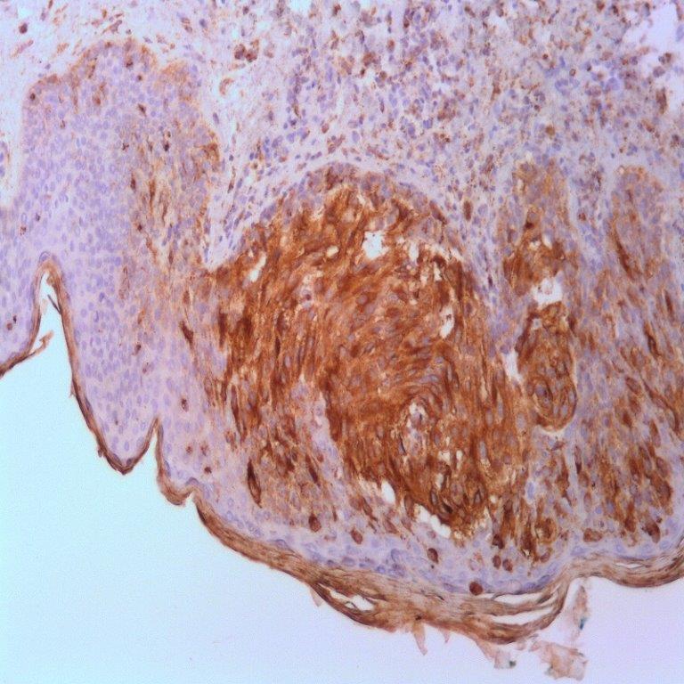
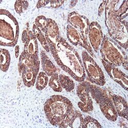
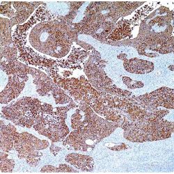
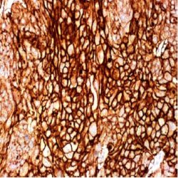
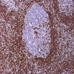
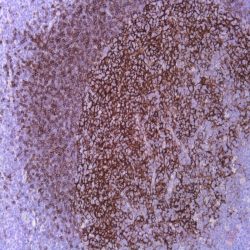
.(Name: Mouse anti-human Cathepsin K Monoclonal Antibody (Clone 3F9

Description and Applications: Cathepsin K is a lysosomal papain–like cystine proteinase and can be expressed in normal osteoblasts, fibroblasts, and skin. Recently, cathepsin K has been reported as useful in distinguishing translocation renal cell carcinoma (RCC) from other subtypes of RCC. Mortignoni et al. tested 17 cases of translocation RCC and found that cathepsin K stained 6 of 8 cases of translocation RCC with t(X;1), all seven cases of translocation RCC with t(6;11), but was negative for one case with t(X;3) and one with t(X;17).1 Additionally, the study compared IHC detection of TFE3 antibody in the 17 cases and demonstrated that all translocation RCC with t(6;11) were completely negative for TFE3, but positive for cathepsin K; all other translocation RCC with different chromosomal tranlocations other than t(X;1) expressed TFE3. Thus, it is recommended that antibodies against both TFE3 and cathepsin K should be used for evaluating translocation renal cell carcinomas. None of the 305 other renal cell neoplasms representing the most common renal cell neoplasm subtypes were positive for cathepsin K. Zheng et al. reported only 2.7% of 1,140 carcinomas from various organs exhibited cathepsin K labeling, indicating that among carcinomas cathepsin K labeling is highly specific for translocation RCC and can be used to help differentiate translocation RCC from other carcinomas.

COMPOSITION: Anti-human Cathepsin K mouse monoclonal antibody purified from serum and prepared in 10mM PBS, pH 7.4, with 0.2% BSA and 0.09% sodium azide.

حجم موجود: 3 / 7 / 12 میلی‌لیتر

مورد استفاده: ایمونوهیستوشیمی روی بافت پارافین. بر روی بافتهای فروزن یا وسترن بلات تست نشده است.

کلون: 3F9

ایزوتایپ: IgG2b

کنترل مثبت IHC: پوست ، ملانوما ، انتقال RCC

قابلیت دیده شدن: سیتوپلاسمی

روش پیشنهادی IHC:

  • برش تهیه شده با ضخامت μm4 باید روی لام شارژ شده در طول شب و در دمای 60 درجه سانتی گراد خشک شود.
  • دپارافینه کردن، رهیدراته کردن و استفاده از حرارت جهت بازیابی اپی توپ : بافت را با استفاده از بافر EDTA pH 8 برای بیست دقیقه در دمای 95 درجه سانتی گراد بجوشانید. سپس 5-3 بار با استفاده از آب مقطر یا دیونیزه شده شسته و سپس بیست دقیقه در دمای اتاق خنک کنید.
  • بلاک پروکسیدازهای درونی : بلاک با استفاده از محلول پروکسیداز به مدت ده دقیقه
  • آنتی بادی اولیه: برای 10 دقیقه در انکوبه کنید پروتکل ممکن است بسته به تهیه نمونه و کاربرد خاص متفاوت باشد. شرایط استاندارد باتوجه به آزمایشگاه و کاربر متفاوت می باشد.
  • برای رنگ آمیزی ، می توانید از کیت Master Polymer Plus Detection یا کیتهای دیتکشن سایر کمپانی ها استفاده کنید.
  • رنگ میزی نهایی با هماتوکسیلین و فیکس نهایی اسلاید.

شرایط نگهداری و پایداری : حداکثر 18 ماه؛ در دمای 2-8 درجه سانتیگراد نگهداری شود. فریز نشود.


این آنتی‌بادی متعلق به پنل‌های تشخیصی DERMATOPATHOLOGY و UROGENITAL است.

دانلود کاتالوگ

Icon

Cathepsin K (3F9) 224.60 KB 8 downloads

...
.well.c2a3 .btn { padding: 11px 30px; font-size: 11pt; } .well.c2a3 .media-body { font-size: 11pt; } .well.c2a3 .wpdm_icon { height: 42px; width: auto; }

نقد و بررسی‌ها

هنوز بررسی‌ای ثبت نشده است.

اولین کسی باشید که دیدگاهی می نویسد “آنتی بادی Cathepsin K (3F9)”

نشانی ایمیل شما منتشر نخواهد شد. بخش‌های موردنیاز علامت‌گذاری شده‌اند *

اسکرول به بالا