دسته: مونوکلونال
نمایش 321–340 از 412 نتیجه
فیلتر ها-
Quartett
آنتی بادی C-Myc کلون QR061 برند Quartett
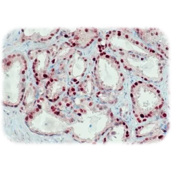
نمره 0 از 5c-Myc is a transcription factor required for cell cycle progression and cell proliferation. Burkitt lymphoma is almost uniformly associated with chromosomal translocations involving c-Myc gene. Additionally, oncogene c-Myc is involved in tumorigenesis of multiple cancers, including breast, liver, pancreas and prostate cancer. [1-5]
-
Quartett
آنتی بادی CD34 کلون QR093 برند Quartett
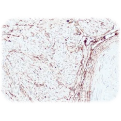
نمره 0 از 5CD34 is a single-pass type I transmembrane glycoprotein and functions as a cell to cell adhesion factor. It is expressed on endothelial cells, hematopoietic progenitor cells, fibroblasts and other stromal components. The protein is thought to mediate the stem cell attachment to the bone marrow, ECM or stromal cells. CD34 is a known marker for quantifying and purifying hematopoietic progenitor cells. In identification of tumors with endothelial or lymphoid differentiation CD34 plays a useful role. It also supports in detection of gastrointestinal stromal tumors.
-
Quartett
آنتی بادی CD56(NCAM-1) کلون QR044 برند Quartett
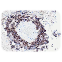
نمره 0 از 5CD56, also known as neural cell adhesion molecule (NCAM), is a part of the immunoglobulin superfamily of adhesion molecules and mediates homophilic interactions through its extracellular region. It is used as a marker fordetection of T-LGL (T-Large Granular Lymphocyte) lymphoproliferative disorders and NK cells. CD56 is also known as a marker of neural lineage due to its discovery site. For example, astrocytoma, neuroblastoma or small cell carcinoma of lung are CD56-positive. [1-6]
-
Quartett
آنتی بادی Chromogranin A کلون QR096 برند Quartett
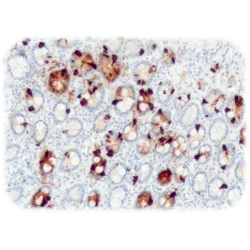
نمره 0 از 5Chromogranin A is a member of the granin family of neuroendocrine secretory proteins and is located in secretory vesicles of neurons and endocrine cells. It regulates function of secretory granula, is involved in hormone processing and secretion, and is disassembled to biologically active peptides.
Chromogranin A is the most important and most common granin in neuroendocrine tumors. Antibody Chromogranin A is the single most specific marker for such tumors including anterior pituitary, gastrointestinal tract, pancreas, thyroid and lung as well as neuronal tumors and merkel cell carcinomas. Co-expression of chromogranin A and neuron specific enolase (NSE) is common in neuroendocrine neoplasms. -
Quartett
آنتی بادی CD8 کلون QR068 برند Quartett
نمره 0 از 5CD8 is a transmembrane glycoprotein and co-receptor that functions together with T cell receptor (TCR) in adaptive immune response. CD8 is mainly expressed on cytotoxic T cells as well as natural killer cells, cortical thymocytes and dendritic cells. Furthermore, CD8 is expressed in T cell LGL leukemia and together with CD4 in some T lymphoblastic lymphoma.CD8 can be detected in colorectal carcinoma and ovarian, renal, hepatocellular and esophageal tumors.
-
Quartett
آنتی بادی CD99 کلون QR067 برند Quartett
نمره 0 از 5CD99 is an O-glycosylated transmembrane protein and the gene product of MIC2 [1]. It is expressed on Ewing’s sarcoma/primitive neuroectodermal tumor (ES/PNET) cells, Sertorli cells, pancreatic islet cells, thymocytes and other hematopoietic cells. CD99 plays a role in the T-cell adhesion, differentiation of primitive neuroectodermal cells and leukocyte migration.
-
Quartett
آنتی بادی Calretinin کلون QR059 برند Quartett
نمره 0 از 5Calretinin is a calcium binding protein abundantly expressed in neurons. Outside the nervous system, calretinin is found in mesothelial cells, steroid producing cells, testicular Sertoli cells, rete testis, ovarian surface epithelium, some neuroendocrine cells, breast glands, eccrine sweat glands, hair follicular cells, thymic epithelial cells, endometrial stromal cells and fat cells.
-
Quartett
آنتی بادی L1CAM(CD171) کلون QR039 برند Quartett
نمره 0 از 5CD171 (Cluster of Differentiation), also known as L1 cell adhesion molecule isoform 1 precursor (L1CAM), is a transmembrane protein and a member of the immunoglobulin superfamily. This cell adhesion molecule has an important role in the development of the nervous system, including neuronal migration and differentiation.CD171 is involved in neuron-neuron adhesion, neurite fasciculation, outgrowth of neuritis, and others.
-
Quartett
آنتی بادی Cytokeratin 14 کلون QR057 برند Quartett
نمره 0 از 5Keratins are cytoplasmic intermediate filament proteins expressed by epithelial cells. Cytokeratin 14 is a type I keratin that is found in basal cells of squamous epithelia, some glandular epithelia, myoepithelium and mesothelial cells in various tissues including prostate and breast. A cocktail of CK14, its corresponding partner CK5 and p63 is useful to detect the basal-like phenotype of breast carcinoma and to differentiate normal and prostate cancer.
-
Quartett
آنتی بادی Caldesmon کلون QR063 برند Quartett
نمره 0 از 5Caldesmon is a regulatory protein that interacts with actin, myosin, tropomyosin, and calmodulin. It is found in smooth muscle and other tissues. Caldesmon is useful to distinguish smooth muscle and it is more specific than desmin and muscle specific actin. Caldesmon antibody is a marker to identify epitheloid mesothelioma and it can be used to differentiate uterus leiomyoma from endometrial stroma tumor.
-
Quartett
آنتی بادی CD21 کلون QR076 برند Quartett
نمره 0 از 5CD21 (Cluster of Differentiation 21) is a transmembrane protein involved in the complement system. It is the complement receptor for C3d and the Epstein-Barr virus. This antibody recognizes follicular dendritic cells, mature marginal and mantle B-cells as well as neoplasms derived from follicular dendritic cells.
-
Quartett
آنتی بادی CD71 کلون QR073 برند Quartett
نمره 0 از 5CD71 (Cluster of Differentiation 71) is required for iron delivery from transferrin to cells and is highly expressed on placental syncytiotrophoblasts, myocytes, basal keratinocytes, hepatocytes, endocrine pancreas, spermatocytes and erythroid precursors. This antibody detects erythroid precursors in bone marrow, acute myeloid leukemia and myelodysplastic syndrome (MDS).
-
Quartett
آنتی بادی CD31(PECAM-1) کلون QR034 برند Quartett
نمره 0 از 5CD31 is an integral membrane glycoprotein located on the surface of endothelial cells, platelets and some hematopoietic cells. It plays a key role in removing aged neutrophils from the body. CD31 is often used as an endothelial cell marker. The antibody recognizes cells from arteries, arterioles, venules, veins, non-sinusoidal capillaries of various tissues as well as tumors of endothelial origin and vascular invasions of tumors. CD31 reacts with normal, benign and malignant endothelial cells that form the lining of the blood vessels. The extent of CD31 expression can help determine the degree of tumor angiogenesis.
-
Quartett
آنتی بادی CD138 کلون QR102 برند Quartett
نمره 0 از 5CD138, also known as syndecan-1, is a transmembrane heparan sulfate proteoglycan and is a member of the syndecan proteoglycan family, which mediate cell binding, cell signaling and cytoskeletal organization. The syndecan 1 protein participates in cell proliferation, cell migration and cell-matrix interactions. This antibody is a useful marker for labeling normal mature plasma cells and early pre B-cells, while other haematolymphoid cells are negative. Various types of epithelial cells are also CD138 positive. Among haematolymphoid neoplasms, CD138 is expressed in practically all cases of plasma cell malignancies. Among non-haematolymphoid neoplasms, the expression of CD138 is found in various types of carcinomas.
-
Quartett
آنتی بادی Cytokeratin PAN کلون MNF-116 برند Quartett
نمره 0 از 5Anti-human antibody for immunohistochemical use. The primary antibody is intended for qualitative detection of antigens in formalin-fixed, paraffin-embedded (FFPE) tissue sections. The antibody may be used manually or with any automated staining platform. Authorized and skilled personnel may only use the product. The clinical interpretation of any test results should be evaluated within the context of the patient’s medical history and other diagnostic laboratory test results. A qualified pathologist must perform evaluation.
-
Quartett
آنتی بادی Cadherin 17 کلون QR098 برند Quartett
نمره 0 از 5Cadherins are calcium dependent cell adhesion proteins that play a role in cell recognition and segregation, morphogenetic regulation, and tumor suppression. Cadherin 17 may have a role in the morphological organization of liver and intestine and may be involved in intestinal peptide transport. Expression is found in the gastrointestinal tract and pancreatic duct.
-
Quartett
آنتی بادی CD45(LCA) کلون QR106 برند Quartett
نمره 0 از 5CD45, also known as leucocyte common antigen, is a transmembrane protein-tyrosine phosphatase, which is found in almost all hematolymphoid cells including lymphocytes, monocytes, macrophage and granulocytes. This antibody recognizes hematolymphoid neoplasms/tumors, it is non-reactive with some lymphoid neoplasms like Hodgkin’s disease, some T-cell lymphoma and some leukemias.CD45 is an important marker in the primary tumor screening panel in order to identify hematolymphoid differentiation. Loss of CD45 in precursor B-cell neoplasmsis a negative prognostic parameter.
-
Quartett
آنتی بادی CD5 کلون QR111 برند Quartett
نمره 0 از 5CD5, also known as Lymphocyte Antigen T1/Leu-1, is a transmembrane glycoprotein involved in regulation of Tcell proliferation. CD5 is a pan T-cell marker that is also found on a subset of B cells, but not on the majority of peripheral blood B cells. Furthermore, T cell malignancies, B cell chronic lymphocytic leukemia (CLL)/small lymphocytic lymphoma(SLL) and mantle-cell lymphomas are CD5 positive.
-
Quartett
آنتی بادی CD30 کلون QR109 برند Quartett
نمره 0 از 5CD30 is a cell membrane protein, that regulates activation of NF-kappa B and apoptosis. It is expressed by Reed-Sternberg cells of classical Hodgkin’s disease. CD30 is a member of the tumor necrosis factor receptor (TNF-R)superfamily, which comprises more than 10 different members. CD30 has an extra cytoplasmic domain, transmembrane region, and a cytoplasmic domain. The protein is heavily glycosylated within the Golgi apparatus(120 kDa). This antibody detects Hodgkin‘s lymphoma, anaplastic large cell lymphomas, primary cutaneousCD30+ T-cell lymphoproliferative disorders and embryonal carcinomas.
-
Quartett
آنتی بادی Cytokeratin 8 کلون QR112 برند Quartett
نمره 0 از 5Keratins are cytoplasmic intermediate filament proteins expressed by epithelial cells. Cytokeratin 8 is a type II keratin that is expressed in early embryogenesis. It is found in simple epithelia in respiratory, gastrointestinal and reproductive tract as well as thyroid. This antibody detectsmost non-squamous epithelial tumors andadenocarcinomas of the breast, ovary, gastrointestinal tract, thyroid, pancreas, bile duct and salivary glands. It is often co-expressed with cytokeratin 18, and the major keratin pair in simple-type epithelia, as found in the liver, pancreas, and intestine.

