دسته: مونوکلونال
نمایش 441–460 از 477 نتیجه
فیلتر ها-
آنتی بادیهای ایمونوهیستوشیمی
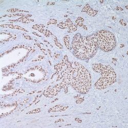
آنتی بادی PR (Progesterone Receptor) کلون Y85 برند Patho Sage
نمره 0 از 5Specification:
This anti-Progesterone Receptor antibody reacts with progesterone receptor forms alpha and beta. This antibody stains nuclei in breast, ovarian and endometrial epithelia, as well as myometrial nuclei. Since the early 1990’s the immunohistochemical (IHC) assay determination of progesterone receptor status has replaced the dextran-coated charcoal method as a prognostic indicator in breast carcinoma. IHC has shown to be superior in prognostic significance when using any one of several available methods of quantitation using this technique.
Presentation:
Anti-Progesterone Receptor is a rabbit monoclonal from tissue culture supernatant diluted in tris buffered saline, pH 7.3-7.7, with protein base, and preserved with sodium azide
-
آنتی بادیهای ایمونوهیستوشیمی
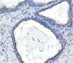
آنتی بادی Prostein کلون ZR9 برند Patho Sage
نمره 0 از 5Specification:
Human prostein is a 553 aa protein identified by cDNA library substraction abd subsequent high throughput microarray a screening of prostate cancer. Prostein has multiple transmembrane domains. Prostein has been shown to be uniquely expressed in normal and cancerous prostatic tissues. By immunohistochemistry, prostein is expressed in the vast majority of normal and malignant prostatic tissues, regardless of grade and metastatic status. No protein expression is detected in normal and malignant tissue samples representing the great majority of essential tissues and tumors. Prostein is expressed in most of poorly differentiated prostatic carcinoma, including small cell prostate carcinoma. Prostein is more specific and sensitive for prostatic carcinomas than PSA and PSAP.
Immunogen:
Synthesized peptides to the N-terminus of human prostein
Presentation:
Tissue culture supernatant with 0.2% BSA and 15mM sodium azide
-
آنتی بادیهای ایمونوهیستوشیمی
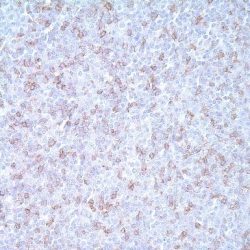
آنتی بادی CD5 کلون 4C7 برند Patho Sage
نمره 0 از 5Specification:
CD5 is a T-cell marker that also reacts with a range of neoplastic B-cells, e.g. B-cell chronic lymphocytic leukemia (B-CLL), Bcell small lymphocytic lymphoma (B-SLL), and mantle cell lymphoma. CD5 is expressed in T lymphocyte subsets and is modulated during cellular activation. CD5 does not react with granulocytes or monocytes.
Presentation:
Anti-CD5 is a mouse monoclonal antibody from supernatant diluted in tris buffered saline, pH 7.3-7.7, with protein base, and preserved with sodium azide
-
آنتی بادیهای ایمونوهیستوشیمی
آنتی بادی Podoplanin کلون 1433 برند Patho Sage
نمره 0 از 5Specification:
It recognizes a muco-protein of 38-43kDa, which is identified as Podoplanin (PDPN). It localizes in stromal cells of peripheral lymphoid tissue and thymic epithelial cells. As a regulator of the lymphatic endothelium, Podoplanin probably plays a role inthe maintaining the unique shape of podocytes. It is selectively expressed in lymphatic endotheliomas well as lymphoangiomas, Kaposi sarcomas and in the subset of angiosarcomas with probable lymphatic differentiation. Recent studies have also shown Podoplanin to be a highly sensitive and relatively specific marker for epithelioid mesothelioma. Therefore, it can be used in a panel to distinguish mesotheliomas or mesothelial cells from pulmonary carcinomas.
Presentation:
Bioreactor Concentrate with 0.05% BSA and 0.05% Azide
-
آنتی بادیهای ایمونوهیستوشیمی
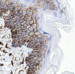
آنتی بادی CD44 کلون 156-3C11 برند Patho Sage
نمره 0 از 5Specification:
This antibody recognizes a cell surface glycoprotein of 80-95kDa (CD44) on lymphocytes, monocytes, and granulocytes (Leucocyte Typing Workshop V). Its epitope is resistant to digestion by trypsin and chymotrypsin. This MAb selectively interferes with lymphocyte binding to lymph node, mucosal and synovial endothelium. The CD44 family of glycoproteins exists in several variant isoforms, the most common being the standard 85-95kDa or hematopoietic variant (CD44s). Higher molecular weight isoforms are described in epithelial cells (CD44v), which are believed to function in intercellular adhesion and stromal binding. CD44 immunostaining is commonly used for the discrimination of urothelial transitional cell carcinoma in-situ from non-neoplastic changes in the urothelium.
Immunogen:
Stimulated human leukocytes
Presentation:
Protein A/G purified antibody from Bioreactor Concentrate with 0.05% BSA and 0.05% Azide.
-
آنتی بادیهای ایمونوهیستوشیمی
آنتی بادی Mammaglobin کلون 304-1A5 برند Patho Sage
نمره 0 از 5Specification:
Mammaglobin is a breast-associated glycoprotein distantly related to secretoglobin family that includes human uteroglobin and lipophilin. Unlike other secretoglobin family members, mammaglobin mRNA expression is breast specific, which has been shown to be a very sensitive marker of occult breast cancer cells in sentinel lymph nodes and peripheral blood. By paraffin immunohistochemistry, the overall sensitivity of mammaglobin for breast cancers was reported about 80%. When combined with other breast-restricted markers such as GCDFP-15, an overall sensitivity of 84% could be achieved. Mammaglobin can play a contributing role in the identification of primary sites of carcinomas presenting at metastatic sites. Positive control: normal human breast tissue. In normal breast tissue, 304-1A5 labels breast ductal and lobular epithelial cells. In tumor cells, it is reactive with all types of breast adenocarcinoma regardless tumor differentiation and types. Adenocarcinomas from other organs rarely express mammaglobin.
Presentation:
Purified antibody with 0.2% BSA and 15mM sodium azide
-
آنتی بادیهای ایمونوهیستوشیمی
آنتی بادی Podoplanin کلون D2-40 برند Patho Sage
نمره 0 از 5Specification:
Podoplanin is a transmembrane mucoprotein (38 kDa) recognized by the D2-40 monoclonal antibody. Podoplanin is selectively expressed in lymphatic endothelium as well as lymphangiomas, Kaposi sarcomas and in a subset of angiosarcomas with probable lymphatic differentiation. Podoplanin has also been shown to be expressed in epithelioid mesotheliomas hemangioblastomas and seminomas.
Presentation:
Anti-Podoplanin is a mouse monoclonal antibody from supernatant diluted in tris buffered saline, pH 7.3-7.7, with protein base, and preserved with sodium azide.
-
آنتی بادیهای ایمونوهیستوشیمی
آنتی بادی PRAME 1 کلون QR005 برند Patho Sage
نمره 0 از 5Specification:
PRAME (PReferentially expressed Antigen in MElanoma), also known as Preferentially expressed antigen in Melanoma, is a tumor-associated antigen that is preferentially expressed in human melanomas and that is recognized by cytolytic T lymphocytes. Normal tissues show low or no expression except in testis, ovary, placenta, adrenals and endometrium. Therefore, it is a member of the family of cancer testis antigens. PRAME is also expressed in malignant cells, including leukaemias, Hodgkin’s lymphoma and breast cancer. This antibody may be used as melanoma marker to differentiate benign from malign tumors.
Immunogen:
Synthetic peptide of human PRAME
Presentation:
Bioreactor Concentrate with 0.05% Azide
-
آنتی بادیهای ایمونوهیستوشیمی
آنتی بادی Inhibin Alpha کلون R1 برند Patho Sage
نمره 0 از 5Specification:
Anti-Inhibin alpha is an antibody against a peptide hormone which has demonstrated utility in the differentiation between adrenal cortical tumors and renal cell carcinoma. Sex cord stromal tumors of the ovary as well as trophoblast tumors also demonstrate cytoplasmic positivity with this antibody. This antibody has been used to make the differential diagnosis of intra-uterine vs. ectopic pregnancy in endometrial currettings.
Immunogen:
Synthetic peptide comprised of amino acids 1-32 of human inhibin alpha
Presentation:
Anti-Inhibin alpha is a mouse monoclonal antibody from supernatant, preserved with sodium azide
-
آنتی بادیهای ایمونوهیستوشیمی
آنتی بادی Glypican 3 کلون 1G12 برند Patho Sage
نمره 0 از 5Specification:
Glypican-3 (GPC3) is a glycosylphospatidyl inositol-anchored membrane protein, which may also be found in a secreted form. Recently, GPC3 was identified to be useful tumour marker for the diagnosis of HCC, hepatoblastoma, melanoma, testicular germ cell tumours, and Wilms tumour. In patients with HCC, GPC3 was overexpressed in neoplastic liver tissue and elevated in serum but was undetectable in normal liver, benign liver, and the serum of healthy donors. GPC3 expression was also found to be higher in HCC liver tissue than in cirrhotic liver or liver with focal lesions such as dysplastic nodules and areas of hepatic adenoma (HA) with malignant transformation. In the context of testicular germ cell tumours, GPC3 expression is up regulated in certain histologic subtypes, specifically yolk sac tumours and choriocarcinoma. A high level of GPC3 expression has also been found in some types of embryonal tumours, such as Wilms tumour and hepatoblastoma, with a low or undetectable expression in normal adjacent tissue. Together these studies indicate that GPC3 is an important tumour marker.
Immunogen:
A recombinant fragment containing amino acids 511-580 of human glypican-3
Presentation:
Bioreactor Concentrate with 0.05% Azide
-
آنتی بادیهای ایمونوهیستوشیمی
آنتی بادی MITF (Microphthalmia-Associated Transcription Factor) کلون C5+D5 برند Patho Sage
نمره 0 از 5Specification:
Mi is a transcription factor implicated in pigmentation, bone development and in mast cells. Various forms of Mi exits ranging from 50-70 kD in size. This antibody targets the 52-56 kD range. This antibody has been useful in identifying malignant, melanoma, and distinguishing mast cell lesions from lesions of myeloid derivation. A relatively rare class of tumors known as PEComas (tumors showing perivascular epithelioid cell differentiation) express MiTF in a high percentage of cases (~90%).
Presentation:
Anti-MiTF is a cocktail of two mouse monoclonal antibodies from tissue culture supernatant diluted in tris buffered saline, pH 7.3-7.7, with protein base, and preserved with sodium azide
-
آنتی بادیهای ایمونوهیستوشیمی
آنتی بادی CD7 کلون EP132 برند Patho Sage
نمره 0 از 5Specification:
CD7 antigen is a cell surface glycoprotein of 40 kD expressed on the surface of immature and mature T-cells as well as natural Killer (NK) cells. It is a member of the immunoglobulin gene superfamily and is the first T-cell lineage associated antigen to appear in T-cell ontogeny, being expressed in T-cell precursors (preceding CD2 expression), and in myeloid precursors, in fetal liver and bone marrow, and persisting in circulating mature T-cells. While its precise function is not known, it is suggested that the molecule functions as an Fc receptor for IgM. CD7 is the most consistently expressed T-cell antigen in lymphoblastic lymphomas/ leukemias and is therefore anti-CD7 is a useful marker in the identification of such neoplastic proliferations. In mature post-thymic T-cell neoplasms, CD7 is the most common pan-T antigen to be aberrantly expressed, which is a useful pointer to a neoplastic T-cell process. CD7 has been shown to be immunoexpressed in 85% of mature peripheral T-cells, the majority of post-thymic T-cells, NK cells, T-cell lymphoblastic leukemia/lymphoma, acute myeloid leukemia, and chronic myelogenous leukemia, CD7 is conspicuously absent in adult T-cell leukemia/lymphoma and is not expressed in mycosis fungoides.
Immunogen:
A synthetic peptide corresponding to residues of human CD7 protein
Presentation:
Antibody in Tris Buffer, pH 7.3-7.7, with 1% BSA and <0.1% Sodium Azide
-
آنتی بادیهای ایمونوهیستوشیمی
آنتی بادی MLH1 کلون ES05 برند Patho Sage
نمره 0 از 5Specification:
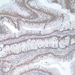
MLH1, a mismatch repair protein involved in maintaining the integrity of genetic information, alongside MSH2, MSH6 and PMS2. During DNA replication, strand misalignment can occur resulting in alterations to microsatellite repeats, often referred to as microsatellite instability (MSI). These defects in DNA repair pathways have been linked to human carcinogenesis. Mutations in the MLH1 gene have been reported to be found in tumors with MSI, such as some forms of colon cancer e.g. Hereditary nonpolyposis colon cancer (HNPCC), a subset of sporadic carcinomas and breast cancer. Loss of expression of MLH1 has also been reported in acute lymphoblastic leukemia, endometrial carcinoma, gastric carcinoma and ovarian carcinoma.
Immunogen:
Prokaryotic recombinant protein corresponding to 210 amino acids of human MLH1
Presentation:
purified immunoglobulin fraction diluted in PBS with 1% BSA containing 15 mM sodium azide as a preservative
-
آنتی بادیهای ایمونوهیستوشیمی
آنتی بادی MLH1 کلون G168-728 برند Patho Sage
نمره 0 از 5Specification:
MLH1 is a mismatch repair gene that is deficient in a high proportion of patients with microsatellite instability (MSI-H). This finding is associated with the autosomal dominant condition known as Hereditary Non-Polyposis Colon Cancer (HNPCC). The anti-MLH1 antibody is useful in screening patients and families for this condition. Colon cancers that are microsatellite unstable have a better prognosis than their microsatellite stable counterparts.
Presentation:
Anti-MLH1 is a mouse monoclonal antibody from supernatant diluted in tris buffered saline, pH 7.3-7.7, with protein base, and preserved with sodium azide
-
آنتی بادیهای ایمونوهیستوشیمی
آنتی بادی RCC کلون PN-15 برند Patho Sage
نمره 0 از 5Specification:
Anti-Renal Cell Carcinoma antibody recognizes a 200 kD glycoprotein localized in the brush border of the proximal renal tubule. This antibody immune-reacts with approximately 90% of primary renal cell carcinomas and approximately 85% of metastatic renal cell carcinomas. Other tumors that react with this antibody are parathyroid adenoma, an occasional breast carcinoma. Nephroblastoma, oncocytoma, mesoblastic nephroma, transitional cell carcinoma, and angiomyolipoma are not labeled with this antibody.
Immunogen:
Microsomal fraction of human renal cortical tissue homogenate
Presentation:
Bioreactor Concentrate with 0.05% Azide
-
آنتی بادیهای ایمونوهیستوشیمی
آنتی بادی P40 کلون ZR8 برند Patho Sage
نمره 0 از 5Specification:
p63 consists of two major isoforms -TAp63 and ΔNp63. These isoforms differ in the structure of the N-terminal domains. The TAp63 isoform (identified by anti-p63 antibody) contains a transactivation- competent ‘TA’domain with homology to p53, which regulates the expression of the growth -inhibitor y genes. In contrast, ΔNp63 isoform (identified by anti-p40 antibody) contains an alternative transcriptionally- inactive ‘ΔN’ domain, which antagonizes the activity of TAp63 and p53. The p40 (clone ZR8) recognizes exclusively ΔNp63 but not TAp63. p40 is a squamous cell carcinoma ‘specific’ antibody. It reacts with the vast majority of cases of squamous cell carcinomas of various origins, but not with adenocarcinomas. It is particularly useful in differentiating lung squamous cell carcinoma from lung adenocarcinoma. p40 antibody can also be used as an alternative basal cell/myoepithelial cell marker, which has similar sensitivity and specificity as that of p63 antibody. Therefore, p40 antibody may also be used as an alternative immunohistochemical marker for determining prostate adenocarcinoma vs. benign prostate glands and for determining breast intraductal carcinoma v s. invasive breast ductal carcinoma.
Immunogen:
Synthesized polypeptides from N-terminal domain of p63
Presentation:
Purified antibody from in 0.2% BSA and 15mM sodium azide.
-
آنتی بادیهای ایمونوهیستوشیمی
آنتی بادی CD33 کلون PWS44 برند Patho Sage
نمره 0 از 5Specification:
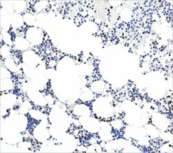
CD33 (gp67, or siglec-3) is a 67 kDa glycosylated transmembrane protein that is a member of the sialic acid–binding immunoglobulin-like lectin (siglec) family. The genomic locus of this protein has been mapped to chromosome 19q13.1-3.5. In maturing granulocytic cells, there is progressive downregulation of CD33 from the blast stage to mature neutrophils. However, in monocytes and macrophages/histiocytes, strong expression of CD33 is maintained throughout maturation. Therefore, the positive control tissue should be bone marrow myeloid cells (especially myeloid precursors), liver Kupffer cells, lung alveolar macrophages, or placental syncytiotrophoblasts. Detection of CD33 using monoclonal antibodies has been a critical component in immunophenotyping acute leukemias, particularly acute myeloid leukemias. This anti-CD33 may be particularly advantageous for cases of acute myeloid leukemia, minimally differentiated (AML-M0) and acute monocytic leukemia (AML-M5), in which other paraffin section markers of myeloid differentiation (such as antimyeloperoxidase) may be negative. However, anti-CD33 staining cannot be used in isolation and must be correlated with other myeloid and lymphoid markers because cases of myeloid antigen–positive acute lymphoblastic leukemia may show bona fide CD33 expression.
Presentation:
Antibody diluted in Tris Buffer, pH 7.3-7.7, with 1% BSA and <0.1% Sodium Azide
-
آنتی بادیهای ایمونوهیستوشیمی
آنتی بادی Oct3/4 کلون C-10 برند Patho Sage
نمره 0 از 5Specification:
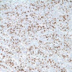
Transcription factors containing the POU homeo domain have been shown to be important regulators of tissue-specific gene expression in lymphoid and pituitary differentiation and in early mammalian development. POU domain proteins contain a bipartite DNA-binding domain divided by a flexible linker that enables them to adopt various monomer configurations on DNA. The versatility of POU protein operation is additionally conferred at the dimerization level. Oct-3 (also known as Oct-4) is a mammalian POU transcription factor expressed by early embryo cells and germ cells. Oct-3/4 is essential for the identity of the pluripotential founder cell population in the mammalian embryo. A critical amount of Oct3/4 is required to sustain stem-cell self renewal, and up or down regulation induce divergent developmental programs. Two isoforms of Oct-3,termed Oct-3A and Oct-3B, are generated by alternative splicing. The gene which encodes Oct-3/4 maps to human chromosome 6p21.3. Oct-3/4 (C-10) is recommended for detection of Oct-3A (Oct-4) and Oct-3B of mouse, rat and human origin by Western Blotting, immunoprecipitation, immunofluorescence, and paraffin immunohistochemistry.
Immunogen:
Amino acids1-134 of Oct-3/4 of human origin
Presentation:
Purified antibody in 0.2 % BSA and 15mM sodium azide
-
آنتی بادیهای ایمونوهیستوشیمی
آنتی بادی GFAP کلون EP672Y برند Patho Sage
نمره 0 از 5Specification:
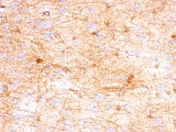
Anti-GFAP antibody detects astrocytes, Schwann cells, satellite cells, enteric glial cells and some groups of ependymal cells. This marker is mainly used to distinguish neoplasms of astrocytic origin from other neoplasms in the central nervous system.
Presentation:
Anti-GFAP is a rabbit monoclonal from tissue culture supernatant diluted in tris buffered saline, pH 7.3-7.7, with protein base, and preserved with sodium azide
-
آنتی بادیهای ایمونوهیستوشیمی
آنتی بادی GFAP کلون GA5 برند Patho Sage
نمره 0 از 5Specification:
This antibody recognizes a protein of ~50kDa which is identified as Glial Fibrillary Acidic Protein (GFAP, also known as Astrocyte or Intermediate Filament Protein). It shows no cross-reaction with other intermediate filament proteins. GFAP is specifically found in astroglia. GFAP is a very popular marker for localizing benign astrocyte and neoplastic cells of glial origin in the central nervous system.
Immunogen:
GFAP isolated from pig spinal cord
Presentation:
Bioreactor Concentrate with 0.05% Azide

