دسته: پلی کلونال
نمایش 21–40 از 511 نتیجه
فیلتر ها-
آنتی بادیهای ایمونوهیستوشیمی
آنتی بادی Lysozyme (Polyclonal)
نمره 0 از 5Name: Lysozyme Antibody (Polyclonal)
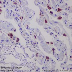
Description and applications: Lysozyme is one of the anti-microbial agents found in human milk, and is also present in spleen, lung, kidney, white blood cells,
plasma, saliva, and tears. Missense mutations in LYZ have been identified in heritable renal amyloidosis. Lysozyme is synthesized predominantly in reactive histiocytes rather than in resting, unstimulated phagocytes. Anti-lysozyme stains myeloid cells, histiocytes, granulocytes, macrophages, and monocytes. It is an important marker that may demonstrate the myeloid or monocytic nature of acute leukemia. Anti-lysozyme may aid in the identification of histiocytic neoplasias, large lymphocytes, and classifying lymphoproliferative disorders.Composition: anti-human Lysozyme rabbit polyclonal antibody purified from serum and prepared in 10mM PBS, pH 7.4, with 0.2% BSA and 0.09% sodium azide
-
آنتی بادیهای ایمونوهیستوشیمی
آنتی بادی Osteocalcin (G5)
نمره 0 از 5Name: Osteocalcin Antibody-Clone G5
Description and applications: Bone γ carboxyglutamic acid (Gla) protein, known as BGLAP, BGP or osteocalcin, is an abundant, non-collagenous protein component of bone that is produced by osteoblasts. In mice, osteocalcin is composed of a cluster of three genes known as OG1, OG2 and ORG, all of which can be found within a 23 kb span of genomic DNA. Human osteocalcin is a highly conserved, 46-50 amino acid, single chain protein that contains three vitamin K-dependent γcarboxyglutamic acid residues. Osteocalcin appears transiently in embryonic bone at the time of mineral deposition, where it binds to hydroxyapatite in a calcium-dependent manner. In addition, osteocalcin is one of the most abundant, non-collagenous proteinsfound in mineralized adult bone. Genetic variation at the osteocalcin locus on chromosome 1q impacts postmenopause bone mineral density (BMD) levels and may predispose some women to osteoporosis.
Composition: Anti-human Osteocalcin rabbit polyclonal antibody purified from serum and prepared in PBS with < 0.1% sodium azide and 0.1% gelatin.
IMMUNOGEN: antibody raised against amino acids 1-100 representing full length osteocalcin of human origin.
-
آنتی بادیهای ایمونوهیستوشیمی
آنتی بادی Pancreatic Polypeptide (Polyclonal)
نمره 0 از 5Name:Pancreatic Polypeptide Antibody
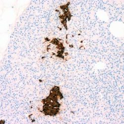
Description and applications: This gene belongs to the NPY family and it encodes a protein that is synthesized as a 95 aa polypeptide precursor in the pancreatic islets of Langerhans. It is cleaved into two peptide products; the active hormone of 36 aa and an icosapeptide of unknown function. The hormone acts as a regulator of pancreatic and gastrointestinal functions and may be important in the regulation of food intake. Plasma level of this hormone has been shown to be reduced in conditions associated with increased food intake and elevated in anorexia nervosa. In addition, infusion of this hormone in obese rodents has shown to decrease weight gain.
Composition: anti-human Pancreatic Polypeptide goat polyclonal antibody purified from serum and prepared in 10mM PBS, pH 7.4, with 0.2% BSA and 0.09% sodium azide
Immunogen: Synthetic peptide sequence (TRPRYGKRHKEDT) corresponding to the internal amino acids of PPY
-
آنتی بادیهای ایمونوهیستوشیمی
آنتی بادی ACTH (Adrenocorticotropic Hormone) (Polyclonal)
نمره 0 از 5Name: Rabbit anti-human ACTH (Adrenocorticotrophic Hormone) Polyclonal Antibody
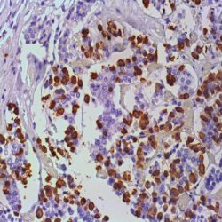
Description and applications: Anti-ACTH is a useful marker in classification of pituitary tumors and the study of pituitary disease. It reacts with ACTH-producing cells (corticotrophs). It also may react with other tumors (e.g., some small cell carcinomas of the lung) causing paraneoplastic syndromes by secreting ACTH.
Composition: anti-human ACTH rabbit monoclonal antibody purified from serum and prepared in 10mM PBS, pH 7.4, with 0.2% BSA and 0.09% sodium azide
Intended use : Immunohistochemistry (IHC) on paraffin embedded tissues. Not tested on frozen tissues or Western-Blotting
Immunogen: Synthetic peptide derived from Nterminus of human ACTH.
-
آنتی بادیهای ایمونوهیستوشیمی
آنتی بادی Gastrin (Polyclonal)
نمره 0 از 5Name: Rabbit anti-human Gastrin Antibody
Description and applications: Gastrin is a 17 amino acid peptide hormone and neurotransmitter widely distributed throught the gastrointestinal (GI) tract and the central nervous system (CNS). Antibodies that react specifically with gastrin may be used to study the differential tissue expression and intracellular and subcellular localization of gastrin in neuroendocrine cells of the gastrointestinal tract, and in the CNS. Antibodies to gastrin are also useful for the identification and detection of gastrin in normal and neoplastic tissue.
Available Volume: 3ml / 7ml / 12ml / Concentrated
Composition: anti-human Gastrin rabbit polyclonal antibody purified from serum and prepared in 10mM PBS, pH 7.4, with 0.2% BSA and 0.09% sodium azide
Intended use: Immunohistochemistry (IHC) on paraffin embedded tissues. Not tested on frozen tissues or Western-Blotting
Immunogen: Synthetic peptide corresponding to Nterminus of Gastrin 1.
-
آنتی بادیهای ایمونوهیستوشیمی
آنتی بادی GLUT 1 (Polyclonal)
نمره 0 از 5Name: glut-1 Antibody
Description and applications: Glucose transporter type I (GLUT1), a prototype member of GLUT super family, reacts with a 55 kD protein, is a membrane-associated erythrocyte glucose transport protein. It is a major glucose transporter in the mammalian blood-brain barrier, and also mediates glucose transport in endothelial cells of the vasculature, adipose tissue and cardiac muscle. GLUT1 is detectable in many human tissues including those of colon, lung, stomach, esophagus, and breast. GLUT1 is overexpressed in malignant cells and in a variety of tumors that include the breast, pancreas, cervix, endometrium, lung, mesothelium, colon, bladder, thyroid, bone, soft tissues, and oral cavity. Immuohistochemical detection of GLUT1 can discriminate between reactive mesothelium and malignant mesothelioma. Anti-GLUT1 with anti-Claudin1, and anti-EMA are “perineurial” markers in diagnosis of perineuriomas. Anti-GLUT1 is also useful in distinguishing benign endometrial
hyperplasia from atypical endometrial hyperplasia and adenocarcinoma. GLUT1 expression has been associated with increased malignant potential, invasiveness, and a poor prognosis in general. Expression of GLUT1 is a late event in colorectal cancer and expression in a high proportion of cancer cells is associated with a high incidence of lymph node metastasesComposition: anti-human GLUT 1 rabbit polyclonal antibody purified from serum and prepared in 10mM PBS, pH 7.4, with 0.2% BSA and 0.09% sodium azide
-
آنتی بادیهای ایمونوهیستوشیمی
آنتی بادی Alpha-1-Antichymotrypsin (Polyclonal)
نمره 0 از 5Name: Alpha-1-Antichymotrypsin (Polyclonal)
Description and applications: This antibody reacts with the human alpha-1- antichymotrypsin, a surfactant protein in acute phase of the inflamation, plus a serpin that inactivates preferably the chymotrypsin, the cathepsin G and the chymase. As an inhibitor protein of serine protease, it is closely related to the neuritic plaques in the Alzheimer’s disease and in human and monkey brains with normal aging signs. The regulation of serine proteases and their inhibitors have an important function in the neuromuscular pathology differential diagnosis. The alpha-1-antichymotrypsin binds primarily to the prostate specific antigen (PSA), a serine protease similar to the chymotrypsin, to form a complex. Furthermore, this antibody can be used to identify hematopoietic cells of the monocyte-macrophage series and neoplasias derived from them.
Composition: anti-human Alpha-1-Antichymotrypsin rabbit polyclonal antibody purified from serum and prepared in 10mM PBS, pH 7.4, with 0.2% BSA and 0.09% sodium azide
Clone: Polyclonal
Ig isotype: Rabbit IgG
IHC positive control: Macrophages in the tonsils.
Visualization: Cytoplasmic.
-
آنتی بادیهای ایمونوهیستوشیمی
آنتی بادی C3d Complement Component (Polyclonal)
نمره 0 از 5Name: C3d Antibody
Description and applications: This antibody recognises the C3d fragment of the complement. Component C3 can be activated through both classical and alternative complement pathways. Among the different complement proteins, C3 and C4 opsonins have a protective thioester radical. This molecule enables C3 (and C4) complement fragments to form covalent bonds when activated by the target molecule, generating C3b. The proteolytic cleavage of C3b generates the C3d fragment, which remains covalently attached to the target structure, while the C3c fragment is released. The C3d fragment is stable and it can be identified in paraffin-embedded biopsies. Under normal conditions, renal glomerular capillaries are stained. In transplants, the identification of diffuse deposits of C3d and C4d in renal peritubular capillaries or cardiac capillaries supports the diagnosis of acute humoral rejection of the graft. Deposits of C3d have been identified in grafts with chronic rejection but its significance has yet to be clarified. Deposition of C3d on paraffin-embedded biopsies from skin inflammatory pathologies has been demonstrated by different authors. Magro and Dyrsen identified prominent granular C3d deposits along the dermoepidermal junction for all discoid lupus erythematosus (20/20) and systemic LE (5/5) cases. In systemic LE, vascular C3d or C4d deposits were shown. All cases of urticarial (5), leukocytoclastic (6), and lymphocytic (1) vasculitis exhibited prominent mural C3d and C4d deposits in vessels, whereas Henoch-Schönlein purpura (10/10) showed only C3d deposits. Pfaltz et al found deposits of C3d at the epidermal basement membrane in 97% (31/32) of bullous pemphigoid cases.
Composition: anti-human C3d Complement Component rabbit polyclonal antibody purified from serum and prepared in 10mM PBS, pH 7.4, with 0.2% BSA and 0.09% sodium azide
IHC positive control: Kidney tissue section with acute humoral rejection.
-
آنتی بادیهای ایمونوهیستوشیمی
آنتیبادی Herpes Simplex Virus I (Polyclonal)
نمره 0 از 5Name: Herpes Simplex Virus I Antibody
Description and aplications: Herpes simplex virus is quite ubiquitous and is quite variable in it’s presentation in human disease. Type I usually infects the non-genital mucosal surfaces. It may affect the skin or internal organs (typically brain, lung, liver, adrenal gland, or GI tract) of immunocompromised individuals. This polyclonal antibody reacts with Type I Herpes viruses. There may be cross-reactivity with varicella zoster virus at higher concentrations. Crossreactivity with CMV or Epstein-Barr virus is not seen with this antibody.
Composition: anti-human Herpes Simplex Virus Irabbit polyclonal antibody purified from serum andprepared in 10mM PBS, pH 7.4, with 0.2% BSA and0.09% sodium azide
Intended use: Immunohistochemistry (IHC) on paraffin embedded tissues. Not tested on frozen tissues or Western-Blotting
-
آنتی بادیهای ایمونوهیستوشیمی
آنتی بادی Herpes Simplex Virus II (Polyclonal)
نمره 0 از 5Name: Herpes Simplex Virus II Antibody
Description and aplications: Herpes viruses type 1 and 2 (HSV-1 and HSV-2) belong to the family of herpesviridae. Its genome consists of a linear double molecule of DNA of approximately 100,000 kD encoding more than 75 gene products. The homology between the genomic structure of HSV-1 and 2 is of approximately 50%, so that while most of the polypeptides encoded for each type of herpes virus have a similar structure and are antigenically related, there are numerous proteins of both viruses that condition particular responses of the host’s immune system and allow its serological and immunohistochemical differentiation. The monoclonal antibody described herein reacts with the herpes simplex virus type 2 and is useful in detecting HSV II induced lesions. At high concentrations, cross-reactivity has been observed against some strains of the varicella zoster virus. The antibody does not react against CMV or Epstein-Barr virus cultures.
Composition: anti-human Herpes Simplex Virus II rabbit polyclonal antibody purified from serum and prepared in 10mM PBS, pH 7.4, with 0.2% BSA and 0.09% sodium azide
-
آنتی بادیهای ایمونوهیستوشیمی
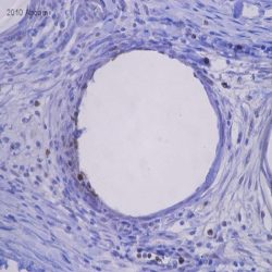
آنتی بادی Phosphohistone H3 (PHH3) (Polyclonal)
نمره 0 از 5Name: Phospho-Histone H3 (PHH3) Antibody
Description and aplications: The phosphohistone H3 (phh3) is the phosphorylated form of nuclear histone H3, which together with the latter one forms the major protein component of the chromatin in eukaryotic cells. In mammalian cells the phosphohistone H3 is negligible during interphase, but reaches its maximum expression at mitosis, more precisely at the transition between the late G2 and M phases of the cell cycle. Immunohistochemical studies have shown that the PHH3 antibody specifically detects the nuclear histone H3 only when protein phosphorylation occurs at the level of serine 10 or serine 28. These studies have also shown that this phosphorylation during apoptosis does not occur on the histone H3, so that the presence of phh3 molecule can serve as a good marker for separating mitotic figures from the apoptotic bodies and nuclear karyorrhexis detritus and is likely to constitute a useful tool for tumor grading, treatment stratification and tumor follow-up especially in CNS, skin, lung, esophagus, breast, soft tissue, stromal gastrointestinal and gynecologic tumors. In fact, recent studies granted PHH3 with greater importance and relevance than Ki- 67 in identifying mitotic figures and a higher intra -and inter reproducibility in cases of melanoma.
Composition: anti- PHH3 rabbit polyclonal antibody obtained from supernatant culture and prediluted in a Tris buffered solution pH 7.4 containing 0.375mM sodium azide solution as bacteriostatic and bactericidal. The quantity of the active antibody was not determined.
Intended use : Immunohistochemistry (IHC) on paraffin embedded tissues. Not tested on frozen tissues or Western-Blotting
-
آنتی بادیهای ایمونوهیستوشیمی
آنتی بادی P501S (Polyclonal)
نمره 0 از 5Name: P501S antibody
Description and applications: Prostein is a novel prostate-specific protein expressed in normal and malignant prostate tissues. This protein appears to be a type IIIa plasma membrane protein with a cleavable signal peptide and 11 transmembrane-spanning regions. Prostein expression is androgen responsive since treatment of LNCaP cells with androgen upregulates prostein message and protein expression levels. Thus, prostein is a prostate-specific marker whose detection and quantitation may be useful in diagnosing and monitoring prostate cancer.
Composition: anti-human Prostein P501S rabbit polyclonal antibody purified from serum and prepared in 10mM PBS, pH 7.4, with 0.2% BSA and 0.09% sodium azide
-
آنتی بادیهای ایمونوهیستوشیمی
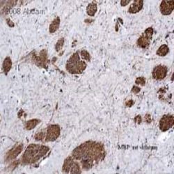
آنتی بادی Myelin Basic Protein (Polyclonal)
نمره 0 از 5Name: Myelin Basic Protein Antibody
Description and applications: MBP is present in the central and peripheral nervous system, the antibody reacts with soft tissue tumors. MBP has been demonstrated in neuromas, neurofibromas, and neurogenic sarcomas; other spindle cell neoplasms do not stain with this antibody. Immunoreactivity for anti-MBP in granular cell tumors strengthens the concept of a Schwann cell derivation of these lesions. Unlike other nervous system proteins e.g., GFAP and S-100, MBP has not been demonstrated in melanocytes or tumors derived from them.
Composition: anti-human Myelin Basic Protein rabbit polyclonal antibody purified from serum and prepared in 10mM PBS, pH 7.4, with 0.2% BSA and 0.09% sodium azide
Intended use : Immunohistochemistry (IHC) on paraffin embedded tissues. Not tested on frozen tissues or Western-Blotting
-
آنتی بادیهای ایمونوهیستوشیمی
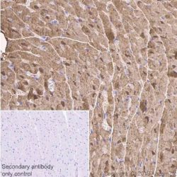
آنتی بادی Myoglobin (Polyclonal)
نمره 0 از 5Name: Myoglobin antibody
Description and applications: Immunostaining with anti-myoglobin provides a specific, sensitive and practical procedure for the identification of tumours of muscle origin. Since myoglobin is found exclusively in skeletal and cardiac muscle and is not present in any other cells of the human body, it may be used to distinguish rhabdomyosarcoma from other soft tissue tumors. Anti-myoglobin staining is also useful when demonstrating rhabdomyoblastic differentiation in other tumours, e.g. neurogenic sarcomas, and malignant mixed mesodermal tumours of the uterus and ovary
Composition: anti-human Myoglobin rabbit monoclonal antibody purified from serum and prepared in 10mM PBS, pH 7.4, with 0.2% BSA and 0.09% sodium azide
Intended use : Immunohistochemistry (IHC) on paraffin embedded tissues. Not tested on frozen tissues or Western-Blotting
-
آنتی بادیهای ایمونوهیستوشیمی
آنتی بادی Myeloperoxidase (Polyclonal)
نمره 0 از 5Name: Myeloperoxidase antibody
Description and application: Myeloperoxidase is an important enzyme used by granulocytes during phagocytic lysis of foreign particles engulfed. In normal tissues and in a variety of myeloproliferative disorders myeloid cells of both neutrophilic and eosinophilic types, at all stages of maturation, exhibit strong cytoplasmic reactivity for MPO. Erythroid precursors, megakaryocytes, lymphoid cells, mast cells, and plasma cells are nonreactive. MPO is not observed in the neoplastic cells of a wide variety of epithelial tumors and sarcomas. MPO is useful in differentiating between myeloid and lymphoid leukemias.
Composition: Anti-human Myeloperoxidase rabbit polyclonal antibody purified from serum and prepared in 10mM PBS, pH 7.4, with 0.2% BSA and 0.09% sodium azide
Intended use: Immunohistochemistry (IHC) on paraffin embedded tissues. Not tested on frozen tissues or Western-Blotting
-
آنتی بادیهای ایمونوهیستوشیمی
آنتی بادی BRST-1/BCA-225 (CU-18)
نمره 0 از 5(NAME: Mouse anti-human BRST-1/BCA-225 Monoclonal Antibody (Clone CU-18
DESCRIPTION AND APPLICATIONS:This antibody recognises BCA-225, a glycoprotein with a molecular mass of 225 kD secreted by the T47D breast carcinoma cell line that has subsequently been identified in various normal and neoplastic tissues
COMPOSITION:Anti-human BRST-1/BCA-225 mouse monoclonal antibody purified from serum and prepared in 10mM PBS, pH 7.4, with 0.2% BSA and 0.09% sodium azide
INTENDED USE: Immunohistochemistry (IHC) on paraffin embedded tissues. Not tested on frozen tissues or Western-Blotting
.IMMUNOGEN: BCA 225 protein secreted by the T47D (clone 11) human breast carcinoma cell line
-
آنتی بادیهای ایمونوهیستوشیمی
آنتی بادی ALDH1A1 (Polyclonal)
نمره 0 از 5Name: ALDH1A1 (Polyclonal)
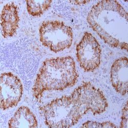
Description and applications: ALDH1 isoform A1, also known as retinal (acetaldehyde) dehydrogenase 1 or aldehyde dehydrogenase 1 (ALDH1) has the mission to convert / oxidize the retinaldehyde in retinoic acid and is codified by a gene on the chromosome region 9q21.13.1.This enzyme belongs to a family of evolutionarily highly conserved ALDH enzymes comprising 19 isoforms localized in the cytoplasm, mitochondria or the nucleus and are involved in the metabolism of aldehydes units, retinol, certain xenobiotic compounds and likewise are involved in ethanol oxidation and Ras GTPase activity. Low levels of cytosolic together with normal levels of mitochondrial ALDH it’s seen in patients with alcoholic fatty liver.
Composition: anti-human ALDH1A1 goat polyclonal antibody purified by protein A/G in PBS/1% BSA buffer pH 7.6 with less than 0.1% sodium azide.
Immunogen: peptide mapping at the N-terminus of ALDH1A1 of human origin.
-
آنتی بادیهای ایمونوهیستوشیمی
آنتی بادی Alpha-1 Antitrypsin (Polyclonal)
نمره 0 از 5Name: Alpha-1 Antitrypsin (Polyclonal)
Description and applications: This antibody reacts with human alpha-1 antitrypsin, a glycoprotein responsible for the antiproteolytic activity of normal serum. Alpha-1-antitrypsin is mainly synthesized by hepatocytes, although human histiocytes and peripheral blood monocytes also express this protein. This antibody may be useful in the study of hereditary alpha-1 antitrypsin deficiency, benign and malignant liver tumours, embryonal carcinomas, and a variety of benign and malignant lesions of histiocytic origin. These antibody cross-reacts with horse, pig, dog, and rat alpha-1 antitrypsin.
Composition: anti-human Alpha-1 Antitrypsin rabbit polyclonal antibody purified from serum and prepared in 10mM PBS, pH 7.4, with 0.2% BSA and 0.09% sodium azide.
Immunogen: Human alpha-1 antitrypsin.
-
آنتی بادیهای ایمونوهیستوشیمی
آنتی بادی Anexin A1 (29)
نمره 0 از 5Name: Anexin A1 (29)
DESCRIPTION AND APPLICATIONS: Annexin I (a.k.a. lipocortin-1 or calpactin II) is a 38kDa protein that is part of a calcium-dependent phospholipid-binding protein family. Members of these family, share a common core domain, but each has a unique Nterminal tail that imparts its functional specificity. It is believed that phosphorylation of this region in
annexin I regulates its functions.COMPOSITION: Anti-human Annexin 1 mouse monoclonal antibody purified from serum and prepared in 10mM PBS, pH 7.4, with 0.2% BSA and 0.09% sodium azide INTENDED USE.
IMMUNOGEN: Aminoacids 1-346 of bovine annexin.
-
آنتی بادیهای ایمونوهیستوشیمی
آنتی بادی ATRX (POLYCLONAL)
نمره 0 از 5Name: ATRX (POLYCLONAL)
Description and aplications: ATRX, also known as ATP-dependent helicase ATRX, Xlinked helicase II,X-linked nuclear protein or Znf-HX, is encoded by a gene located in the chromosomal region Xq21.1, which undergoes an inactivation and encodes a nuclear and homologous NTP protein with several types of helicases II present at the membrane level. It belongs to the superfamily of proteins similar to the SNF2 subgroup and features a C-terminal region rich in glutamine similar to other nuclear transcription factors. Mutations of the gene cause a severe mental retardation syndrome linked to alphathalassemia (ATR-X syndrome) characterized by severe psychomotor retardation, characteristic facial features, urogenital abnormalities and alpha thalassemia with H hemoglobin inclusions at the erythrocyte level. Other syndromes related to mutations of the ATRX gene have been described such as X-linked mental retardation-hypotonic faciessyndrome or alpha thalassemia myelodysplastic syndrome. After interacting with the DAXX domain and potential binding with the EZH2, the ATRX protein plays several roles in mitosis, rearrangement of the chromatin and transcription as well as in the biology of the telomeric regions.
Composition: Rabbit anti-ATRX plyclonal antibody obtained from purified ascitic fluid and prepared in 10mM PBS, pH
7.4, with 0.2% BSA and 0.09% sodium azide.Immunogen: Recombinant protein corresponding to the alpha thalassemia X-linked intellectual disability syndrome (ATR-X) subunit.

