برچسب: فلوسایتومتری
Showing all 19 results
فیلتر ها-
آنتی بادیهای فلوسایتومتری
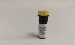
آنتی بادی فلوسایتومتری 7-AAD Viability Staining Solution
نمره 0 از 57-AAD Viability Staining Solution
Company: BioLegend
Catalog: 420403
Size: 200 tests
Storage & Handling: Protect from light. Store between 2°C and 8°C
Description:
7-AAD (7-amino-actinomycin D) has a high DNA binding constant and is efficiently excluded by intact cells. It is useful for DNA analysis and dead cell discrimination during flow cytometric analysis. When excited by 488 laser light, 7- AAD fluorescence is detected in the far red range of the spectrum (650 nm longpass filter).
-
آنتی بادیهای فلوسایتومتری
آنتی بادی فلوسایتومتری APC anti-human CD3 Antibody
نمره 0 از 5APC anti-human CD3
Company: BioLegend
Catalog : 300311, 300312
Size: 25 tests / 100 tests
Clone: HIT3a
Isotype : Mouse IgG2a, κ
Reactivity: Human
Description:
CD3ε is a 20 kD chain of the CD3/T-cell receptor (TCR) complex which is composed of two CD3ε, one CD3γ, one CD3δ, one CD3ζ (CD247), and a T-cell receptor T-cell heterodimer. It is found on all mature T lymphocytes, NK-T cells, and some thymocytes. CD3, also known as T3, is a member of the immunoglobulin superfamily that plays a role in antigen recognition, signal transduction, and T cell activation.
-
آنتی بادیهای فلوسایتومتری
آنتی بادی فلوسایتومتری APC anti-mouse CD8a Antibody
نمره 0 از 5APC anti-mouse CD8a Antibody
Company:BioLegend
Catalog Number: 100711
Size : 25 µg
Clone : 53-6.7
Isotype : Rat IgG2a, κ
Reactivity : Mouse
Description :
CD8, also known as Lyt-2, Ly-2, or T8, consists of disulfide-linked α and β chains that form the α(CD8a)/β(CD8b) heterodimer and α/α homodimer. CD8a is a 34 kD protein that belongs to the immunoglobulin family. The CD8 α/β heterodimer is expressed on the surface of most thymocytes and a subset of mature TCR α/β T cells. CD8 expression on mature T cells is non-overlapping with CD4. The CD8 α/α homodimer is expressed on a subset of γ/δ TCR-bearing T cells, NK cells, intestinal intraepithelial lymphocytes, and lymphoid dendritic cells. CD8 is an antigen co-receptor on T cells that interacts with MHC class I on antigenpresenting cells or epithelial cells. CD8 promotes T cell activation through its association with the TCR complex and protein tyrosine kinase lck.
-
آنتی بادیهای فلوسایتومتری
آنتی بادی فلوسایتومتری FITC anti-human CD2 Antibody
نمره 0 از 5FITC anti-human CD2 Antibody
Company:BioLegend
Catalog : 300206
Size : 100 tests
Clone : RPA-2.10
Isotype : Mouse IgG1, κ
Reactivity : Human, African Green, Baboon, Capuchin Monkey, Chimpanzee, Cynomolgus, Pigtailed Macaque, Rhesus, Swine (Pig, Porcine)
Description:
CD2 is a 50 kD type I transmembrane glycoprotein also known as LFA-2, T11, and sheep red blood cell receptor (SRBC-R). This immunoglobulin superfamily member is expressed on thymocytes, T lymphocytes, NK cells, and thymic B cell subsets. The major ligand for CD2 is CD58 (also known as LFA-3). CD2 has also been reported to bind CD48, CD59, and CD15. CD2 plays a critical role in alternative T cell activation, T cell signaling, and cell-cell adhesion.
-
آنتی بادیهای فلوسایتومتری
آنتی بادی فلوسایتومتری FITC anti-human CD16 Antibody
نمره 0 از 5FITC anti-human CD16 Antibody
Company:BioLegend
Clone: B73.1
Isotype: Mouse IgG1, κ
Reactivity: Human
Cat Number: 360716
Antibody Type: Monoclonal
Description:
CD16 is known as low affinity IgG receptor III (FcγRIII). It is expressed as two distinct forms (CD16a and CD16b). CD16a (FcγRIIIA) is a 50-65 kD polypeptide-anchored transmembrane protein. It is expressed on the surface of NK cells, activated monocytes, macrophages, a subset of T cells and placental trophoblasts in humans. CD16b (FcγRIIIB) is a 48 kD glycosylphosphatidylinositol (GPI)-anchored protein. Its extracellular domain is over 95% homologous to that of CD16a, and it is expressed specifically on neutrophils. CD16 binds aggregated IgG or IgG-antigen complex which functions in NK cell activation, phagocytosis, and antibody-dependent cell-mediated cytotoxicity (ADCC).
-
آنتی بادیهای فلوسایتومتری
آنتی بادی فلوسایتومتری FITC anti-human CD33 Antibody
نمره 0 از 5FITC anti-human CD33 Antibody
Company:BioLegend
Catalog : 303303
Size: 25 tests
Isotype: Mouse IgG1, κ
Reactivity: Human, Cross-Reactivity : Chimpanzee
Description:
CD33 is a 67 kD type I transmembrane glycoprotein also known as Siglec-3, gp67, and p67. It is a sialoadhesion immunoglobulin superfamily member expressed on myeloid progenitors, monocytes, granulocytes, dendritic cells and mast cells. CD33 is absent on normal platelets, lymphocytes, erythrocytes and hematopoietic stem cells. CD33 functions as a sialic acid-dependent cell adhesion molecule with carbohydrate/lectin binding activity.
-
آنتی بادیهای فلوسایتومتری
آنتی بادی فلوسایتومتری FITC anti-mouse CD4 Antibody
نمره 0 از 5FITC anti-mouse CD4 Antibody
Company:BioLegend
Catalog Number : 100405
Size : 50 µg
Clone : GK1.5
Isotype: Rat IgG2b, κ
Reactivity : Mouse
Description :
CD4 is a 55 kD protein also known as L3T4 or T4. It is a member of the Ig superfamily, primarily expressed on most thymocytes, a subset of T cells, and weakly on macrophages and dendritic cells. It acts as a coreceptor with the TCR during T cell activation and thymic differentiation by binding MHC class II and associating with the protein tyrosin kinase, lck.
-
آنتی بادیهای فلوسایتومتری
آنتی بادی فلوسایتومتری FITC anti-mouse CD8a Antibody
نمره 0 از 5FITC anti-mouse CD8a Antibody
Company:BioLegend
Catalog Number : 100705
Size : 50 µg
Clone : 53-6.7
Isotype : Rat IgG2a, κ
Reactivity : Mouse
Description :
CD8, also known as Lyt-2, Ly-2, or T8, consists of disulfide-linked α and β chains that form the α(CD8a)/β(CD8b) heterodimer and α/α homodimer. CD8a is a 34 kD protein that belongs to the immunoglobulin family. The CD8 α/β heterodimer is expressed on the surface of most thymocytes and a subset of mature TCR α/β T cells. CD8 expression on mature T cells is non-overlapping with CD4. The CD8 α/α homodimer is expressed on a subset of γ/δ TCR-bearing T cells, NK cells, intestinal intraepithelial lymphocytes, and lymphoid dendritic cells. CD8 is an antigen co-receptor on T cells that interacts with MHC class I on antigenpresenting cells or epithelial cells. CD8 promotes T cell activation through its association with the TCR complex and protein tyrosine kinase lck.
-
آنتی بادیهای فلوسایتومتری
آنتی بادی فلوسایتومتری FITC anti-mouse CD49b (pan-NK cells) Antibody
نمره 0 از 5FITC anti-mouse CD49b (pan-NK cells) Antibody
Company:BioLegend
Catalog Number : 108905
Size : 50 µg
Clone : DX5
Isotype : Rat IgM, κ
Reactivity : Mouse
Description :
DX5 antigen has been recently characterized as CD49b. It is a 150 kD integrin α chain also known as α integrin, VLA-2 α chain, and integrin α chain. CD49b non-covalently associates with CD29 (β integrin) to form the CD49b/CD29 complex known as VLA-2, a receptor for collagen and laminin. CD49b is expressed on platelets, the majority of NK cells, NKT cells, and a small subset of CD8+ T cells (this population can be significantly increased following viral infection). DX5 is used for the identification and isolation of NK cells, and is especially useful for identifying NK cells in mice lacking the NK1.1 antigen.
-
آنتی بادیهای فلوسایتومتری
آنتی بادی فلوسایتومتری FITC anti-mouse/human CD11b Antibody
نمره 0 از 5FITC anti-mouse/human CD11b Antibody
Company:BioLegend
Catalog Number : 101205
Size : 50 µg
Clone : M1/70
Isotype : Rat IgG2b, κ
Reactivity : Mouse, Human, Cross-Reactivity: Chimpanzee, Baboon, Cynomolgus, Rhesus, Rabbit (Lapine)
Description :
CD11b is a 170 kD glycoprotein also known as αM integrin, Mac-1 α subunit, Mol, CR3, and Ly-40. CD11b is a member of the integrin family, primarily expressed on granulocytes, monocytes/macrophages, dendritic cells, NK cells, and subsets of T and B cells. CD11b non-covalently associates with CD18 (β2 integrin) to form Mac-1. Mac-1 plays an important role in cell-cell interaction by binding its ligands ICAM-1 (CD54), ICAM-2 (CD102), ICAM-4 (CD242), iC3b, and fibrinogen.
-
آنتی بادیهای فلوسایتومتری
آنتی بادی فلوسایتومتری FITC F(ab’)2 Goat anti-human IgG Fcγ Antibody
نمره 0 از 5FITC F(ab’)2 Goat anti-human IgG Fcγ Antibody
Company:BioLegend
Catalog Number: 398006
Size : 100 µg
Clone : Poly23980
Isotype : Goat Polyclonal Ig
Reactivity : Human
Description :
gG Fc is a homodimer that is composed of the constant region of the two heavy chains that form the IgG molecule. The Fc fragment mediates opsonization, antibody dependent cellular cytotoxicity (ADCC), and complement activation through binding to Fc receptors such as CD16, CD32, CD64, and the complement factor C1.
-
آنتی بادیهای فلوسایتومتری
آنتی بادی فلوسایتومتری PE anti-human CD79a (Igα) Antibody
نمره 0 از 5PE anti-human CD79a (Igα) Antibody
Company:BioLegend
Catalog Number : 333503
Size: 25 tests
Clone : HM47
Isotype : Mouse IgG1, κ
Reactivity: Human
Description:
CD79a is a 47 kD type I integral membrane protein, also known as mb-1 or Iga. It is a member of the Ig superfamily and disulphide-associated with CD79b (B29). The interaction of CD79a/CD79b heterodimer with B cell suface Ig forms B cell antigen complex. CD79a is expressed in B cells from early pre-B to plasma cell stage. It has been shown that CD79a is also weakly expressed in some precursors of T- and myeloid cells. CD79 mediates the transport of IgM to B cell surface and transduces signals initiated by BCR aggregation.
-
آنتی بادیهای فلوسایتومتری
آنتی بادی فلوسایتومتری PE anti-mouse CD366 (Tim-3) Antibody
نمره 0 از 5PE anti-mouse CD366 (Tim-3) Antibody
Company:BioLegend
Catalog Number : 119703
Size : 50 µg
Clone : RMT3-23
Isotype : Rat IgG2a, κ
Reactivity : Mouse
Description :
CD366 (Tim-3) is a transmembrane protein also known as T cell immunoglobulin and mucin domain containing protein-3. Tim-3 is expressed at high levels on Th1 lymphocytes and CD11b macrophages. Tim-3 has also been shown to exist as a soluble protein. Cells expressing Tim-3 are present at high levels in the CNS of animals at the onset of experimental autoimmune encephalomyelitis (EAE), a disease mediated by lymphocytes secreting Th1-like cytokines. Tim-3 has been proposed to inhibit Th1-mediated immune responses and promote immunological tolerance.
-
آنتی بادیهای فلوسایتومتری
آنتی بادی فلوسایتومتری PE anti-mouse Ly-6G/Ly-6C (Gr-1) Antibody
نمره 0 از 5PE anti-mouse Ly-6G/Ly-6C (Gr-1) Antibody
Company:BioLegend
Catalog Number : 108407
Size : 50 µg
Clone : RB6-8C5
Isotype : Rat IgG2b, κ
Reactivity : Mouse
Description :
Gr-1 is a 21-25 kD protein also known as Ly-6G/Ly-6C. This myeloid differentiation antigen is a glycosylphosphatidylinositol (GPI)-linked protein expressed on granulocytes and macrophages. In bone marrow, the expression levels of Gr-1 directly correlate with granulocyte differentiation and maturation; Gr-1 is also transiently expressed on bone marrow cells in the monocyte lineage. Immature Myeloid Gr-1+ cells play a role in the development of antitumor immunity.
-
آنتی بادیهای فلوسایتومتری
آنتی بادی فلوسایتومتری PE anti-TCF1 (TCF7) Antibody
نمره 0 از 5PE anti-TCF1 (TCF7) Antibody
Company:BioLegend
Catalog Number : 655208
Size : 100 tests
Clone : 7F11A10
Isotype : Mouse IgG1, κ
Reactivity : Human
Description :
TCF1 is the first identified member of the T-cell-specific transcription factor family. It plays an important role in T cell development and differentiation. TCF1 is inactivated by association with the transcriptional repressor TLE proteins. During Wnt signaling, the transcriptional coactivator CTNNB1 accumulates and, in turn, replaces the transcriptional repressor associated with TCF1. Interaction with CTNNB1 results in transactivation of TCF1 target genes. Deletion of TCF1 causes massive apoptosis of double positive thymocyte, suggesting that TCF1 is required for thymocyte survival during T cell development. In addition to its function in thymus, TCF1 promotes T cell differentiation to Th2 cells in the periphery through transcriptional activation of GATA3.
-
آنتی بادیهای فلوسایتومتری
آنتی بادی فلوسایتومتری PE/Cyanine5 anti-human CD3 Antibody
نمره 0 از 5PE/Cyanine5 anti-human CD3 Antibody
Company:BioLegend
Clone: HIT3a
Isotype: Mouse IgG2a, κ
Cat Number: 300309
Reactivity: Human
Antibody Type: Monoclonal
Description:
CD3ε is a 20 kD chain of the CD3/T-cell receptor (TCR) complex which is composed of two CD3ε, one CD3γ, one CD3δ, one CD3ζ (CD247), and a T-cell receptor (α/β or γ/δ) heterodimer. It is found on all mature T lymphocytes, NK-T cells, and some thymocytes. CD3, also known as T3, is a member of the immunoglobulin superfamily that plays a role in antigen recognition, signal transduction, and T cell activation.
-
آنتی بادیهای فلوسایتومتری
آنتی بادی فلوسایتومتری PE/Cyanine5 anti-human CD19 Antibody
نمره 0 از 5PE/Cyanine5 anti-human CD19 Antibody
Company:BioLegend
Catalog Numbeer: 302209
Size : 25 tests
Clone : HIB19
Isotype: Mouse IgG1, κ
Reactivity: Human, Chimpanzee, Rhesus
Description :
CD19 is a 95 kD type I transmembrane glycoprotein also known as B4. It is a member of the immunoglobulin superfamily expressed on B-cells (from pro-B to blastoid B cells, absent on plasma cells) and follicular dendritic cells. CD19 is involved in B cell development, activation, and differentiation. CD19 forms a complex with CD21 (CR2) and CD81 (TAPA-1), and functions as a BCR coreceptor.
-
آنتی بادیهای فلوسایتومتری
آنتی بادی فلوسایتومتری PerCP/Cyanine5.5 anti-mouse CD279 (PD-1) Antibody
نمره 0 از 5PerCP/Cyanine5.5 anti-mouse CD279 (PD-1) Antibody
Company:BioLegend
Catalog Number : 135207
Size : 25 µg
Clone : 29F.1A12
Isotype : Rat IgG2a, κ
Reactivity : Mouse
Description :
CD279, also known as programmed death-1 (PD-1), is a 50-55 kD glycoprotein belonging to the CD28 family of the Ig superfamily. PD-1 is expressed on activated splenic T and B cells and thymocytes. It is induced on activated myeloid cells as well. PD-1 is involved in lymphocyte clonal selection and peripheral tolerance through binding its ligands, B7-H1 (PD-L1) and B7-DC (PD-L2). It has been reported that PD-1 and PD-L1 interactions are critical to positive selection and play a role in shaping the T cell repertoire. PD-L1 negative costimulation is essential for prolonged survival of intratesticular islet allografts.

