دسته: ایمونو هیستوشیمی
نمایش 401–420 از 540 نتیجه
فیلتر ها-
آنتی بادیهای ایمونوهیستوشیمی
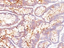
آنتی بادی CEA (Carcinoembryonic Antigen) کلون CEA31 برند Patho Sage
نمره 0 از 5Specification:
This antibody recognizes proteins of 80-200kDa, identified as different members of CEA family. CEA is synthesized during development in the fetal gut and is re-expressed in increased amounts in intestinal carcinomas and several other tumors. This MAb does not react with nonspecific cross-reacting antigen (NCA) and with human polymorphonuclear leucocytes. It shows no reaction with a variety of normal tissues and is suitable for staining of formalin/paraffin tissues. CEA is not found in benign glands, stroma, or malignant prostatic cells. Antibody to CEA is useful in detecting early foci of gastric carcinoma and in distinguishing pulmonary adenocarcinomas (60-70% are CEA+) from pleural mesotheliomas (rarely or weakly CEA+).
Immunogen:
Human colon carcinoma extract
Presentation:
Bioreactor Concentrate with 0.05% Azide
-
آنتی بادیهای ایمونوهیستوشیمی
آنتی بادی CEA (Carcinoembryonic Antigen) کلون Col-1 برند Patho Sage
نمره 0 از 5Specification:
CEA (carcinoembryonic antigen, CD66) consists of a heterogeneous family of related oncofetal 200 kD glycoproteins that is secreted into the glycocalyx surface of gastrointestinal cells. The staining of CEA is considered important because: 1. In breast carcinomas, CEA staining correlated well with clinical outcome, independent of histologic type of tumors (with or without metastases). 2. A CEA-staining primary tumor usually shows CEA elevations when there is disease recurrence or progression. 3. Before proceeding with extensive or repeated blood monitoring of CEA as an index of biological activity of diseases, it is important to find out if the substance is present in the original malignant lesion, preferably at the time of biopsy. 4. Excellent correlation between CEA staining of primary and metastatic tumors (including micrometastases to the regional lymph nodes) has been observed with respect to cervical, colonic and ovarian carcinomas. Usually CEA is demonstrated as a linear labeling of the apical poles of cells lining the glandular lumen and, occasionally, as weak staining near the apex of colonic epithelial cells. More uniform cytoplasmic staining is a feature of more aggressive tumors. CEA, however, should not be used as a marker of differentiation because many colon and lung tumors actually show increased staining with differentiation. Pancreatic carcinomas, testicular tumor, gall bladder neoplasms and granular cell Myo blastomas stain positive, whereas malignant tumors of brain, prostate, skin, lymphoreticular tissues, hepatocellular carcinomas, esophageal squamous cell carcinomas, and mesothelioma fail to stain for CEA.
Immunogen:
Human colon carcinoma extract
Presentation:
Bioreactor Concentrate with 0.05% Azide
-
آنتی بادیهای ایمونوهیستوشیمی
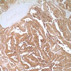
آنتی بادی DOG1 (Anoctamin 1) کلون DG1/1484 برند Patho Sage
نمره 0 از 5Specification:
Expression of DOG-1 protein is elevated in the gastrointestinal stromal tumors (GIST’s), KIT signaling-driven mesenchymal tumors of the GI tract. DOG-1 is rarely expressed in other soft tissue tumors, which, due to appearance, may be difficult to diagnose. Immunoreactivity for DOG-1 has been reported in 97.8 percent of scorable GIST’s, including all KIT negative GIST’s. Overexpression of DOG-1 has been suggested to aid in the identification of GISTs, including Platelet-Derived Growth Factor Receptor Alpha mutants that fail to express KIT antigen. The overall sensitivity of DOG1 and KIT in GIST’s is nearly identical: 94.4% vs. 94.7%.
Immunogen:
Recombinant human DOG-1 protein
Presentation:
Bioreactor Concentrate with 0.05% Azide
-
آنتی بادیهای ایمونوهیستوشیمی
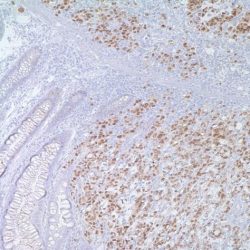
آنتی بادی P53 کلون DO7 برند Patho Sage
نمره 0 از 5Specification:
Anti-p53 Tumor Suppressor Protein antibody recognizes a 53 kDa phosphoprotein, identified as p53 suppressor gene product. It reacts with the mutant as well as wild form of p53. Positive nuclear staining with this antibody has been shown to be a negative prognostic factor in Breast carcinoma, Lung carcinoma, colorectal, and Urothelial carcinoma. p53 positivity has also been used to differentiate Uterine serous carcinoma from endometrioid carcinoma as well as to detect Intratubular germ cell neoplasia.
Immunogen:
Recombinant human wild type p53 protein expressed in E. coli.
Presentation:
Bioreactor Concentrate with 0.05% Azide
-
آنتی بادیهای ایمونوهیستوشیمی
آنتی بادی DOG1 (Anoctamin 1) کلون DOG1.1 برند Patho Sage
نمره 0 از 5Specification:
Expression of DOG-1 protein is elevated in the gastrointestinal stromal tumors (GIST’s), KIT signaling-driven mesenchymal tumors of the GI tract. DOG-1 is rarely expressed in other soft tissue tumors, which, due to appearance, may be difficult to diagnose. Immunoreactivity for DOG-1 has been reported in 97.8 percent of scorable GIST’s, including all KIT negative GIST’s. Overexpression of DOG-1 has been suggested to aid in the identification of GISTs, including Platelet-Derived Growth Factor Receptor Alpha mutants that fail to express KIT antigen. The overall sensitivity of DOG1 and KIT in GIST’s is nearly identical: 94.4% vs. 94.7%.
Immunogen:
A synthetic peptide from human DOG-1 protein, conjugated to a carrier protein
Presentation:
Bioreactor Concentrate with 0.05% Azide
-
آنتی بادیهای ایمونوهیستوشیمی
آنتی بادی CD19 کلون EP169 برند Patho Sage
نمره 0 از 5Specification:
CD19 is expressed only on B-cells and follicular dendritic cells. It is a specific and sensitive marker of B-cells widely expressed from early pre-B stages, normal B-cells and normal plasma cells. It is considered a positive regulator of both intrinsic and stimulus-dependent pathways in B-lymphocytes. CD19 is useful in identification of B-cell lineage of majority of B-cell neoplasms but appears to be less useful in subclassifying of B-cell neoplasms in histological material. It appears to be potentially useful additional marker of follicular dendritic cell tumors.
Presentation:
Phosphate buffered Saline (PBS), pH 7.2 with 1% BSA and <0,1% Sodium Azide
-
آنتی بادیهای ایمونوهیستوشیمی
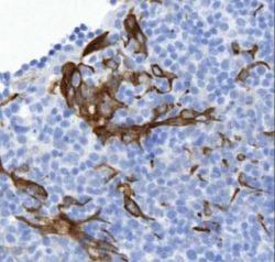
آنتی بادی Factor 13a کلون EP3372 برند Patho Sage
نمره 0 از 5Specification:
Factor 13 (plasma transglutaminase, fibrin stabilizing factor) is a plasma protein that plays an important role in the final stages of blood coagulation and fibrinolysis. It circulates in blood as a tetramer consisting of two “A” and two “B” subunits. The amino acid sequence of the enzymatically active subunit, Factor 13a, is unique and does not exhibit internal homology, but its active center is like that of the thiol proteases. Factor 13a is activated by thrombin and calcium ion to a transglutaminase that catalyzes the cross linking of fibrin molecules, forming intermolecular isopeptide bonds, thus stabilizing blood clots. In two diseases that share some histological resemblance (Lymphocyte-poor graft-versus-hostreaction and toxic epidermal necrolysis), Factor-13a-positive dendrocytes show some morphological changes, probably as a response to altered cytokine environment. Factor-13a-positive dendrocytes thus are reported to possibly play a role in the regulation of the connective tissue remodeling that may accompany epidermal destruction.
Immunogen:
synthetic peptide corresponding to residues in human Factor XIIIa
Presentation:
Rabbit Monoclonal antibody in Tris glycerine Buffer, pH 7.4, containing 0,15M NaCl, 40% Glycerol, 0,05% BSA and 0.01% sodium azide.
-
آنتی بادیهای ایمونوهیستوشیمی
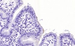
آنتی بادی Helicobacter Pylori کلون HPLY/7225 برند Patho Sage
نمره 0 از 5Specification:
The spiral shaped bacterium Helicobacter pylori is strongly associated with inflammation of the stomach and is also implicated in the development of gastric malignancy. H. pylori is known to cause peptic ulcers and chronic gastritis in human. It is associated with duodenal ulcers and may be involved in development of adenocarcinoma and low-grade lymphoma of mucosa associated lymphoid tissue in the stomach. This antibody stains the individual H. pylori bacterium when it presents on the surface of the epithelium or in the cytoplasm of the epithelial cells in biopsy tissue sections from the antrum and body of the stomach.
Immunogen:
Recombinant Helicobacter pylori Catalase protein fragment (around aa323-445) (Exact sequence is proprietary)
Presentation:
Bioreactor Concentrate with 0.05% Azide
-
آنتی بادیهای ایمونوهیستوشیمی
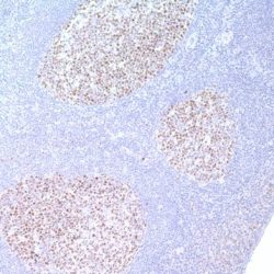
آنتی بادی BCL6 کلون IG191E/A8 برند Patho Sage
نمره 0 از 5Specification:
Bcl-6 is a transcriptional regulator gene which codes for a 706-amino-acid nuclear zinc finger protein. Antibodies to this protein stain the germinal center cells in lymphoid follicles, the follicular cells and interfollicular cells in Follicular Lymphoma, Diffuse Large B-Cell Lymphomas, and Burkitt’s lymphoma, and most of the Reed-Sternberg cells in Nodular Lymphocyte Predominant Hodgkin’s Disease. In contrast, anti-BCL6 rarely stains Mantle Cell Lymphoma and MALT Lymphoma. BCL6 expression is seen in approximately 45% of CD30+ Anaplastic Large Cell Lymphomas but is consistently absent in other peripheral T-cell lymphomas.
Immunogen:
Recombinant BCL-6 protein
Presentation:
Bioreactor Concentrate with 0.05% Azide
-
آنتی بادیهای ایمونوهیستوشیمی
آنتی بادی Inhibin Alpha کلون INHA/4265 برند Patho Sage
نمره 0 از 5Specification:
It recognizes a 47kDa protein, which is identified as alpha sub-unit of Inhibin. It is a gonadal protein that preferentially suppresses the secretion of pituitary follicle-stimulating hormone (FSH). Inhibin comprises two subunits, Inhibin A and Inhibin B. Each subunit consists of the same subunit, covalently linked to 1 of 2 distinct subunits, – or -. Originally isolated from ovarian follicular fluid and characterized as a disulphide-linked dimeric glycoprotein, inhibin belongs to the transforming growth factor (TFG) superfamily. Antibodies against Inhibin are useful in making a differentiation between adrenal cortical tumors and renal cell carcinoma. Sex cord stromal tumors of the ovary as well as trophoblastic tumors also demonstrate cytoplasmic positivity. Inhibin antibody is also used to make the differential diagnosis of intra-uterine vs. ectopic pregnancy in endometrial curetting.
Immunogen:
A synthetic peptide (around aa 73-96) of α-subunit of human inhibin A protein (exact sequence is proprietary)
Presentation:
Bioreactor Concentrate with 0.05% Azide
-
آنتی بادیهای ایمونوهیستوشیمی
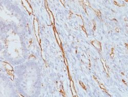
آنتی بادی CD31 (PECAM-1) کلون JC/70A برند Patho Sage
نمره 0 از 5Specification:
CD31 (PECAM-1) is a transmembrane glycoprotein member of the immunoglobulin supergene family of adhesion molecules. It is widely used as an endothelial cell marker. CD31 Ab reacts with normal, benign, and malignant endothelial cells which make up blood vessel lining. The level of CD31 expression can help to determine the degree of tumor angiogenesis, and a high level of CD31 expression may imply a rapidly growing tumor and potentially a predictor of tumor recurrence.
Immunogen:
Membrane preparation of a spleen from a patient with hairy cell leukemia
Presentation:
Bioreactor Concentrate with 0.05% Azide
-
آنتی بادیهای ایمونوهیستوشیمی
آنتی بادی Cytokeratin 8/18 کلون K.8 + DC10 برند Patho Sage
نمره 0 از 5Specification:
Cytokeratin 8 (CK8) belongs to the type II (or B or basic) subfamily of high molecular weight cytokeratin’s and exists in combination with cytokeratin 18 (CK18). This MAb cocktail recognizes all simple epithelia including glandular epithelium, for example thyroid, female breast, gastrointestinal tract, respiratory tract, and urogenital tract including transitional epithelium. All adenocarcinomas and most squamous carcinomas are positive but keratinizing squamous carcinomas are usually negative. This antibody is useful in demonstrating the presence of Paget cells; there is very little keratin 18 in the normal epidermis so only Paget cells are stained. Immunohistochemical staining with this MAb is indistinguishable from that obtained with monoclonal antibody 5D3.
Immunogen:
Keratin preparation from a human carcinoma (K8.8); PMC-42 human breast carcinoma cells (DC10)
Presentation:
Protein A/G purified antibody from Bioreactor Concentrate with 0.05% BSA and 0.05% Azide.
-
آنتی بادیهای ایمونوهیستوشیمی
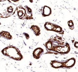
آنتی بادی Cytokeratin PAN کلون LU5 برند Patho Sage
نمره 0 از 5Specification:
Monoclonal antibody Lu-5 reacts with an epitope which is common to all cytokeratins respectively with a cytokeratin associated protein. The antigen recognized by anti-cytokeratin pan is present in all types of epithelia. It recognizes most subtypes of cytokeratins in cryofixed and paraffin sections and is ideally suited as a first order pan-epithelial marker. The epitope recognized by Lu-5 is a formalin-resistant marker of great value in tumor diagnosis, located on the surface of cytokeratin filaments. It has been preserved during vertebrate evolution and can be shown in all species from amphibia to man. The epitope is present in most cytokeratin polypeptides of both the acidic (type I) and basic (type II) subfamily but does not occur in other cytoskeletal proteins. The epithelial specificity and the broad tissue and species cross reactivity provide an excellent probe for the differential diagnosis of epithelial versus mesenchymal tumors, large cell lymphomas and neural tumors.
Immunogen:
Lung tumor cell lines A549 and A2182
Presentation:
Concentrated cell culture supernatant in phosphate buffered saline pH 7.2 (PBS), and 0.05% sodium azide as a preservative
-
آنتی بادیهای ایمونوهیستوشیمی
آنتی بادی MELAN A (MART1) کلون M2-9E3 برند Patho Sage
نمره 0 از 5Specification:
This antibody recognizes a protein doublet of 20-22kDa, identified as MART-1 (Melanoma Antigen Recognized by T cells 1) or Melan-A. MART-1 is a newly identified melanocyte differentiation antigen recognized by autologous cytotoxic T lymphocytes. Seven other melanoma associated antigens recognized by autologous cytotoxic T cells include MAGE-1, MAGE-3, tyrosinase, gp100, gp75, BAGE-1, and GAGE-1. Subcellular fractionation shows that MART-1 is present in melanosomes and endoplasmic reticulum. This MAb labels melanomas and other tumors showing melanocytic differentiation. It is also a useful positive marker for angiomyolipoma’s. It does not stain tumor cells of epithelial, lymphoid, glial, or mesenchymal origin.
Immunogen:
Recombinant hMART-1 protein
Presentation:
Bioreactor Concentrate with 0.05% Azide
-
آنتی بادیهای ایمونوهیستوشیمی
آنتی بادی CDX2 کلون MonoPoly/6271R برند Patho Sage
نمره 0 از 5Specification:
The specificity of this monoclonal antibody to its intended target was validated by HuProt TM Array, containing more than 19.000 full-length human proteins. The intestine-specific transcription factors CDX1 and CDX2 are important for directing intestinal development, differentiation, proliferation and maintenance of the intestinal phenotype. CDX2 protein expression has been seen in GI carcinomas. Anti-CDX2 has been useful to establish GI origin of metastatic adenocarcinoma and carcinoids and is especially useful in distinguish metastatic colorectal adenocarcinoma from lung adenocarcinoma. However, mucinous carcinomas of the ovary also expressed CDX2 protein. It limits the usefulness of this marker in the distinction of metastatic colorectal adenocarcinoma from mucinous of the ovary.
Immunogen:
Recombinant fragment and/of synthetic peptides of human CDX2 protein
Presentation:
Bioreactor Concentrate with 0.05% Azide
-
آنتی بادیهای ایمونوهیستوشیمی
آنتی بادی CD43 کلون MT1 برند Patho Sage
نمره 0 از 5Specification:
CD43 antigen is expressed on all T cells, NK cells, myeloid cells and monocytes as well as on CD5 positive mature B cells. It is also present on all immature hematopoietic cells in the bone marrow. The antigen is deficient in patients with Wiskott-Aldrich syndrome. A soluble form of CD43 is present in human serum.CD43, clone MT1 antibodies are used to identify B cell lines and myeloma cells. It may be used in the diagnosis of chronic lymphocytic leukemias (CLL), as an alternative to CD5. The antigen detected by MT1 is the only marker expressed on neoplasms of the very early precursor cells. In immunohistochemistry it reacts with T cells, macrophages, myeloid cells and B cells (weak). It is used for the typing of lymphomas in paraffin sections. CD43 may function as an adhesion molecule via interaction with CD54 although this has not been definitely established. It may also inhibit leucocyte interactions with other cells. The antigen is involved in the activation of T-cells, B cells, NK cells and monocytes. The membrane proximal portion of the cytoplasmic domain mediates an association with the cytoskeleton.
Presentation:
Antibodies are supplied in 0.01M sodium phosphate, 0.15 M NaCl, pH 7.3, 0.2% BSA, 0.09% sodium azide
-
آنتی بادیهای ایمونوهیستوشیمی
آنتی بادی Helicobacter Pylori کلون MX014 برند Patho Sage
نمره 0 از 5Specification:
Anti-H.pylori antibody reacts with H.pylori typically found on the surface of pyloric and stomach mucosa. Studies have shown that H.pylori plays an important role in the ethology of chronic gastritis and the development of peptic ulcer disease.
Presentation:
Tissue culture supernatant containing 15mM sodium azide.
-
آنتی بادیهای ایمونوهیستوشیمی
آنتی بادی CD19 کلون MX016 برند Patho Sage
نمره 0 از 5Specification:
CD19 is expressed only on B-cells and follicular dendritic cells. It is a specific and sensitive marker of B-cells widely expressed from early pre-B stages, normal B-cells and normal plasma cells (staining is weaker than normal B-cells and a subpopulation may lack expression). It is considered a positive regulator of both intrinsic and stimulus-dependent pathways in B-lymphocytes. CD19 is useful in identification of B-cell lineage of majority of B-cell neoplasms but appears to be less useful in subclassifying of B-cell neoplasms in histological material. It appears to be potentially useful additional marker of follicular dendritic cell tumors.
Presentation:
Liquid tissue culture supernatant containing 15mM sodium azide
-
آنتی بادیهای ایمونوهیستوشیمی
آنتی بادی Calretinin کلون MX027 برند Patho Sage
نمره 0 از 5Specification:
Calretinin is a 29 kDa calcium-binding protein, member of the family of so-called EF-hand proteins, to which also the S-100 proteins belong. Calretinin is abundantly expressed in neurons. Outside the nervous system, calretinin is found in mesothelial cells, steroid producing cells (adrenal cortical cells, testicular Leydig cells, ovarian theca internal cells), testicular Sertoli cells, rete testis, ovarian surface epithelium, some neuroendocrine cells, breast glands, eccrine sweat glands, hair follicular cells, thymic epithelial cells, endometrial stromal cells and fat cells. In calretinin positive cells, the protein is generally found in both cytoplasm and nuclei.
Presentation:
Tissue culture supernatant containing 15mM sodium azide
-
آنتی بادیهای ایمونوهیستوشیمی
آنتی بادی CD31 (PECAM-1) کلون MX032 برند Patho Sage
نمره 0 از 5Specification:
CD31 is a transmembrane glycoprotein, 130-140 kDa, also designated platelet-endothelium cell adhesion molecule = PECAM-1, belonging to the immunoglobulin super family. CD31 is ligand for CD38 and plays a role in thrombosis and angiogenesis. CD31 is strongly expressed in endothelial cells and weakly expressed in megakaryocytes, platelets, occasional plasma cells, lymphocytes (esp. marginal zone B-cells, peripheral T-cells) and neutrophils.
Presentation:
Tissue culture supernatant containing 15mM sodium azide.

