دسته: مونوکلونال
نمایش 421–440 از 477 نتیجه
فیلتر ها-
آنتی بادیهای ایمونوهیستوشیمی
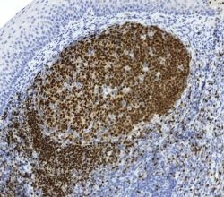
آنتی بادی Cyclin D1 کلون CCND1/3370R برند Patho Sage
نمره 0 از 5Specification:
Recognizes a protein of 36kDa, identified as Cyclin D1, one of the key cell cycle regulators, is a putative protooncogene overexpressed in 2 wide varieties of human neoplasms. This antibody neutralizes the activity of Cyclin D1 in vivo. About 60% og mantle cell lymphomas (MCL) contain at (11;14)(q13;32) translocation resulting in over-expression of cyclin D1. This antibody is useful in identifying mantle cell lymphomas from CLL/SLL and follicular lymphomas. Occasionally, hairy cell leukemia and plasma cell myeloma weakly express Cyclin D1.
Immunogen:
A synthetic peptide corresponding to residues within aa 270-295 of Cyclin D1
Presentation:
Bioreactor Concentrate with 0.05% Azide
-
آنتی بادیهای ایمونوهیستوشیمی
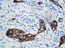
آنتی بادی CA15-3 کلون DF3 برند Patho Sage
نمره 0 از 5Specification:
This antibody has been used for evaluating the primary site of a metastatic carcinoma of unknown origin and distinguishing between benign and malignant lesions. It is believe that CA15-3 reacts primarily with the DF3-antigen, a 300 kDa mucinlike glycoprotein present on the apical border of secretory mammary epithelial cells. CA15-3 has been detected with immunohistochemistry in a wide spectrum of carcinomas, including Breast Carcinomas (ductal and lobular), Sarcomas (Synovial Sarcoma and Malignant Fibrous Histiocytomas), and Lung Carcinomas. CA15-3 can be used as a supplementary marker for epithelial differentiation. CA15-3 does not stain Melanomas or Ewing’s Sarcomas. Approximately 30% of Hepatocellular Carcinomas are positive for CA15-3.
Presentation:
CA15-3 is a mouse monoclonal antibody derived from cell culture supernatant that is concentrated, dialyzed, filter sterilized and diluted in buffer pH 7.5, containing BSA and sodium azide as a preservative.
-
آنتی بادیهای ایمونوهیستوشیمی
آنتی بادی Tryptase کلون G3 برند Patho Sage
نمره 0 از 5Specification:
Tryptases constitute a subfamily of trypsin-like proteinases, stored in mast cell secretory granules and basophils. Upon cellular activation, these enzymes are released into the extracellular environment. Anti-tryptase is a good marker for mast cells, basophils, and their derivatives.
Presentation:
Anti-Tryptase is a mouse monoclonal antibody from ascites diluted in phosphate buffered saline, pH 7.3-7.7, with protein base, and preserved with sodium azide
-
آنتی بادیهای ایمونوهیستوشیمی
آنتی بادی Granzyme B کلون GrB-7 برند Patho Sage
نمره 0 از 5Specification:
Granzymes are serine proteases which are stored in specialized lytic granules of cytotoxic T lymphocytes (CTL) and in natural killer cells. Granzyme B is involved in target cell apoptosis during lymphocyte mediated cyto-toxicity. Exocytosis of granzyme containing granules in the cytoplasm of the target cell will lead to induction of DNA fragmentation and apoptosis of the target cell. Anti-Granzyme B has been useful in diagnosing Natural killer/T cell lymphoma, as well as anaplastic largecell lymphoma. High percentages of cytotoxic T cells have been shown to be an unfavorable prognostic indicator in Hodgkin’s disease. The monoclonal antibody reacts with the 33 kD human serine protease granzyme B. It does not react with human granzyme A.
Presentation:
Tissue culture supernatant with 0.1% BSA and 0.02% sodium azide
-
آنتی بادیهای ایمونوهیستوشیمی
آنتی بادی PD1 کلون NAT105 برند Patho Sage
نمره 0 از 5Specification:
PDCD-1 (Programmed cell death-1 protein), also designated CD279m is a type I transmembrane receptor and member of the immunoglobulin gene superfamily. It is expressed on activated T-cells, B-cells and myeloid cells. Anti-PDCD-1 is a marker of angioimmunoblastic lymphoma and suggest a unique cell of origin for this neoplasm. Unlike CD10 and BCL6, PDCD-1 is expressed by few B-cells, so anti-PDCD-1 expression provides evidence that angioimmunoblastic lymphoma is a neoplasm derived from germinal center-associated T-cells.
Presentation:
Bioreactor Concentrate with 0.05% BSA % 0.05% Azide.
-
آنتی بادیهای ایمونوهیستوشیمی
آنتی بادی CA125 (Mucin 16) کلون OC125 برند Patho Sage
نمره 0 از 5Specification:
Anti CA-125 antibody reacts with epitheloid malignancies of the ovary, papillary serous carcinoma of the cervix, adenocarcinoma of the endometrium, clear cell adenocarcinoma of the bladder, and epitheloid mesothelioma. The antigen is formalin resistant, permitting the detection of ovarian cancer by immunohistochemistry, although serum assays for this protein are widely used to monitor ovarian cancer. Anti CA-125 also reacts with antigens in seminal vesicle carcinoma.
Presentation:
Anti-CA-125 is a mouse monoclonal antibody from ascites diluted in Tris- buffered saline, pH 7.3 – 7.7, with protein base, and preserved with sodium azide.
-
آنتی بادیهای ایمونوهیستوشیمی
آنتی بادی Tyrosinase کلون T311 برند Patho Sage
نمره 0 از 5Specification:
This antibody recognizes a cluster of proteins between 70-80kDa, identified as tyrosinase. Occasionally a minor band at 55kDa is also detected. This MAb shows no cross-reaction with MAGE-1 and tyrosinase-related protein 1, TRP-1/gp75. Tyrosinase is a copper-containing metalloglycoprotein that catalyzes several steps in the melanin pigment biosynthetic pathway: the hydroxylation of tyrosine to L-3,4-dihydroxy-phenylalanine (dopa), and the subsequent oxidation of dopa to dopaquinone. Mutations of the tyrosinase gene occur in various forms of albinism. Tyrosinase is one of the targets for cytotoxic T-cell recognition in melanoma patients. Staining of melanomas with this MAb shows tyrosinase in melanotic as well as amelanotic variants. This MAb is a useful marker for melanocytes and melanomas.
Immunogen:
Recombinant tyrosinase protein
Presentation:
Bioreactor Concentrate with 0.05% Azide
-
آنتی بادیهای ایمونوهیستوشیمی
آنتی بادی CD61 Intergrin Beta3 کلون YR/51 برند Patho Sage
نمره 0 از 5Specification:
Reacts with human integrin beta3 (GPIIIa, vitronectin receptor beta chain). It associates with the αV-chain (CD51) to form vitronectin receptor, or with the αIIb-chain (CD41) to form the GpIIb/GpIIIa complex (CD41/CD61). The CD41/CD61 complex appears early in megakaryocyte maturation. The activated CD41/CD61 complex is a receptor for von Willebrand factor, soluble fibrinogen, fibronectin, vitronectin and thrombospondin. It plays a central role in platelet activation and aggregation. The CD51/CD61 is implicated in tumors metastasis and adenoviral infection. The antibody detects platelets in smears of blood and bone marrow, as well as megakaryocytes in frozen sections and cell smears. The antibody is useful for classification of megakaryoblast leukemia.
Presentation:
Purified antibody from Bioreactor concentrate by Protein A/G. Prepared in 10mM PBS with 0.05% BSA & 0.05% Azide.
-
آنتی بادیهای ایمونوهیستوشیمی
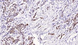
آنتی بادی AR (Androgen Receptor) کلون EP120 برند Patho Sage
نمره 0 از 5Specification:
Androgen Receptor belongs to the super-family of nuclear hormone receptors that employ complex molecular mechanisms to guide the development and physiological functions of their target tissues. Androgen Receptor function plays a pivotal role in normal prostate development and physiology as well as prostrate tumorigenesis. Androgen stimulates results in cell proliferation in both developing prostate and the malignant prostrate. Androgen Receptor is a phosphoprotein and also regulates mitogen-activated protein kinase (MAP Kinase). The inhibition f the MEK1/2 pathway correlates directly with a change in phosphorylation state of the androgen receptor. In prostate cancer, Androgen Receptor has been proposed as a marker of the hormone responsiveness.
Immunogen:
Synthetic peptide derived from near N-terminus of human Androgen Receptor.
Presentation:
Purified antibody is diluted in Tris-HCL buffer containing stabilizing protein and <0,1% Sodium Azide.
-
آنتی بادیهای ایمونوهیستوشیمی
آنتی بادی Cytokeratin کلون MNF116 برند Patho Sage
نمره 0 از 5Specification:
This antibody recognizes cytokeratin polypeptides ranging from 40 through 58kD, corresponding to cytokeratin’s 5, 6, 8,17, and 19. It shows a broad pattern of reactivity with human epithelial tissues from simple glandular epithelia to stratified squamous epithelia, like epidermis, mammary gland ducts, and tracheal epithelium.
Immunogen:
Crude extract of splenic cells from a nude mouse engrafted with MCF-7 cells
Presentation:
This antibody is supplied as tissue culture supernatant containing sodium azide as a preservative
-
آنتی بادیهای ایمونوهیستوشیمی
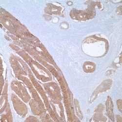
آنتی بادی CD326 (Ep-CAM) کلون VU1D9 برند Patho Sage
نمره 0 از 5Specification:
Ep-CAM (epithelial specific antigen) is an ~40 kDa transmembrane glycoprotein involved in several cellular processes including adhesion, proliferation, maintenance of stemness, migration and invasion (Spizzo, 2012, Denzel, 2012). Ep-CAM is expressed on most, but not all, normal epithelia and their corresponding malignancies. Although the role of Ep-CAM expression in tumorigenesis remains to be fully elucidated, Ep-CAM may downregulate immunity and help tumors actively escape from immune surveillance. Since the 1980s Ep-CAM has been recognized as a pan-carcinoma antigen marker because of its widespread expression on epithelium and their derived tumors (Litnov, 1994; Ruf, 2007). More recently, Ep-CAM has also been identified as a stem cell marker, and it is thought that Ep-CAM antibody positive tumor cells may have stem-like properties (Flatmark, 2011) Ep-CAM antibody is widely used to help distinguish epithelial from non-epithelial neoplasms. Ep-CAM antibody positive tumors are epithelium derived, whereas Ep-CAM antibody negative tumors can originate from either non-epithelial or epithelial tissues. Ep-CAM antibody has also been used for identifying circulating tumor cells (Saif, 2012, de Albuquerque, 2012). Ep-CAM antibody is often used as part of a panel with other tumor or tissue antibody markers when classifying tumors of known, suspected or elusive origin. Ep-CAM expression may be upregulated in tumors and overexpression can most pronounced on tumor initiating cells (Imrich, 2012). Overexpression may be an indication of early malignancy and correlate with a poor prognosis (Ruf, 2007). The VU1D9 Ep-CAM antibody clone has been widely cited in the literature, and researchers are encouraged to review the published literature for additional information.
Immunogen:
Small cell lung carcinoma cells
Presentation:
Ep-CAM antibody in PBS with 15 mM sodium azide
-
آنتی بادیهای ایمونوهیستوشیمی
آنتی بادی ESA (Epithelial Specific Antigen) کلون BER-EP4 برند Patho Sage
نمره 0 از 5Specification:
Ep-CAM/Epithelial Specific Antigen consists of two glycoproteins, 34 and 39 kDa, sometimes designated Epithelial antigen, epithelial specific antigen, and epithelial glycoprotein. In paraffin sections, the protein is detected with mAbs like Ber-EP4 and MOC-31. The glycoproteins are located on the cell membrane surface and in the cytoplasm of virtually all epithelial cells except for most squamous epithelia, hepatocytes, renal proximal tubular cells, gastric parietal cells and myoepithelial cells. However, focal positivity may be seen in the basal layer of squamous cell epithelium of endoderm (e.g., palatine tonsils) and mesoderm (e.g., uterine cervix). In liver lesions like hepatitis and cirrhosis, the hepatocytes frequently become Ep-CAM positive. Normal mesothelial cells are Ep-CAM negative but may express focal reaction when undergoing reactive changes. Mesenchymal cells and cells from the neural crest are negative, except for olfactory neurons. Ep-CAM is found in most adenocarcinomas of most sites (50-100% in various studies; as well as neuroendocrine tumours, including small cell carcinoma. Renal cell carcinoma and hepatocellular carcinoma stain in about 30% of the cases. Squamous cell carcinoma of endoderm and mesoderm is usually Ber-EP4 positive, while that of ectoderm is negative. Basal cell and basosquamous carcinoma are Ber-EP4 positive in almost all cases. Choroid plexus papilloma and carcinoma are usually negative. Malignant mesothelioma (epithelioid and biphasic) is Ep-CAM positive in 4-26% of the cases. The staining is usually focal but may occasionally be widespread. Synovial sarcoma (epithelioid and biphasic) and desmoplastic small round cell tumour stain for Ep-CAM in most cases. Seminoma, embryonal carcinoma, yolk sac tumour and choriocarcinoma reveal Ber-EP4 positivity in a minor proportion of cases. Among neural tumours, Ep-CAM positivity is seen only in olfactory neuroblastoma. Ep-CAM can be of great help in the differential diagnosis of malignant involvement in the peritoneal and pleural cavities. The lack of reactivity in the majority of malignant mesothelioma can in an appropriate panel be utilized in the discrimination between this tumour and adenocarcinoma. As for a series of anti-epithelial antibodies, Ber-EP4 or MOC-31 may be used in the demonstration of epithelial cell differentiation in cases where anti-cytokeratin’s do not produce a clearcut positivity or in cases where a false positivity for cytokeratin cannot be excluded, such as in sub mesothelial cells.
Immunogen:
Breast carcinoma cell line MCF-7
Presentation:
Bioreactor Concentrate with 0.05% Azide
-
آنتی بادیهای ایمونوهیستوشیمی
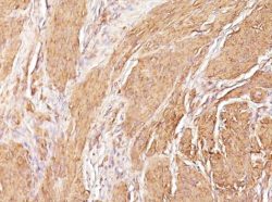
آنتی بادی Actin Muscle Specific کلون HHF35 برند Patho Sage
نمره 0 از 5Specification:
Actin is a major component of the cytoskeleton. This antibody recognizes actin of skeletal, cardiac, and smooth muscle cells. It is not reactive with other mesenchymal cells except for myoepithelium. Actin can be resolved on the basis of its isoelectric points into three distinctive components: alpha, beta, and gamma in order of increasing isoelectric point. Anti Muscle-Specific Actin recognizes alpha and gamma isotypes of all muscle groups. Non-muscle cells such as vascular endothelial cells and connective tissues are non-reactive. Also, neoplastic cells of non-muscle-derived tissue such as carcinomas, melanomas, and lymphomas are negative. This antibody is useful in the identification of rhabdoid cellular elements.
Immunogen:
SDS extract of human myocardium
Presentation:
Bioreactor Concentrate with 0.05% Azide
-
آنتی بادیهای ایمونوهیستوشیمی
آنتی بادی ESA (Epithelial Specific Antigen) کلون MOC-31 برند Patho Sage
نمره 0 از 5Specification:
SCLC-CD2; epithelial antigen, EGP-2. MOC-31 reacts with an epithelial antigen of 40kD present on most normal and malignant epithelia.MOC-31 has been assigned to a group of antibodies known as SCLC-Cluster 2 which react with an epithelial antigen determined at the Second International Workshop on Small Cell Lung Cancer (SCLC)Antigens. In normal tissues, clone MOC-31 reacts with glands and hair shafts in skin, pancreatic exocrine and endocrine glands, epithelia of the digestive and respiratory tract, including sero-mucous glands. MOC-31 stains epithelia of kidney, endometrium, breast, liver, prostate, pancreatic acini, slets and ducts. No reactivity is observed with adrenal, ovary, brain, peripheral nerve, ganglion cells, peripheral blood and bone marrow. Lung-derived tumours such as squamous cell carcinomas, adenomas, small cell lung carcinomas, carcinoids, adenocystic carcinomas, carcinosarcoma, mucoepidermal carcinoma and pleomorphic adenoma stain positively with MOC-31.Also,colon adenocarcinoma, gastric adenocarcinoma, esophagealgastric adenocarcinoma, some bladder transitional cell carcinomas, prostate adenocarcinoma, testicular yolk tumours, uterine adenomatoid tumours, ovary mucinous/endometrial cancers, serous carcinoma of the ovary, some breast carcinomas, thyroid papillary carcinoma and thyroid medullary carcinomas may be positive.
Immunogen:
Neuraminidase treated cells from a variant small cell lung carcinoma cell line (GLS-1)
Presentation:
Bioreactor Concentrate with 0.05% Azide
-
آنتی بادیهای ایمونوهیستوشیمی
آنتی بادی PAX5 کلون 24 برند Patho Sage
نمره 0 از 5Specification:
There are at least nine members of the paired box (PAX) gene family whose protein products are transcription factors involved in development. The conserved paired box DNA-binding domain is found in the N-terminal region of Pax proteins. An octomer and homeodomain sequence are conserved in the centre of the proteins. The Ser/Thr/ Pro-rich region in the Cterminal portion contains a conserved 100 amino acid transactivating domain. One of the best studied Pax family members, Pax-5 influences the expression of several B-cell-specific genes, such as CD 19 and CD 20. Pax-5 is expressed primarily in pro-, pre-, and mature B cells, but not in plasma cells. Interestingly, Pax-5 mRNA is transiently detected in the mesencephalon and spinal cord during embryogenesis. Expression then shifts to the fetal liver and correlates with the onset of B lymphopoiesis. PAX-5 has been found to be important in both B cell and nervous system development.
Immunogen:
Human PAX-5 aa. 151-306
Presentation:
0.25 mg/ml in aqueous buffered solution containing BSA, glycerol, and ≤ 0.09% sodium azide
-
آنتی بادیهای ایمونوهیستوشیمی
آنتی بادی Cytokeratin 7 کلون R17-S برند Patho Sage
نمره 0 از 5Specification:
Cytokeratin 7 antibody reacts with proteins that are found in most ductal, glandular and transitional epithelium of the urinary tract and bile duct epithelial cells. Cytokeratin 7 distinguishes between lung and breast epithelium that stain positive, and colon and prostate epithelial cells that are negative. This antibody also reacts with many benign and malignant epithelial lesions, e.g. adenocarcinomas of the ovary, breast and lung. Transitional cell carcinomas are positive and prostate cancer is negative. This antibody does not recognize intermediate filament proteins.
Immunogen:
Peptide derived from N-terminal sequence of human cytokeratin 7
Presentation:
monospecific rabbit clonal antibody in 20 mM Tris-HCl, pH 8.0 with 20 mg/ml BSA and 0.05% NaN3
-
آنتی بادیهای ایمونوهیستوشیمی
آنتی بادی CD221 کلون 1H7 برند Patho Sage
نمره 0 از 5Specification:
IGF-1 Receptor recognizes human CD221, a 155kD receptor tyrosine kinase, also known as Insulin-like growth factor I receptor (IGF-I Receptor). CD221 is composed of two extracellular alpha-subunits and two transmembrane beta-subunits. Clone 1H7 recognizes an epitope in the alpha subunits of CD221, demonstrated by Western blotting. CD221 is expressed in a range of tissues, including kidney, liver, placenta, mammary gland, brain, ovary and skin. The ligands for CD221 include IGF-I and IGF-II, which bind to CD221 and activate tyrosine kinase activity, resulting in phosphorylation of several intracellular signaling proteins. Clone 1H7 is reported to partically block binding of IGF-I and IGF-II to CD221.
Immunogen:
Purified human placental CD221 IGF-1 receptor
Presentation:
Purified IgG prepared by affinity chromatography on Protein G from tissue culture supernatant
-
آنتی بادیهای ایمونوهیستوشیمی
آنتی بادی TRPS1 کلون 8131R برند Patho Sage
نمره 0 از 5Specification:
Transcriptional repressor binds specifically to GATA sequences and repress expression of GATA-regulated genes at selected sites and stages in vertebrate development. Regulates chondrocyte proliferation and differentiation. Executes multiple functions in proliferating chondrocytes, expanding the region of distal chondrocytes, activating proliferation in columnar cells and supporting the differentiation of columnar into hypertrophic chondrocytes. Ubiquitously expressed in the adult. Found in fetal brain, lung, kidney, liver, spleen and thymus. More highly expressed in androgen-dependent than in androgen-independent prostate cancer cells.
Immunogen:
Recombinant fragment (around aa900-1100) of human-TRPS1 protein (exact sequence is proprietary)
Presentation:
Bioreactor Concentrate with 0.05% Azide
-
آنتی بادیهای ایمونوهیستوشیمی
آنتی بادی BRST-1 (BCA225) کلون CU-18 برند Patho Sage
نمره 0 از 5Specification:
Anti-BCA-225 antibody recognizes a human breast carcinoma associated glycoprotein BCA-225 (220-225kD). This protein differs in size and distribution from other breast carcinoma antigens. Unlike other antibodies against breast carcinoma antigens, this antibody does not react with benign or malignant colonic, stomach, prostate, liver, pancreas, thyroid, or parotid tissues. Adenocarcinomas of the lung, ovary and endometrium also stain with this antibody.
Immunogen:
A synthetic peptide
Presentation:
Purified antibody with 0.2% BSA and 15mM sodium azide.
-
آنتی بادیهای ایمونوهیستوشیمی
آنتی بادی CD1a کلون EP3622 برند Patho Sage
نمره 0 از 5Specification:
CD1a is a non-polymorphic major histocompatibility complex class I-related cell surface glycoprotein (45 to 55 kDa) and is expressed in association with β-microglobulin. In normal tissues, anti-CD1a reacts with cortical thymocytes, Langerhans cells, interdigitating dendritic cells, and rare antigen-presenting cells of the lymph node. Anti-CD1a labels Langerhans cell histiocytosis (Histiocytosis X), extranodal histiocytic sarcoma, a subset of T-lymphoblastic lymphoma/leukemia, and interdigitating dendritic cell sarcoma of the lymph node. When combined with antibodies against TTF-1 and CD5, anti-CD1a is useful in distinguishing between pulmonary and thymic neoplasms since CD1a is consistently expressed in thymic lymphocytes in both typical and atypical thymomas, but only focally in 1/6 of thymic carcinomas and not in lymphocytes in pulmonary neoplasms. Anti-CD1a was reported to be a new marker for perivascular epithelial cell tumor (PEComa).
Presentation:
Tris Buffer, pH 7.3-7.7, with 1% BSA and <0.1% Sodium Azide


