Name: CD63 Antibody Clone NKI/C3
Description and applications:The CD63 molecule, also called melanoma-associated antigen MLA1 or ME491, granulophysine, or
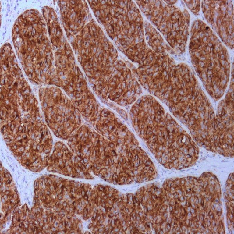
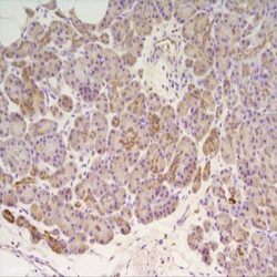
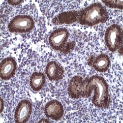
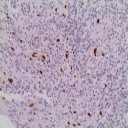
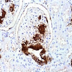
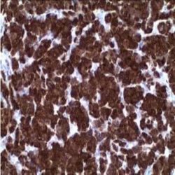
membrane-associated lysosomal glycoprotein 3 (LAMP-3), is a 53-kDa glycoprotein belonging to the transmembrane 4 protein superfamily or tetraspanins. It is made up of 237 amino acids and is commonly expressed in melanoma tumour cells, where it is associated with early stages of tumour progression. The ME491 gene, which encodes it, is located on the chromosomal region 12q13.2 and the lack of this protein has been observed in Hermansky- Pudlak syndrome, which is characterized by the presence of deficient lysosomes with dense granules and accumulation of ceroid material in reticuloendothelial system cells. Functionally, CD63 plays an important role in intracellular transport and is a necessary molecule in the trafficking processes that take place in the PMEL luminal domain, which are essential in the development and maturation of melanocytes. Similarly, CD63 is important for leukocyte-endothelial cell adhesion through activation by P-selectin. Finally, CD63 appears to be involved in degranulation of mast cells in response to their activation through subunit beta of high affinity immunoglobulin receptor’s epsilon chain (Ms4a2/FceRI system). CD63 antigen is widely distributed in normal tissues,being present in platelets’ lysosomal granules, neutrophil granulocytes, and basophil granulocytes, as well as in a small proportion of inactive T cells, endothelial cells, and macrophages. In tumour cells, CD63 is expressed in dysplastic nevi and radial growth phase melanomas, as well as in renal angiomyolipoma, cellular neurothekeoma, atypical fibroxanthoma and dermatofibrosarcoma protuberans, clear cell fibrous papule of the nose, and non-neural granular cell tumour of the skin. Its usefulness has also been proven in the differential diagnosis between renal oncocytomas, which show apical staining, and renal cell carcinomas with eosinophilic cytoplasm, which show diffuse cytoplasmic staining. Other tumours such as breast carcinomas, Merkel cell carcinomas, astrocytomas, and lung adenocarcinomas may also show positive staining, being, in the latter two cases, an indicator of a better prognosis.
Composition: Anti-human CD63 mouse monoclonal antibody purified from serum and prepared in 10mM PBS, pH 7.4, with 0.2% BSA and 0.09% sodium azide.

دیدگاهها
هیچ دیدگاهی برای این محصول نوشته نشده است.