-
 پروبهای فیش
پروب فیش IRF4/DUSP22 Break Apart FISH Probe
INTRODUCTION: Rearrangements and abnormal expression of the IRF4 gene – also known as NF-EM5, MUM1, LSIRF or IRF-4 – and the DUSP22 gene – also called JKAP, JSP-1, JSP1, LMW-DSP2, LMWDSP2, MKP-x, MKPX or VHX – have been observed in multiple myeloma (MM) and other lymphoid malignancies, viral malignancies, skin cancer and lymphomatoid papulosis (LyP), a chronic papulonecrotic or papulonodular skin disease with histologic features suggestive of a malignant lymphoma.
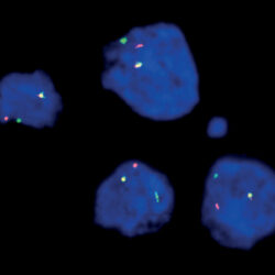
INTENDED USE: The IRF4/DUSP22 Break Apart FISH probe Kit is designed to detect rearrangements in the human IRF4 and DUSP22 genes and the surrounding regions located on chromosome band 6p25.3. In addition to revealing breaks, which can lead to translocation of parts of the genes, inversion, or their fusion to other genes, the probe set can also be used to identify other IRF4 and DUSP22 aberrations such as deletions or amplifications.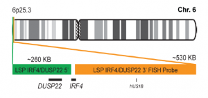
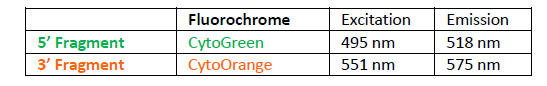
PROBE DESCRIPTION:The 5’ telomeric fragment of the IRF4/DUSP22 Break Apart FISH Probe kit labeled with the fluorochrome CytoGreen covers the entire DUSP22 gene, genomic sequences adjacent to the 5’ (start) and the 3′ (end) portions of the gene, and genomic sequences upstream of the IRF4 gene’s 5′ (start) end. The 3’ centromeric fragment of the IRF4/DUSP22 Break apart FISH Probe Kit labeled with CytoOrange covers some sequences downstream of the 3’ end of the IRF4 gene. The two probes are flanking sequences across the IRF4 and DUSP22 genes in which various breakpoints have been observed.
-
 پروبهای فیش
پروب فیش JAK2 Break Apart FISH Probe
INTRODUCTION: The JAK2 gene provides instructions for making a protein that promotes the growth and division (proliferation) of cells. This protein is part of a signaling pathway called the JAK/STAT pathway, which transmits chemical signals from outside the cell to the cell’s nucleus. The JAK2 protein is especially important for controlling the production of blood cells from hematopoietic stem cells. These stem cells are located within the bone marrow and have the potential to develop into red blood cells, white blood cells, and platelets. Rearrangements and abnormal expression of the JAK2 gene – also known as THCYT3 or JTK10 – have been observed in acute myeloid and lymphoid leukemia’s and other malignancies.
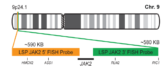
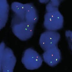
INTENDED USE: The JAK2 Break Apart FISH probe Kit is designed to detect rearrangements in the human JAK2 gene located on chromosome band 9p24.1. In addition to revealing breaks, which can lead to translocation of parts of the gene, inversion, or its fusion to other genes, the probe set can also be used to identify other JAK2 aberrations such as deletions or amplifications.
PROBE DESCRIPTION: The 5’ telomeric fragment of the JAK2 Break Apart FISH Probe kit labeled with the fluorochrome CytoOrange covers the 5’ (start) portion of the JAK2 gene and some adjacent genomic sequences. The 3’ centromeric fragment of the JAK2 Break apart FISH Probe Kit labeled with CytoGreen covers the 3’ (end) part as well as sequences downstream of the gene. The two probes are flanking sequences across the JAK2 gene in which variable breakpoints have been observed.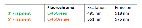
-
 آنتی بادیهای فلوسایتومتری
آنتی بادی مونوکلونال فلوسایتومتری CD1a-FITC ،کلون NA1-34-HLK
Name: Flow Cytometry Antibody CD1a-FITC, Clone NA1-34-HLK
- Antibody CD1a-FITC is a monoclonal antibody (mAb) labelled with fluorescein isothiocyanate (FITC) designed for flow cytometry use as a direct immunofluorescence reagent in the identification and enumeration of CD1a antigen-expressing cells. This reagent must be used by flow cytometry qualified personal.
SUMMARY AND EXPLANATION
CYT-1AF1 MAb recognizes CD1a antigen, a 49KDa membrane glycoprotein expressed non-covalently with β2- microglobulin. This antigen is found on cortical double positive and single positive thymocytes, Langerhans cells, a subset of dendritic cells and a B cell subset. CD1a has structural similarities to the MHC class I antigen and plays a role in antigen presentation. In addition, CD1a has been implicated in thymic T cell development. It is also present on some T cell leukaemias and lymphomas. Analysis of the expression of the CD1a antigen is useful in the characterization of hematologic neoplasia.
REAGENT COMPOSITION
The purified monoclonal CD1a antibody conjugated with fluorescein isothiocyanate (FITC) is supplied in phosphatebuffered saline (PBS) containing 0.09% sodium azide.
- Clone: NA1/34-HLK.
- Isotype: IgG2a.
- Amount per 1 mL vial: 200 tests (5 µL MAb per determination).
- Reagent is considered non-sterile.
-
 آنتی بادیهای فلوسایتومتری
آنتی بادی مونوکلونال فلوسایتومتری CD2-FITC، کلون RPA-2.10
Name: Flow Cytometry Antibody CD2-FITC, Clone RPA-2.10
- Antibody CD2-FITC is a monoclonal antibody (mAb) labelled with fluorescein isothiocyanate (FITC) designed for use as a direct immunofluorescence reagent in the identification and enumeration of cells which express the CD2 antigen by flow cytometry (FC). CD2 can be considered a pan-T cell antigen, as it is expressed on the surface of all normal Tlymphocytes. This reagent must be used by flow cytometry qualified personal.
SUMMARY AND EXPLANATION
The CYT-2F7 mAb recognizes the 50kDa lymphocyte surface antigen expressed on all peripheral blood T lymphocytes, the majority of thymocytes and malignant cells of T cell origin, including T ALL cells. CD2 is a useful marker in the assessment of lymphoid malignancies as it is expressed in the majority of precursor and posthymic lymphomas and leukemias. In some neoplastic T-cell populations e.g. in peripheral T cell lymphomas, CD2 may be aberrantly deleted.
REAGENT COMPOSITION
Purified monoclonal CD2 antibody conjugated with fluorescein isothiocyanate (FITC), supplied in phosphate buffered saline with 0,09% sodium azide.
- Clone: RPA-2.10
- Isotype: IgG1
- Purification: Affinity chromatography
- Amount per 1 mL vial: 200 tests (5 µL mAb to 106 cells)
- Reagents are not considered sterile.
-
 آنتی بادیهای فلوسایتومتری
آنتی بادی مونوکلونال فلوسایتومتری CD3، کلون UCHT-1
+Name: CD3-FITC, Clone (UCHT-1)
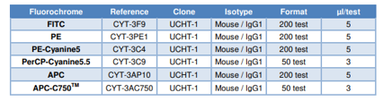
- Antibody CD3 reagent is a monoclonal antibody (mAb) conjugated with different fluorochromes (see table) and designed for use as a direct immunofluorescence reagent in the identification and enumeration of cells which express the CD3 antigen by flow cytometry. This reagent must be used by flow cytometry qualified personnel.
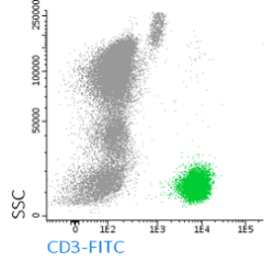
SUMMARY AND EXPLANATION
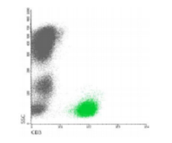
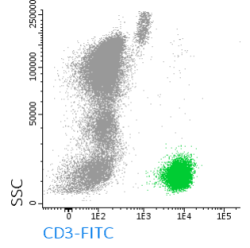
The anti-CD recognizes the CD3 antigen present in T cells and can therefore be used in the characterization studies for immunophenotyping of lymphocytes. These studies are widely applied for monitoring of the immunologic status of posttransplant patients and in the characterization and follow-up of immunodeficiencies, autoimmune diseases, leukemia etc. The T lymphocyte (CD3+) count is generally expressed as a percentage of the total amount of lymphocytes or leucocytes present in the sample which can itself be determined by flow cytometry based on its typical pattern of FSC/SSC (size/granularity or complexity). Because each flow cytometer has different operating characteristics each laboratory must determine its optimal operating procedure.
REAGENT COMPOSITION
Purified monoclonal CD3 antibody conjugated with different fluorochromes (see table above) is supplied in phosphatebuffered saline (PBS) containing 1% (m/v) BSA and 0.09% (m/v) sodium azide.
- Clone: UCHT-1
- Isotypes: Mouse / IgG1
- Purification: Affinity
- chromatography Amount per vial: 50 or 200 tests (3 or 5 µl mAb to 106 cells).
- Reagents are not considered sterile.
-
 آنتی بادیهای فلوسایتومتری
آنتی بادی مونوکلونال فلوسایتومتری CD3-PE، کلون UCHT-1
Name: Flow Cytometry Antibody CD3-PE, Clone UCHT-1
- Antibody CD3 reagent is a monoclonal antibody (mAb) conjugated with different fluorochromes (see table) and designed for use as a direct immunofluorescence reagent in the identification and enumeration of cells which express the CD3 antigen by flow cytometry. This reagent must be used by flow cytometry qualified personnel.
SUMMARY AND EXPLANATION
The anti-CD recognizes the CD3 antigen present in T cells and can therefore be used in the characterization studies for immunophenotyping of lymphocytes. These studies are widely applied for monitoring of the immunologic status of posttransplant patients and in the characterization and follow-up of immunodeficiencies, autoimmune diseases, leukemia etc. The T lymphocyte (CD3+) count is generally expressed as a percentage of the total amount of lymphocytes or leucocytes present in the sample which can itself be determined by flow cytometry based on its typical pattern of FSC/SSC (size/granularity or complexity). Because each flow cytometer has different operating characteristics each laboratory must determine its optimal operating procedure.
REAGENT COMPOSITION
Purified monoclonal CD3 antibody conjugated with different fluorochromes (see table above) is supplied in phosphatebuffered saline (PBS) containing 1% (m/v) BSA and 0.09% (m/v) sodium azide.
- Clone: UCHT-1
- Isotypes: Mouse / IgG1
- Purification: Affinity chromatography
- Amount per vial: 50 or 200 tests (3 or 5 µl mAb to 106 cells) (see table above).
- Reagents are not considered sterile.
-
 آنتی بادیهای فلوسایتومتری
آنتی بادی فلوسایتومتری CD3-FITC، کلون 33-2A3 (Cris-7)
Name: CD3-FITC, (Clone 33-2A3 (Cris-7))
Antibody CD3-FITC is a monoclonal antibody (mAb) labelled with fluorescein isothiocyanate (FITC) designed for use as a direct immunofluorescence reagent in the identification and enumeration of cells which express the human CD3 antigen by flow cytometry. This reagent must be used by flow cytometry qualified personal.
SUMMARY AND EXPLANATION
The CYT-3F8 mAb recognizes the CD3 antigen present in T cells and can therefore be used in the characterization studies for immunophenotyping of lymphocytes. These studies are widely applied for monitoring of the immunologic status of posttransplant patients and in the characterization and follow-up of immunodeficiencies, autoimmune diseases, leukemia etc. The T lymphocyte (CD3+) count is generally expressed as a percentage of the total amount of lymphocytes or leucocytes present in the sample which can itself be determined by flow cytometry based on its typical pattern of FSC/SSC (size/granularity or complexity). Because each flow cytometer has different operating characteristics each laboratory must determine its optimal operating procedure.
REAGENT COMPOSITION
Purified monoclonal CD3 Antibody conjugated with fluorescein isothiocyanate (FITC), supplied in phosphate buffered saline with 0,09% (m/v) sodium azide.
- Clone: 33-2A3 (Cris-7).
- Isotype: IgG2a.
- Purification: Affinity chromatography.
- Size: 200 tests (1 ml per vial)
- Usage: 5 µl mAb per determination
- Reagents are not considered sterile.
-
 آنتی بادیهای فلوسایتومتری
آنتی بادی مونوکلونال فلوسیتومتری CD2-FITC، کلون RPA-2.10
Name: Flow Cytometry Antibody CD2-FITC, Clone RPA-2.10
- Antibody CD2-FITC is a monoclonal antibody (mAb) labelled with fluorescein isothiocyanate (FITC) designed for use as a direct immunofluorescence reagent in the identification and enumeration of cells which express the CD2 antigen by flow cytometry (FC). CD2 can be considered a pan-T cell antigen, as it is expressed on the surface of all normal Tlymphocytes. This reagent must be used by flow cytometry qualified personal.
SUMMARY AND EXPLANATION
The CYT-2F7 mAb recognizes the 50kDa lymphocyte surface antigen expressed on all peripheral blood T lymphocytes, the majority of thymocytes and malignant cells of T cell origin, including T ALL cells. CD2 is a useful marker in the assessment of lymphoid malignancies as it is expressed in the majority of precursor and posthymic lymphomas and leukemias. In some neoplastic T-cell populations e.g. in peripheral T cell lymphomas, CD2 may be aberrantly deleted.
REAGENT COMPOSITION
Purified monoclonal CD2 antibody conjugated with fluorescein isothiocyanate (FITC), supplied in phosphate buffered saline with 0,09% sodium azide.
- Clone: RPA-2.10
- Isotype: IgG1
- Purification: Affinity chromatography
- Amount per 1 mL vial: 200 tests (5 µL mAb to 106 cells)
- Reagents are not considered sterile.
-
 فلوسایتومتری
آنتی بادی مونوکلونال فلوسایتومتری CD3-APC، کلون UCHT-1
Name: Flow Cytometry Antibody CD3-APC, Clone UCHT-1
- Antibody CD3 reagent is a monoclonal antibody (mAb) conjugated with different fluorochromes (see table) and designed for use as a direct immunofluorescence reagent in
- the identification and enumeration of cells which express the CD3 antigen by flow cytometry. This reagent must be used by flow cytometry qualified personnel.
SUMMARY AND EXPLANATION
The anti-CD recognizes the CD3 antigen present in T cells and can therefore be used in the characterization studies for immunophenotyping of lymphocytes. These studies are widely applied for monitoring of the immunologic status of posttransplant patients and in the characterization and follow-up of immunodeficiencies, autoimmune diseases, leukemia etc. The T lymphocyte (CD3+) count is generally expressed as a percentage of the total amount of lymphocytes or leucocytes present in the sample which can itself be determined by flow cytometry based on its typical pattern of FSC/SSC (size/granularity or complexity). Because each flow cytometer has different operating characteristics each laboratory must determine its optimal operating procedure.
REAGENT COMPOSITION
Purified monoclonal CD3 antibody conjugated with different fluorochromes (see table above) is supplied in phosphatebuffered saline (PBS) containing 1% (m/v) BSA and 0.09% (m/v) sodium azide.
- Clone: UCHT-1
- Isotypes: Mouse / IgG1
- Purification: Affinity chromatography
- Amount per vial: 50 or 200 tests (3 or 5 µl mAb to 106 cells.
- Reagents are not considered sterile.
-
 آنتی بادیهای فلوسایتومتری
آنتی بادی مونوکلونال فلوسایتومتری CD3-APC-C750 ، کلون UCHT-1
Name: Flow Cytometry AntibodyCD3-APC-C750™, Clone UCHT-1
- Antibody CD3 reagent is a monoclonal antibody (mAb) conjugated with different fluorochromes (see table) and designed for use as a direct immunofluorescence reagent in the identification and enumeration of cells which express the CD3 antigen by flow cytometry. This reagent must be used by flow cytometry qualified personnel.
SUMMARY AND EXPLANATION
The anti-CD recognizes the CD3 antigen present in T cells and can therefore be used in the characterization studies for immunophenotyping of lymphocytes. These studies are widely applied for monitoring of the immunologic status of posttransplant patients and in the characterization and follow-up of immunodeficiencies, autoimmune diseases, leukemia etc. The T lymphocyte (CD3+) count is generally expressed as a percentage of the total amount of lymphocytes or leucocytes present in the sample which can itself be determined by flow cytometry based on its typical pattern of FSC/SSC (size/granularity or complexity). Because each flow cytometer has different operating characteristics each laboratory must determine its optimal operating procedure.
REAGENT COMPOSITION
Purified monoclonal CD3 antibody conjugated with different fluorochromes (see table above) is supplied in phosphatebuffered saline (PBS) containing 1% (m/v) BSA and 0.09% (m/v) sodium azide.
- Clone: UCHT-1
- Isotypes: Mouse / IgG1
- Purification: Affinity chromatography
- Amount per vial: 50 or 200 tests (3 or 5 µl mAb to 106 cells).
- Reagents are not considered sterile.
-
 آنتی بادیهای فلوسایتومتری
آنتی بادی مونوکلونال CD3-Pacific Blue، کلون UCHT-1
Name: Flow Cytometry Antibody CD3-Pacific Blue, Clone UCHT-1
- Antibody CD3-Pacific BlueTM is a monoclonal antibody (mAb) labelled with Pacific BlueTM designed for use as a direct immunofluorescence reagent in the identification and enumeration of cells which express the CD3 antigen by flow cytometry. This reagent must be used by flow cytometry qualified personal.
SUMMARY AND EXPLANATION
Human lymphocytes may be classified in three main populations according to their biological function and their cell surface antigen expression: T lymphocytes, B lymphocytes and natural killer cells (NK). T lymphocytes (CD3+), the precursors of which originate in the bone marrow and then migrate and mature in the thymus, can be subdivided as well in functionally different populations. The most clearly defined of these are helper/inducer T cells (CD3+CD4+) and suppressor/cytotoxic T cells (CD3+CD8+). T cells produce no antibodies and are the mediators of cell immunity. CYT-3PBZ mAb recognizes the CD3 antigen present in T cells and can therefore be used in the characterization studies for immunophenotyping of lymphocytes. These studies are widely applied for monitoring of the immunologic status of post-transplant patients and in the characterization and follow-up of immunodeficiencies, autoimmune diseases, leukemia etc. Pacific BlueTM, is excited with the violet laser (405nm) and emits at 550nm. This fluorochrome provides maximum resolution and narrow emission peaks, which results in little spectral overlap and minimal compensation requirements. In multicolor panel it is recommended the use of this fluorochrome combined with Orange Cytognos 515, which is also excited with the violet laser and emits at 515nm.
REAGENT COMPOSITION
Purified monoclonal CD3 antibody antibody conjugated with Pacific BlueTM, supplied in phosphate buffered saline with 0, 09% sodium azide.
- Clone: UCHT1.
- Isotype: Mouse / IgG1.
- Amount per 0,5 mL vial: 100 tests (5 µL mAb per determination).
- Reagents are not considered sterile.
-
آنتی بادیهای فلوسایتومتری
آنتی بادی مونوکلونال فلوسایتومتری CD3-PerCP-Cyanine 5.5 ،کلون UCHT-1
Name: CD3-PerCP- Cyanine 5.5, Clone UCHT-1
- Antibody CD3 reagent is a monoclonal antibody (mAb) conjugated with different fluorochromes (see table above) and designed for use as a direct immunofluorescence reagent in the identification and enumeration of cells which express the CD3 antigen by flow cytometry. This reagent must be used by flow cytometry qualified personnel.
SUMMARY AND EXPLANATION
The anti-CD recognizes the CD3 antigen present in T cells, and can therefore be used in the characterization studies for immunophenotyping of lymphocytes. These studies are widely applied for monitoring of the immunologic status of posttransplant patients and in the characterization and follow-up of immunodeficiencies, autoimmune diseases, leukemia etc. The T lymphocyte (CD3+) count is generally expressed as a percentage of the total amount of lymphocytes or leucocytes present in the sample which can itself be determined by flow cytometry based on its typical pattern of FSC/SSC (size/granularity or complexity). Because each flow cytometer has different operating characteristics each laboratory must determine its optimal operating procedure
REAGENT COMPOSITION
Purified monoclonal CD3 antibody conjugated with different fluorochromes (see table above) is supplied in phosphatebuffered saline (PBS) containing 1% (m/v) BSA and 0.09% (m/v) sodium azide.
- Clone: UCHT-1
- Isotypes: Mouse / IgG1
- Purification: Affinity chromatography
- Amount per vial: 50 or 200 tests (3 or 5 µl mAb to 106 cells).
- Reagents are not considered sterile.
-
 آنتی بادیهای فلوسایتومتری
آنتی بادی مونوکلونال فلوسیتومتری CD4-APC ، کلون HP2/6
Name: Flow Cytometry Antibody CD4-APC, Clone HP2\6
Ø Antibody CD4-APC is a monoclonal antibody (mAb) labelled with the Allophycocyanine (APC) designed for use as a direct immunofluorescence reagent in the identification and enumeration of cells which express the CD4 antigen by flow cytometry (FC). This reagent must be used by flow cytometry qualified personal.
SUMMARY AND EXPLANATION
Human lymphocytes may be classified in three main populations according to their biological function and their cell surface antigen expression: T lymphocytes, B lymphocytes and natural killer cells (NK). T lymphocytes (CD3+), the precursors of which originate in the bone marrow and then migrate and mature in the thymus, can be subdivided as well in functionally different populations. The most clearly defined of these are helper/inducer T cells (CD3+CD4+) and suppressor/cytotoxic T cells (CD3+CD8+). T cells produce no antibodies and are the mediators of cell immunity. The CYT-4AP3 MAb recognizes the CD4 antigen present in helper/inducer T cells (CD3+CD4+) and can therefore be used in the characterization studies for immunophenotyping of lymphocytes. These studies are widely applied for monitoring of the immunologic status of post-transplant patients and in the characterization and follow-up of immunodeficiencies, autoimmune diseases, leukemia etc. Individuals with HIV typically exhibit a steady decrease of helper/inducer T lymphocyte counts as the infection progresses. The CD4 antigen is also detected on some monocytes.
REAGENT COMPOSITION
Purified monoclonal CD4 Antibody Allophycocyanine (APC) conjugated, supplied in phosphate buffered saline with 0,09% sodium azide.
-
Clone: HP2/6
-
Isotype: IgG2a
-
Purification: Affinity chromatography
-
Amount per 1 ml vial: 200 tests (5 µL mAb to 106 cells)
-
Reagents are not considered sterile.
-
-
آنتی بادیهای فلوسایتومتری
آنتی بادی مونوکلونال فلوسایتومتری CD4-Pacific Blue™، کلون RPA-T4
Name: Flow Cytometry Antibody CD4-Pacific Blue™, Clone RPA-T4
- Antibody CD4 is a monoclonal antibody (mAb) labelled with different fluorochromes (see table), designed for use as a direct immunofluorescence reagent in the identification and enumeration of cells which express the CD4 antigen by flow cytometry (FC). This reagent must be used by flow cytometry qualified personal.
SUMMARY AND EXPLANATION
Human lymphocytes may be classified in three main populations according to their biological function and their cell surface antigen expression: T lymphocytes, B lymphocytes and natural killer cells (NK). T lymphocytes (CD3+), the precursors of which originate in the bone marrow and then migrate and mature in the thymus, can be subdivided as well in functionally different populations. The most clearly defined of these are helper/inducer T cells (CD3+CD4+) and suppressor/cytotoxic T cells (CD3+CD8+). T cells produce no antibodies and are the mediators of cell immunity. CD4 mAb recognizes the CD4 antigen present in helper/inducer T cells (CD3+CD4+) and can therefore be used in the characterization studies for immunophenotyping of lymphocytes. These studies are widely applied for monitoring of the immunologic status of post-transplant patients and in the characterization and follow-up of immunodeficiencies, autoimmune diseases, leukemia etc. Individuals with HIV typically exhibit a steady decrease of helper/inducer T lymphocyte counts as the infection progresses. The CD4 antigen is also detected on some monocytes.
REAGENT COMPOSITION
Purified monoclonal CD4 antibody conjugated with different fluorochromes (see table) is supplied in phosphatebuffered saline (PBS) containing 1% (m/v) BSA and 0,09% (m/v) sodium azide.
- Purification: Affinity chromatography
- Amount per vial: 50, 100 or 200 tests (3 or 5 µl mAb to 106 cells) (see table).
- Reagents are not considered sterile.
-
آنتی بادیهای فلوسایتومتری
آنتی بادی مونوکلونال فلوسایتومتری CD4-FITC ، کلون RPA-T4
Name: Flow Cytometry Antibody CD4-FITC, Clone RPA-T4
- Antibody CD4 is a monoclonal antibody (mAb) labelled with Fluorescein Isothicyanate, designed for use as a direct immunofluorescence reagent in the identification and enumeration of cells which express the CD4 antigen by flow cytometry (FC). This reagent must be used by flow cytometry qualified personal.
SUMMARY AND EXPLANATION
Human lymphocytes may be classified in three main populations according to their biological function and their cell surface antigen expression: T lymphocytes, B lymphocytes and natural killer cells (NK). T lymphocytes (CD3+), the precursors of which originate in the bone marrow and then migrate and mature in the thymus, can be subdivided as well in functionally different populations. The most clearly defined of these are helper/inducer T cells (CD3+CD4+) and suppressor/cytotoxic T cells (CD3+CD8+). T cells produce no antibodies and are the mediators of cell immunity. CD4 mAb recognizes the CD4 antigen present in helper/inducer T cells (CD3+CD4+) and can therefore be used in the characterization studies for immunophenotyping of lymphocytes. These studies are widely applied for monitoring of the immunologic status of post-transplant patients and in the characterization and follow-up of immunodeficiencies, autoimmune diseases, leukemia etc. Individuals with HIV typically exhibit a steady decrease of helper/inducer T lymphocyte counts as the infection progresses. The CD4 antigen is also detected on some monocytes.
REAGENT COMPOSITION
Purified monoclonal CD4 antibody with Fluorescein Isothicyanate, is supplied in phosphate-buffered saline (PBS) containing 1% (m/v) BSA and 0.09% (m/v) sodium azide.
- Purification: Affinity chromatography
- Clone: RPA-T4
- Isotype: Mouse / IgG1
- Amount per vial: 200 tests (5 µl mAb to 106 cells).
- Reagents are not considered sterile.
-
آنتی بادیهای فلوسایتومتری
آنتی بادی مونوکلونال فلوسایتومتری CD4-PE ، کلون Edu-2
Name: Flow Cytometry Antibody CD4-PE, Clone Edu-2
- Antibody CD4 is a monoclonal antibody (mAb) labelled with R-Phycoerythrin (PE), designed for use as a direct immunofluorescence reagent in the identification and enumeration of cells which express the CD4 antigen by flow cytometry (FC). This reagent must be used by flow cytometry qualified personal.
SUMMARY AND EXPLANATION
Human lymphocytes may be classified in three main populations according to their biological function and their cell surface antigen expression: T lymphocytes, B lymphocytes and natural killer cells (NK). T lymphocytes (CD3+), the precursors of which originate in the bone marrow and then migrate and mature in the thymus, can be subdivided as well in functionally different populations. The most clearly defined of these are helper/inducer T cells (CD3+CD4+) and suppressor/cytotoxic T cells (CD3+CD8+). T cells produce no antibodies and are the mediators of cell immunity. CD4 mAb recognizes the CD4 antigen present in helper/inducer T cells (CD3+CD4+) and can therefore be used in the characterization studies for immunophenotyping of lymphocytes. These studies are widely applied for monitoring of the immunologic status of post-transplant patients and in the characterization and follow-up of immunodeficiencies, autoimmune diseases, leukemia etc. Individuals with HIV typically exhibit a steady decrease of helper/inducer T lymphocyte counts as the infection progresses. The CD4 antigen is also detected on some monocytes.
REAGENT COMPOSITION
Purified monoclonal CD4 antibody with R-Phycoerythrin, is supplied in phosphate-buffered saline (PBS) containing 1% (m/v) BSA and 0.09% (m/v) sodium azide.
- Purification: Affinity chromatography
- Clone: Edu-2
- Isotype: IgG2a
- Amount per vial: 200 tests (5 µl mAb to 106 cells).
- Reagents are not considered sterile.
-
آنتی بادیهای فلوسایتومتری
آنتی بادی مونوکلونال فلوسایتومتری CD4-PE-Cyanine5,، کلون RPA-T4
Name: Flow Cytometry Antibody CD4-PE-Cyanine5, Clone RPA-T4
- CD4 is a monoclonal antibody (mAb) labelled with different fluorochromes (see table), designed for use as a direct immunofluorescence reagent in the identification and enumeration of cells which express the CD4 antigen by flow cytometry (FC). This reagent must be used by flow cytometry qualified personal.
SUMMARY AND EXPLANATION
Human lymphocytes may be classified in three main populations according to their biological function and their cell surface antigen expression: T lymphocytes, B lymphocytes and natural killer cells (NK). T lymphocytes (CD3+), the precursors of which originate in the bone marrow and then migrate and mature in the thymus, can be subdivided as well in functionally different populations. The most clearly defined of these are helper/inducer T cells (CD3+CD4+) and suppressor/cytotoxic T cells (CD3+CD8+). T cells produce no antibodies and are the mediators of cell immunity. CD4 mAb recognizes the CD4 antigen present in helper/inducer T cells (CD3+CD4+) and can therefore be used in the characterization studies for immunophenotyping of lymphocytes. These studies are widely applied for monitoring of the immunologic status of post-transplant patients and in the characterization and follow-up of immunodeficiencies, autoimmune diseases, leukemia etc. Individuals with HIV typically exhibit a steady decrease of helper/inducer T lymphocyte counts as the infection progresses. The CD4 antigen is also detected on some monocytes.
REAGENT COMPOSITION
Purified monoclonal CD4 antibody conjugated with different fluorochromes (see table above) is supplied in phosphatebuffered saline (PBS) containing 1% (m/v) BSA and 0,09% (m/v) sodium azide.
- Purification: Affinity chromatography
- Amount per vial: 50, 100 or 200 tests (3 or 5 µl mAb to 106 cells) (see table).
- Reagents are not considered sterile
-
 آنتی بادیهای فلوسایتومتری
آنتی بادی مونوکلونال فلوسایتومتری CD4-PerCP-Cyanine5.5، کلون RPA-T4
Name: CD4-PerCP-Cyanine5.5, Clone RPA-T4
- Antibody CD4 is a monoclonal antibody (mAb) labelled with different fluorochromes (see table), designed for use as a direct immunofluorescence reagent in the identification and enumeration of cells which express the CD4 antigen by flow cytometry (FC). This reagent must be used by flow cytometry qualified personal.
SUMMARY AND EXPLANATION
Human lymphocytes may be classified in three main populations according to their biological function and their cell surface antigen expression: T lymphocytes, B lymphocytes and natural killer cells (NK). T lymphocytes (CD3+), the precursors of which originate in the bone marrow and then migrate and mature in the thymus, can be subdivided as well in functionally different populations. The most clearly defined of these are helper/inducer T cells (CD3+CD4+) and suppressor/cytotoxic T cells (CD3+CD8+). T cells produce no antibodies and are the mediators of cell immunity. CD4 mAb recognizes the CD4 antigen present in helper/inducer T cells (CD3+CD4+) and can therefore be used in the characterization studies for immunophenotyping of lymphocytes. These studies are widely applied for monitoring of the immunologic status of post-transplant patients and in the characterization and follow-up of immunodeficiencies, autoimmune diseases, leukemia etc. Individuals with HIV typically exhibit a steady decrease of helper/inducer T lymphocyte counts as the infection progresses. The CD4 antigen is also detected on some monocytes.
REAGENT COMPOSITION
Purified monoclonal CD4 antibody conjugated with different fluorochromes (see table) is supplied in phosphatebuffered saline (PBS) containing 1% (m/v) BSA and 0,09% (m/v) sodium azide.
- Purification: Affinity chromatography
- Amount per vial: 50, 100 or 200 tests (3 or 5 µl mAb to 106 cells) (see table).
- Reagents are not considered sterile.
-
 آنتی بادیهای فلوسایتومتری
آنتی بادی مونوکلونال فلوسایتومتری CD5FITC ، کلون MCD5
Name: Flow Cytometry Antibody CD5-FITC, Clone MCD5
- Antibody CD5-FITC is a monoclonal antibody (mAb) labelled with fluorescein isothiocyanate (FITC) designed for use as a direct immunofluorescence reagent in the identification and enumeration of cells which express the CD5 antigen by flow cytometry (FC). CD5 is a useful marker for the identification of certain lymphoprolipherative syndromes when is used within an extended immunophenotyping panel. This reagent must be used by flow cytometry qualified personal.
SUMMARY AND EXPLANATION
The CYT-5F4 mAb recognizes a 67 kDa antigen expressed by a majority of thymocytes and mature T cells and a subset of B cells. CD5 is expressed at low density on thymocytes and at high density on all mature T lymphocytes. The majority of T cell malignancies expresses CD5, and almost 85% of T cell acute lymphoblastic leukemias are CD5 positive. Further, CD5 is expressed at low density on a small subset of mature B lymphocytes, which is expanded in several autoimmune disorders as well as in some B-cell derived lymphoproliferative disorders such as chronic lymphocytic leukemia (CLL) and centrocytic leukemia.
REAGENT COMPOSITION
Purified monoclonal CD5 antibody conjugated with fluorescein isothiocyanate (FITC), supplied in phosphate buffered saline with 0,09% sodium azide.
- Clone: MCD5
- Isotype: IgG2b
- Purification: Affinity chromatography
- Amount per 1 ml vial: 200 tests (5 ml mAb to 106 cells)
- Reagents are not considered sterile
-
 آنتی بادیهای فلوسایتومتری
آنتی بادی مونوکلونال فلوسایتومتری CD5-PE، کلون MCD5
Name: Flow Cytometry Antibody CD5-PE, Clone MCD5
- Antibody CD5-PE is a monoclonal antibody (mAb) labelled with R-phycoerythrin (PE) designed for flow cytometry (FC) use as a direct immunofluorescence reagent in the identification and enumeration of CD5 antigen-expressing cells. This reagent must be used by flow cytometry qualified personal.
SUMMARY AND EXPLANATION
Human lymphocytes may be classified in three main populations according to their biological function and their cell surface antigen expression (T lymphocytes, B lymphocytes and natural killer (NK) cells). CYT-5PE4 mAb recognizes a 67 kDa antigen expressed by a majority of thymocytes and mature T cells and a subset of B cells CD5 is expressed at low density on thymocytes and at high density on all mature T lymphocytes. The majority of T cell malignancies express CD5, and almost 85% of T cell acute lymphoblastic leukemias are CD5 positive. Further, CD5 is expressed at low density on a small subset of mature B lymphocytes, which is expanded in several autoimmune disorders as well as in some B-cell derived lymphoproliferative disorders such as chronic lymphocytic leukemia (CLL) and centrocytic leukemia .
REAGENT COMPOSITION
The purified monoclonal CD5 antibody conjugated with R-phycoerythrin (PE) is supplied in phosphate-buffered saline (PBS) containing 0.09% sodium azid.
- Clone: MCD5.
- Isotype: IgG2b.
- Amount per 1 mL vial: 200 tests (5 µL mAb per determination).
- Reagent is considered non-sterile.
برای تماس کلیک کنید
سما تشخیص آریا ، عرضه کننده متنوع ترین و کامل ترین محصولات کیت های آزمایشگاهی