-
 آنتی بادیهای فلوسایتومتری
آنتی بادی مونوکلونال فلوسایتومتری CD16-FITC ، کلون 3G8
Name: Flow Cytometry Antibody CD16-FITC, Clone 3G8
- Antibody CD16-FITC is a monoclonal antibody (mAb) labelled with fluorescein isothiocyanate (FITC) designed for use as a direct immunofluorescence reagent in the identification and enumeration of cells which express the CD16 antigen by flow cytometry (FC).This reagent must be used by flow cytometry qualified personal.
SUMMARY AND EXPLANATION
Human lymphocytes may be classified in three main populations according to their biological function and their cell surface antigen expression: T lymphocytes, B lymphocytes and natural killer cells (NK). NK cells (CD3-CD16+) mediate cytotoxicity against certain tumors and virus-infected cells. NK-mediated cytotoxicity does not require class I or class II major histocompatibility complex (MHC) molecules to be present on the target cells. The CYT-16F6 mAb recognizes the CD16 antigen present on NK cells, macrophages and neutrophils, and can therefore be used in the characterization studies for immunophenotyping of leucocytes. These studies are widely applied for monitoring of the immunologic status of post-transplant patients and in the characterization and follow-up of immunodeficiencies, autoimmune diseases, leukemia etc.
REAGENT COMPOSITION
Purified monoclonal CD16 Antibody conjugated with fluorescein isothiocyanate (FITC), is supplied in phosphate-buffered saline (PBS) containing 1% (m/v) BSA and 0.09% (m/v) sodium azide.
- Clone: 3G8
- Isotype: IgG1
- Purification: Affinity chromatography
- Amount per 1 ml vial: 200 tests (5 ml mAb to 106 cells)
- Reagents are not considered sterile.
-
 آنتی بادیهای فلوسایتومتری
آنتی بادی مونوکلونال فلوسایتومتری CD16-PE ، کلون 3G8
Name: Flow Cytometry Antibody CD16-PE, Clone 3G8
- Antibody CD16-PE is a monoclonal antibody (mAb) labelled with R-phycoerythrin (PE) designed for use as a direct immunofluorescence reagent in the identification and enumeration of cells which express the CD16 antigen by flow cytometry.
SUMMARY AND EXPLANATION
Flow Cytometry is a powerful tool for the analytical and quantitative characterization of cells which provides rapid, quantitative and multiparametric analysis of heterogeneous cell populations on a cell-by-cell basis. Flow cytometry is performed on cells in liquid suspension that have been incubated with fluorescently labeled antibodies directed against specific cellular proteins. The relative fluorescence intensity of the positive cells indicates the amount of antibody bound to specific binding sites on the cells, and therefore provides a relative measure of antigen expression. Human lymphocytes may be classified in three main populations according to their biological function and their cell surface antigen expression: T lymphocytes, B lymphocytes and natural killer cells (NK). NK cells (CD3-CD16+) mediate cytotoxicity against certain tumors and virus-infected cells. NK-mediated cytotoxicity does not require class I or class II major histocompatibility complex (MHC) molecules to be present on the target cells. The CYT-16PE7 mAb recognizes the CD16 antigen present on NK cells, macrophages, and neutrophils, and can therefore be used in the characterization studies for immunophenotyping of leucocytes. These studies are widely applied for monitoring of the immunologic status of post-transplant patients and in the characterization and follow-up of immunodeficiencies, autoimmune diseases, leukemia etc.
REAGENT COMPOSITION
Purified monoclonal CD16 Antibody conjugated with R-phycoerythrin (PE), supplied in phosphate buffered saline with 0,1% sodium azide.
- Clone: 3G8
- Isotype: IgG1
- Purification: Affinity chromatography
- Amount per 1 ml vial: 200 tests (5 µl mAb to 106 cells)
- Reagents are not considered sterile.
-
 آنتی بادیهای فلوسایتومتری
آنتی بادی مونوکلونال فلوسایتومتری CD19-FITC ، کلون HIB19
Name: Flow Cytometry Antibody CD19-FITC (HIB19), Clone HIB19
- Antibody CD19 reagent is a monoclonal antibody (mAb) conjugated with different fluorochromes (see table) and designed for use as a direct immunofluorescence reagent in the identification and enumeration of cells which express the CD19 antigen by Flow Cytometry (FC). This reagent must be used by FC qualified personal.
SUMMARY AND EXPLANATION
The anti-CD19 mAb recognizes the CD19 antigen present in B cells and can therefore be used in the characterization studies for immunophenotyping of lymphocytes. The CD19 antigen appears early during B cell maturation, it is expressed continuously throughout the remainder of B lymphocyte differentiation including the resting and the activated mature peripheral blood or lymphoid tissue B lymphocyte, and it is lost at the last stage of B lymphocyte differentiation, the plasma cell. The CD19 expression is maintained in B-lineage cells that have undergone neoplastic transformation. Antibodies to CD19 are widely applied in the characterization and follow-up of acute and chronic lymphoproliferative disorders. The B lymphocyte (CD19+) count is generally expressed as a percentage of the total amount of lymphocytes or leucocytes present in the sample which can itself be determined by FC based on its typical pattern of FSC/SSC (size/granularity or complexity). Because each flow cytometer has different operating characteristics each laboratory must determine its optimal operating procedure.
REAGENT COMPOSITION
Purified monoclonal CD19 antibody conjugated with different fluorochromes (see table above) is supplied in phosphatebuffered saline (PBS) containing 0,09% (m/v) sodium azide.
- Purification: Affinity chromatography
- Size: 200 or 50 tests per vial (see table above)
- Usage: 5 or 3 µl mAb per determination (see table above)
- Reagents are not considered sterile.
-
 آنتی بادیهای فلوسایتومتری
آنتی بادی مونوکلونال فلوسایتومتری (CD19-PE (SA287 ، کلون SA287
Name: Flow Cytometry Antibody CD19-PE (SA287), Clone SA287
- Antibody CD19 reagent is a monoclonal antibody (mAb) conjugated with R-phycoerythrin (PE) and designed for use as a direct immunofluorescence reagent in the identification and enumeration of cells which express the CD19 antigen by Flow Cytometry (FC). This reagent must be used by FC qualified personal.
SUMMARY AND EXPLANATION
The anti-CD19 mAb recognizes the CD19 antigen present in B cells and can therefore be used in the characterization studies for immunophenotyping of lymphocytes. The CD19 antigen appears early during B cell maturation, it is expressed continuously throughout the remainder of B lymphocyte differentiation including the resting and the activated mature peripheral blood or lymphoid tissue B lymphocyte, and it is lost at the last stage of B lymphocyte differentiation, the plasma cell. The CD19 expression is maintained in B-lineage cells that have undergone neoplastic transformation. The B lymphocyte (CD19+) count is generally expressed as a percentage of the total amount of lymphocytes or leucocytes present in the sample which can itself be determined by FC based on its typical pattern of FSC/SSC (size/granularity or complexity). Because each flow cytometer has different operating characteristics each laboratory must determine its optimal operating procedure.
REAGENT COMPOSITION
Purified monoclonal CD19 antibody conjugated with R-phycoerythrin (PE) is supplied in phosphate-buffered saline (PBS) containing 0,09% (m/v) sodium azide.
- Clone: SA287.
- Isotype: Mouse/ IgG1.
- Purification: Affinity chromatography
- Amount per 1 mL vial: 200 tests (5 µL mAb to 106 cells)
- Reagents are not considered sterile.
-
 آنتی بادیهای فلوسایتومتری
آنتی بادی مونوکلونال فلوسایتومتری cyTCRβ-APC ، کلون 8A3
Name: Flow Cytometry Antibody cyTCRβ-APC, Clone 8A3
- Antibody cyTCRβ-APC is a monoclonal antibody (mAb) labelled with allophycocyanine (APC) designed for use as a direct immunofluorescence reagent in the identification and enumeration of cells which express the β chain of the T-cell Receptor (TCR -βF1) complex by flow cytometry (FC).
SUMMARY AND EXPLANATION
FC is a powerful tool for the analytical and quantitative characterization of cells which provides rapid, quantitative and multiparametric analysis of heterogeneous cell populations on a cell-by-cell basis. FC is performed on cells in liquid suspension that have been incubated with fluorescently labeled antibodies directed against specific cellular proteins. The relative fluorescence intensity of the positive cells indicates the amount of antibody bound to specific binding sites on the cells, and therefore provides a relative measure of antigen expression. The 8A3 antibody is specific for human TCR β chain constant region which is expressed by αβ TCR-expressing thymocytes and peripheral T lymphocytes. It does not react with γδ TCR-bearing T cells. In the fetal and adult thymus, the TCR β-chain may form homodimers or pair with the pre-TCR α-chain on the surface of immature thymocytes before expression of the TCR α-chain. The 8A3 antibody has been cited in several research papers involving the study of lymphoma. It is most often used for tissue staining and has been used to stain both frozen sections and paraffin-embedded tissue but is also suitable for immunoprecipitation and FC.
REAGENT COMPOSITION
Purified monoclonal TCR -βF1 antibody conjugated with allophycocyanine (APC), supplied in phosphate buffered saline with ≤0,09% (m/v) sodium azide.
- Clone: 8A3.
- Isotype: Mouse / IgG1.
- Amount per vial: 100 tests (5 µl mAb per determination).
- Suggested working dilutions are given as a guide only. It is recommended that the user titrates the antibody for use in their own system using appropriate negative/positive controls.
- Reagents are not considered sterile.
-
آنتی بادیهای فلوسایتومتری
آنتی بادی مونوکلونال فلوسایتومتری CYT-MPOF3، کلون 2C7
Name: Flow Cytometry Antibody CYT-MPOF3, Clone 2C7
- Antibody CYT-MPOF3 is a monoclonal antibody (mAb) anti human Myeloperoxidase labelled with fluorescein isothiocyanate (FITC) designed for use as a direct immunofluorescence reagent in the identification and enumeration of myelomonocytic cells by flow cytometry. This reagent must be used by flow cytometry qualified personal.
REAGENT COMPOSITION
Purified monoclonal cyMPO antibody conjugated with fluorescein isothiocyanate (FITC), supplied in phosphate buffered saline with ≤0,1 % (m/v) sodium Azide.
- Clone: 2C7
- Isotype: Mouse / IgG1
- Size: 100 tests (0,5 ml per vial) Usage: 5 µl/ test
- Reagents are not considered sterile.
-
آنتی بادیهای فلوسایتومتری
آنتی بادی مونوکلونال فلوسایتومتری Cytokeratin18-FITC ، کلون Ks18.04
Name: Flow Cytometry Antibody Cytokeratin18-FITC, Clone Ks18.04
- Antibody Ks 18.04 represents an excellent marker to discriminate simple epithelia from those of different origin. Tumors specifically detected: all adenocarcinoma; mammary carcinoma, urinary bladder carcinoma, undifferentiated carcinoma, cervix carcinoma, hepatocellular carcinoma. Polypeptide reacting: Mr 45 000 polypeptide (human cytokeratin 18) of all simple type epithelium and basal cells of many squamous, nonepidermal epithelium.
Suitable for:
– Immunohistochemistry of frozen and paraffinembedded tissue and cytological material (Work dilution 1:10)
– Cell labeling for later analysis by flow cytometry – This reagent must be used by flow cytometry qualified personal.
REAGENT COMPOSITION
Purified mouse monoclonal antibody to cytokeratin 18-FITC conjugate, supplied in phosphate-buffered saline (PBS) containing 1% (m/v) BSA and ≤0.09% (m/v) sodium azide.
- Clone: Ks18.04
- Isotype: IgG1
- Purification: Protein A affinity chromatography
- Amount per vial: 0.25 ml
-
آنتی بادیهای فلوسایتومتری
آنتی بادی مونوکلونال فلوسایتومتری FMC7-FITC ، کلون FMC7
Name: Flow Cytometry Antibody FMC7-FITC, Clone FMC7
- Antibody FMC7-FITC is a monoclonal antibody (mAb) labelled with fluorescein isothiocyanate (FITC) designed for flow cytometry (FC) use as a direct immunofluorescence reagent in the identification and enumeration of FMC7 antigen-expressing cells.
SUMMARY AND EXPLANATION
FC is a powerful tool in analytical and quantitative characterization of cells which provides rapid and multiparametric analysis of heterogeneous cell populations on a cell-by-cell basis. Flow cytometry is performed on cell suspension after incubating it with fluorescentlabelled antibodies directed against specific cellular proteins. Positive cells relative fluorescence intensity indicates the amount of antibody bonded to specific cell sites providing information about antigen expression. The FMC-7 antibody detects a glycoprotein of 105kD found on circulating B-lymphocytes. Studies on normal lymphocytes, leukaemic cells and cell lines indicate that FMC-7 is a marker for a limited segment of the B cell maturation pathway. This glycoprotein is expressed in varying degrees on normal circulating B-cells, chronic B-cell leukaemia (B-CLL), prolymphocytic leukaemia (PLL) and other B cell neoplasias. It stains peripheral blood B lymphocytes and tonsil B lymphocytes. No reaction with granulocytes, monocytes, platelets, erythrocytes, T lymphocytes or null cells. The FMC-7 antibody reacts with HRIK and Raji cell lines.
REAGENT COMPOSITION
The purified monoclonal FMC7 antibody conjugated with fluorescein isothiocyanate (FITC) is supplied in phosphate-buffered saline (PBS) containing 0.1% sodium azide.
- Clone: FMC7
- Isotype: IgM
- Amount per 1 ml vial: 200 tests (5 µl mAb per determination)
- Reagent is considered non-sterile.
-
آنتی بادیهای فلوسایتومتری
آنتی بادی مونوکلونال فلوسایتومتری HLA-B7-PE ، کلون BB7.1
Name: Flow Cytometry Antibody HLA-B7-PE, Clone BB7.1
- HLA-B7-PE is a monoclonal antibody (mAb) labelled with R-phycoerythrin (PE) designed for flow cytometry (FC) use as a direct immunofluorescence reagent in the identification and enumeration of HLA-B7 antigen-expressing cells.
SUMMARY AND EXPLANATION
FC is a powerful tool in analytical and quantitative characterization of cells which provides rapid and multiparametric analysis of heterogeneous cell populations on a cell-by-cell basis. Flow cytometry is performed on cell suspension after incubating it with fluorescentlabelled antibodies directed against specific cellular proteins. Positive cells relative fluorescence intensity indicates the amount of antibody bonded to specific cell sites providing information about antigen expression. CYT-HLAIB7PE binds to the HLA-B7 antigen and does not cross react with HLAB27 or other related HLA antigens. HLAB27 and HLA-B7 antigens are expressed in 7% and 22% of individuals of Caucasian origin. This mixture of antibodies permits the characterization of HLAB27 specificity in the HLA class I allotype in patients suffering from inflammatory disorders affecting the sacroiliac and intervertebral joints. HLAB27-PE antibody can be used to distinguish true HLA-B27 positives (HLA-B27+ /HLA-B7- ) from false HLA-B27 positives (HLAB7dim+/HLA-B7+).
REAGENT COMPOSITION
The purified monoclonal HLA-B7 Antibody conjugated with R-phycoerythrin (PE) is supplied in phosphate-buffered saline (PBS) containing 0.1% sodium azide.
- Clone: BB7.1
- Isotype: IgG1.
- Amount per 1 ml vial: 200 tests (5 µl mAb per determination).
- Reagent is considered non-sterile.
-
آنتی بادیهای فلوسایتومتری
آنتی بادی مونوکلونال فلوسایتومتری HLA-B27-FITC ، کلون HLA–ABC–m3
Name: Flow Cytometry Antibody HLA-B27-FITC, Clone HLA–ABC–m3
- HLA-B27-FITC is a monoclonal antibody (mAb) labelled with fluorescein isothiocyanate (FITC) designed for flow cytometry (FC) use as a direct immunofluorescence reagent in the identification and enumeration of HLA-B27 antigen-expressing cells.
SUMMARY AND EXPLANATION
FC is a powerful tool in analytical and quantitative characterization of cells which provides rapid and multiparametric analysis of heterogeneous cell populations on a cell-by-cell basis. Flow cytometry is performed on cell suspension after incubating it with fluorescentlabelled antibodies directed against specific cellular proteins. Positive cells relative fluorescence intensity indicates the amount of antibody bonded to specific cell sites providing information about antigen expression. Human Leukocyte Antigen (HLA) B27 is a class I surface antigen encoded by the B locus in the major histocompatibility complex (MHC) on chromosome 6 and presents antigenic peptides to T cells. HLA-B27 and HLAB7 antigens are expressed in 7% and 22% of individuals of Caucasian origin. This mixture of antibodies permits the characterization of HLA-B27 specificity in the HLA class I allotype in patients suffering from inflammatory disorders affecting the sacroiliac and intervertebral joints. This finding aids in the diagnosis of ankylosing spondylitis 90% of sufferers of which express the HLA-B27 antigen, versus 7% in the normal population
REAGENT COMPOSITION
The purified monoclonal HLA-B27 Antibody conjugated with fluorescein isothiocyanate (FITC) is supplied in phosphate-buffered saline (PBS) containing 0.1% sodium azide.
- Clone: HLA–ABC–m3.
- Isotype: IgG2a
- Amount per 1 ml vial: 200 tests (5 µl mAb per determination).
- Reagent is considered non-sterile.
-
آنتی بادیهای فلوسایتومتری
آنتی بادی مونوکلونال فلوسایتومتری HLA-DR-FITC ، کلون L243
Name: Flow Cytometry Antibody HLA-DR-FITC, Clone L243
- Antibody HLA-DR-FITC is a monoclonal antibody (mAb) labelled with fluorescein isothiocyanate (FITC) designed for flow cytometry (FC) use as a direct immunofluorescence reagent in the identification and enumeration of HLA-DR antigenexpressing cells. This reagent must be used by flow cytometry qualified personal.
SUMMARY AND EXPLANATION
The main function of human leucocyte antigen (HLA) molecules is to present antigenic peptides to the T-cell receptor, thereby regulating the induction of the immune response. The HLA molecules are encoded by a cluster of tightly linked genes located on the short arm of chromosome 6. Three classes of HLA molecules (I, II and III) have been denoted. Human class II genes are located in the HLA-D region, consisting of three families called DQ, DP and DR. The products of class II genes form a heterodimeric transmembrane protein, consisting of a heavy α-chain and a light βchain. The DR α-chain is expressed from one non-polymorphic gene, whereas the DR β- chain originates from nine highly polymorphic genes. HLA-DR antigen is constitutively expressed on antigen-presenting cells, such as B lymphocytes, monocytes and dendritic cells but can also be detected on activated T lymphocytes and activated granulocytes. The antigen has been found expressed in cases of different types of acute lymphoblastic leukaemias, acute myeloid leukaemias, chronic lymphoblastic leukaemias, chronic myeloid leukaemias and B- and T-cell non-Hodgkin’s leukaemias. However, the antigen is normally not present on non-haematopoietic tumours and multiple myelomas.
REAGENT COMPOSITION
The purified monoclonal HLA-DR antibody conjugated with fluorescein isothiocyanate (FITC), is supplied in phosphatebuffered saline (PBS) containing 1% (m/v) BSA and 0.09% (m/v) sodium azide.
- Clone: L243
- Isotype: IgG2a.
- Amount per 1 mL vial: 200 tests (5 µL mAb per determination).
- Reagent is considered non-sterile
-
آنتی بادیهای فلوسایتومتری
آنتی بادی مونوکلونال فلوسایتومتری HLA-DR-Pacific BlueTM، کلون L243
Name: Flow Cytometry Antibody HLA-DR-Pacific BlueTM, Clone L243
- HLA-DR is a monoclonal antibody (mAb) labelled with Pacific BlueTM designed for flow cytometry (FC) use as a direct immunofluorescence reagent in the identification and enumeration of HLA-DR antigen-expressing cells.
SUMMARY AND EXPLANATION
FC is a powerful tool in analytical and quantitative characterization of cells which provides rapid and multiparametric analysis of heterogeneous cell populations on a cell-by-cell basis. FC is performed on cell suspension after incubating it with fluorescent-labelled antibodies directed against specific cellular proteins. Positive cells relative fluorescence intensity indicates the amount of antibody bonded to specific cell sites providing information about antigen expression. The main function of human leucocyte antigen (HLA) molecules is to present antigenic peptides to the T-cell receptor, thereby regulating the induction of the immune response. The HLA molecules are encoded by a cluster of tightly linked genes located on the short arm of chromosome 6. Three classes of HLA molecules (I, II and III) have been denoted. Human class II genes are located in the HLA-D region, consisting of three families called DQ, DP and DR. The products of class II genes form a heterodimeric transmembrane protein, consisting of a heavy α-chain and a light βchain. The DR α-chain is expressed from one non-polymorphic gene, whereas the DR β- chain originates from nine highly polymorphic genes. HLA-DR antigen is constitutively expressed on antigen-presenting cells, such as B lymphocytes, monocytes and dendritic cells but can also be detected on activated T lymphocytes and activated granulocytes. The antigen has been found expressed in cases of different types of acute lymphoblastic leukaemias, acute myeloid leukaemias, chronic lymphoblastic leukaemias, chronic myeloid leukaemias and B- and T-cell non-Hodgkin’s leukaemias. However, the antigen is normally not present on non-haematopoietic tumours and multiple myelomas.
REAGENT COMPOSITION
The purified monoclonal HLA-DR Antibody conjugated with Pacific BlueTM is supplied in phosphate-buffered saline (PBS) containing 1% (m/v) BSA and 0.09% (m/v) sodium azide.
- Clone: L243
- Isotype: IgG2a.
- Amount per vial: 100 tests (5 µl mAb per determination).
- Reagent is considered non-sterile.
-
آنتی بادیهای فلوسایتومتری
آنتی بادی مونوکلونال فلوسایتومتری HLA-DR-PE، کلون L243
Name: Flow Cytometry Antibody HLA-DR-PE, Clone L243
- HLA-DR-PE is a monoclonal antibody (mAb) labelled with R-phycoerythrin (PE) designed for flow cytometry (FC) use as a direct immunofluorescence reagent in the identification and enumeration of HLA-DR antigen-expressing cells. This reagent must be used by flow cytometry qualified personal.
SUMMARY AND EXPLANATION
The main function of human leucocyte antigen (HLA) molecules is to present antigenic peptides to the T-cell receptor, thereby regulating the induction of the immune response. The HLA molecules are encoded by a cluster of tightly linked genes located on the short arm of chromosome 6. Three classes of HLA molecules (I, II and III) have been denoted. Human class II genes are located in the HLA-D region, consisting of three families called DQ, DP and DR). The products of class II genes form a heterodimeric transmembrane protein, consisting of a heavy α-chain and a light βchain. The DR α-chain is expressed from one non-polymorphic gene, whereas the DR β- chain originates from nine highly polymorphic genes. HLA-DR antigen is constitutively expressed on antigen-presenting cells, such as B lymphocytes, monocytes and dendritic cells but can also be detected on activated T lymphocytes and activated granulocytes. The antigen has been found expressed in cases of different types of acute lymphoblastic leukaemias, acute myeloid leukaemias, chronic lymphoblastic leukaemias, chronic myeloid leukaemias and B- and T-cell non-Hodgkin’s leukaemias. However, the antigen is normally not present on non-haematopoietic tumours and multiple myelomas.
REAGENT COMPOSITION
The purified monoclonal HLA-DR Antibody conjugated with R-phycoerythrin (PE) is supplied in phosphate-buffered saline (PBS) containing 1% (m/v) BSA and 0.09% (m/v) sodium azide.
- Clone: L243
- Isotype: IgG2a.
- Amount per 1 mL vial: 200 tests (5 µL mAb per determination).
- Reagent is considered non-sterile.
-
آنتی بادیهای فلوسایتومتری
آنتی بادی مونوکلونال فلوسایتومتری Kappa-APC ، کلون Polyclonal
Name: Flow Cytometry Antibody Kappa-APC, Clone Polyclonal
- Antibody Kappa-APC is a polyclonal antibody (pAb) labelled with allophycocyanine (APC) and designed for use as a direct immunofluorescence reagent in the identification and enumeration of cells which express Kappa immunoglobulin Light Chains by flow cytometry. This reagent must be used by FC qualified personal.
SUMMARY AND EXPLANATION
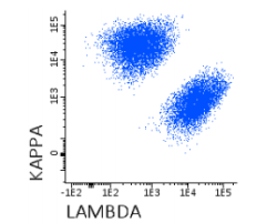
Human lymphocytes may be classified in three main populations according to their biological function and their cell surface antigen expression: T lymphocytes, B lymphocytes and Natural Killer cells (NK). B lymphocytes are the producers of antibodies and mediate humoral immunity particularly effective against toxins, whole bacteria, and free viruses. Most B cells, with the exception of pre-B progenitors, pre B- cells, and mature plasma cells, express immunoglobulin on their surface. Each cell expresses only one light chain type (Kappa or Lambda). In normal peripheral blood and lymph nodes, there is a mixture of Kappa positive and Lambda positive cells, with two-thirds of the cells expressing Kappa and one-third expressing Lambda. Since lymphoid neoplasms are usually clonal expansions of a single cell, malignant cells uniformly express the same Light Chain isotype. Neoplastic B cell lymphoprolipherative disorders can frequently be suspected based on the demonstration of a marker predominance of cells expressing a single Light Chain type. The Kappa immunoglobulin Light Chain count is generally expressed as a percentage of B cells present in the sample that can be determined by flow cytometry based on its positive expression of CD19.
REAGENT COMPOSITION
Purified polyclonal antibody Anti-human Kappa Light Chains, Goat F(ab’)2, conjugated with allophycocyanine (APC), supplied in phosphate buffered saline with 0,09% sodium azide and 1% (w/v) bovine serum albumin (BSA).
- Clone: Polyclonal
- Isotype: Goat / Polyclonal
- Amount per vial: 100 tests (5 µL/Test).
- Reagents are not considered sterile.
-
 آنتی بادیهای فلوسایتومتری
آنتی بادی مونوکلونال فلوسایتومتری ™Kappa-APC-C750 ،کلون Polyclonal
Name: Flow Cytometry Antibody Kappa-APC-C750™, Clone Polyclonal
- Antibody Kappa-APC-C750TM is a polyclonal antibody (pAb) labelled with the tandem allophycocyanine-C750 (APC-C750TM) and designed for use as a direct immunofluorescence reagent in the identification and enumeration of cells that express human Kappa immunoglobulin Light Chains by flow cytometry (FC). This reagent must be used by FC qualified personnel.
SUMMARY AND EXPLANATION
Human lymphocytes may be classified in three main populations according to their biological function and their cell surface antigen expression: T lymphocytes, B lymphocytes and Natural Killer cells (NK). B lymphocytes are the producers of antibodies and mediate humoral immunity particularly effective against toxins, whole bacteria, and free viruses. Most B cells, with the exception of pre-B progenitors, pre B-cells, and mature plasma cells, express immunoglobulin on their surface. Each cell expresses only one light chain type (Kappa or Lambda). In normal peripheral blood and lymph nodes, there is a mixture of Kappa positive and Lambda positive cells, with two-thirds of the cells expressing Kappa and one-third expressing Lambda. Since lymphoid neoplasms are usually clonal expansions of a single cell, malignant cells uniformly express the same Light Chain isotype. Neoplastic B cell lymphoprolipherative disorders can frequently be suspected based on the demonstration of a marker predominance of cells expressing a single Light Chain type. The Lambda immunoglobulin Light Chain count is generally expressed as a percentage of B cells present in the sample which can be determined by FC based on its positive expression of CD19.
APC-C750TM is a tandem dye with a maximum emission peak at 779 nm, which grants bright signal, low unspecific noise and high photostability. When excited by light from a red laser, the APC fluorochrome can transfer energy to C750 molecule, which then emits at a longer wavelength. It is recommended to use a 780/60 nm longpass filter along with a red sensitive detector to use in conjunction antibodies conjugated with APC and APC-C750TM.
REAGENT COMPOSITION
Purified polyclonal antibody anti-human Kappa Light Chains, Goat F(ab’)2, conjugated with tandem allophycocyanineC750 (APC-C750TM), supplied in phosphate buffered saline with 0,09% (m/v) sodium azide and 0,2% (w/v) bovine serum albumin (BSA).
- Clone: Polyclonal
- Isotype: Goat / Polyclonal
- Amount per vial: 50 tests (3µL/Test).
Reagents are not considered sterile
-
آنتی بادیهای فلوسایتومتری
آنتی بادی مونوکلونال فلوسایتومتری Kappa-FITC ، کلون Polyclonal
Name: Flow Cytometry Antibody Kappa-FITC, Clone Polyclonal
- Kappa-FITC is a polyclonal antibody (pAb) labelled with fluorescein isothiocyanate (FITC) and designed for use as a direct immunofluorescence reagent in the identification and enumeration of cells that express human Kappa immunoglobulin Light Chains by flow cytometry. Antibodies to Kappa Light Chains are useful for the identification of clonal excess in B-cell lymphoproliferative disorders together with a panel of other antibodies. Anti-Kappa Light Chains reacts with human free Kappa chains as well as Kappa chains in intact immunoglobulin molecules. This reagent must be used by flow cytometry qualified personal.
SUMMARY AND EXPLANATION
Human lymphocytes may be classified in three main populations according to their biological function and their cell surface antigen expression: T lymphocytes, B lymphocytes and Natural Killer cells (NK). B lymphocytes are the producers of antibodies and mediate humoral immunity particularly effective against toxins, whole bacteria, and free viruses. Most B cells, with the exception of pre-B progenitors, pre-B cells, and mature plasma cells, express immunoglobulin on their surface. Each cell expresses only one light chain type (Kappa or Lambda). In normal peripheral blood and lymph nodes, there is a mixture of Kappa positive and Lambda positive cells, with two-thirds of the cells expressing Kappa and one-third expressing Lambda. Since lymphoid neoplasms are usually clonal expansions of a single cell, malignant cells uniformly express the same Light Chain isotype. Neoplastic B cell lymphoprolipherative disorders can frequently be suspected based on the demonstration of a marker predominance of cells expressing a single Light Chain type. The Kappa immunoglobulin Light Chain count is generally expressed as a percentage of B cells present in the sample which can be determined by flow cytometry based on its positive expression of CD19. Since every flow cytometer has different operating characteristics each laboratory must determine its optimal operating procedure and the combination of markers used to identify the target cell population
REAGENT COMPOSITION
Purified polyclonal antibody anti-human Kappa Light Chains, Goat F(ab’)2, conjugated with fluorescein isothiocyanate (FITC), supplied in phosphate buffered saline with 0,09% (m/v) sodium azide.
Amount per 0,5 ml vial: 100 tests (5µl pAb to 106 cells).
Reagents are not considered sterile.
-
 آنتی بادیهای فلوسایتومتری
آنتی بادی مونوکلونال فلوسایتومتری Kappa-FITC ، کلون Polyclonal
Name: Flow Cytometry Antibody Kappa-FITC, Clone Polyclonal
- Kappa-FITC is a polyclonal antibody (pAb) labelled with fluorescein isothiocyanate (FITC) and designed for use as a direct immunofluorescence reagent in the identification and enumeration of cells that express human Kappa immunoglobulin Light Chains by flow cytometry. Antibodies to Kappa Light Chains are useful for the identification of clonal excess in B-cell lymphoproliferative disorders together with a panel of other antibodies. Anti-Kappa Light Chains reacts with human free Kappa chains as well as Kappa chains in intact immunoglobulin molecules. This reagent must be used by flow cytometry qualified personal.
SUMMARY AND EXPLANATION
Human lymphocytes may be classified in three main populations according to their biological function and their cell surface antigen expression: T lymphocytes, B lymphocytes and Natural Killer cells (NK). B lymphocytes are the producers of antibodies and mediate humoral immunity particularly effective against toxins, whole bacteria, and free viruses. Most B cells, with the exception of pre-B progenitors, pre-B cells, and mature plasma cells, express immunoglobulin on their surface. Each cell expresses only one light chain type (Kappa or Lambda). In normal peripheral blood and lymph nodes, there is a mixture of Kappa positive and Lambda positive cells, with two-thirds of the cells expressing Kappa and one-third expressing Lambda. Since lymphoid neoplasms are usually clonal expansions of a single cell, malignant cells uniformly express the same Light Chain isotype. Neoplastic B cell lymphoprolipherative disorders can frequently be suspected based on the demonstration of a marker predominance of cells expressing a single Light Chain type. The Kappa immunoglobulin Light Chain count is generally expressed as a percentage of B cells present in the sample which can be determined by flow cytometry based on its positive expression of CD19. Since every flow cytometer has different operating characteristics each laboratory must determine its optimal operating procedure and the combination of markers used to identify the target cell population
REAGENT COMPOSITION
Purified polyclonal antibody anti-human Kappa Light Chains, Goat F(ab’)2, conjugated with fluorescein isothiocyanate (FITC), supplied in phosphate buffered saline with 0,09% (m/v) sodium azide.
Amount per 0,5 ml vial: 100 tests (5µl pAb to 106 cells).
Reagents are not considered sterile.
-
آنتی بادیهای فلوسایتومتری
آنتی بادی مونوکلونال فلوسایتومتری Kappa-PerCP-Cyanine5.5 ، کلون Polyclonal
Name: Flow Cytometry Antibody Kappa-PerCP-Cyanine5.5, Clone Polyclonal
- Antibody Kappa-PerCP-Cyanine5.5 is a polyclonal antibody (pAb) labelled with tandem Peridin chlorophyll protein-Cyanine 5.5 (PerCP-Cyanine5.5) and designed for use as a direct immunofluorescence reagent in the identification and enumeration of cells that express human Kappa immunoglobulin Light Chains by Flow Cytometry (FC). Antibodies to Kappa Light Chains are useful for the identification of clonal excess in B-cell lymphoproliferative disorders together with a panel of other antibodies. Anti-Kappa Light Chains reacts with human free Kappa chains as well as Kappa chains in intact immunoglobulin molecules. This reagent must be used by FC qualified personal.
SUMMARY AND EXPLANATION
Human lymphocytes may be classified in three main populations according to their biological function and their cell surface antigen expression: T lymphocytes, B lymphocytes and Natural Killer cells (NK). B lymphocytes are the producers of antibodies and mediate humoral immunity particularly effective against toxins, whole bacteria, and free viruses. Most B cells, with the exception of pre-B progenitors, pre-B cells, and mature plasma cells, express immunoglobulin on their surface. Each cell expresses only one light chain type (Kappa or Lambda). In normal peripheral blood and lymph nodes, there is a mixture of Kappa positive and Lambda positive cells, with two-thirds of the cells expressing Kappa and one-third expressing Lambda. The Kappa immunoglobulin Light Chain count is generally expressed as a percentage of B cells present in the sample which can be determined by flow cytometry based on its positive expression of CD19. Since every flow cytometer has different operating characteristics each laboratory must determine its optimal operating procedure and the combination of markers used to identify the target cell population.
REAGENT COMPOSITION
Purified polyclonal antibody anti-human Kappa Light Chains, Goat F(ab’)2, conjugated with tandem Peridin chlorophyll protein-Cyanine 5.5 (PerCP-Cyanine5.5), supplied in phosphate buffered saline with 0,09% (m/v) sodium azide.
- Amount per vial: 50 tests (3µL pAb to 106 cells).
- Reagents are not considered sterile.
-
آنتی بادیهای فلوسایتومتری
آنتی بادی مونوکلونال فلوسایتومتری Lambda-APC-C750TM ، کلون Polyclonal
Name: Flow Cytometry Antibody Lambda-APC-C750TM, Clone Polyclonal
- Antibody Lambda-APC-C750TM is a polyclonal antibody (pAb) labelled with R-Phycoerythrin (PE) and designed for use as a direct immunofluorescence reagent in the identification and enumeration of cells that express human Lambda immunoglobulin Light Chains by Flow Cytometry (FC). Antibodies to Lambda Light Chains are useful for the identification of clonal excess in B-cell lymphoproliferative disorders together with a panel of other antibodies. Anti-Lambda Light Chains reacts with free Lambda chains as well as Lambda chains in intact immunoglobulin molecules. This reagent must be used by FC qualified personal.
SUMMARY AND EXPLANATION
Human lymphocytes may be classified in three main populations according to their biological function and their cell surface antigen expression: T lymphocytes, B lymphocytes and Natural Killer cells (NK). B lymphocytes are the producers of antibodies and mediate humoral immunity particularly effective against toxins, whole bacteria, and free viruses. Most B cells, with the exception of pre-B progenitors, pre B-cells, and mature plasma cells, express immunoglobulin on their surface. Each cell expresses only one light chain type (Kappa or Lambda). In normal peripheral blood and lymph nodes, there is a mixture of Kappa positive and Lambda positive cells, with two-thirds of the cells expressing Kappa and one-third expressing Lambda. Since lymphoid neoplasms are usually clonal expansions of a single cell, malignant cells uniformly express the same Light Chain isotype. Neoplastic B cell lymphoprolipherative disorders can frequently be suspected based on the demonstration of a marker predominance of cells expressing a single Light Chain type. The Lambda immunoglobulin Light Chain count is generally expressed as a percentage of B cells present in the sample that can be determined by FC based on its positive expression of CD19. Since every flow cytometer has different operating characteristics each laboratory must determine its optimal operating procedure and the combination of markers used to identify the target cell population.
REAGENT COMPOSITION
Purified polyclonal antibody Anti-human Lambda Light Chains, Goat F(ab’)2, conjugated with R-Phycoerythrin (PE), supplied in phosphate buffered saline with 0.09% (m/v) sodium azide.
- Amount per 0,5 ml vial: 100 tests (5 µl pAb to 106 cells).
- Reagents are not considered sterile.
-
آنتی بادیهای فلوسایتومتری
آنتی بادی مونوکلونال فلوسایتومتری Lambda-FITC ، کلون Polyclonal
Name: Flow Cytometry Antibody Lambda-FITC, Clone Polyclonal
- Antibody Lambda-FITC in a goat polyclonal antibody (pAb) labelled with fluorescein isothiocyanate (FITC) and designed for use as a direct immunofluorescence reagent in the identification and enumeration of cells that express human Lambda immunoglobulin Light Chains by flow cytometry. Antibodies to Lambda Light Chains are useful for the identification of clonal excess in B-cell lymphoproliferative disorders together with a panel of other antibodies. Anti-Lambda Light Chains reacts with free Lambda chains as well as Lambda chains in intact immunoglobulin molecules. This reagent must be used by flow cytometry qualified personal.
SUMMARY AND EXPLANATION
Human lymphocytes may be classified in three main populations according to their biological function and their cell surface antigen expression: T lymphocytes, B lymphocytes and Natural Killer cells (NK). B lymphocytes are the producers of antibodies and mediate humoral immunity particularly effective against toxins, whole bacteria, and free viruses. Most B cells, with the exception of pre-B progenitors, pre B-cells, and mature plasma cells, express immunoglobulin on their surface. Each cell expresses only one light chain type (Kappa or Lambda). In normal peripheral blood and lymph nodes, there is a mixture of Kappa positive and Lambda positive cells, with two-thirds of the cells expressing Kappa and one-third expressing Lambda. Since lymphoid neoplasms are usually clonal expansions of a single cell, malignant cells uniformly express the same Light Chain isotype. Neoplastic B cell lymphoprolipherative disorders can frequently be suspected based on the demonstration of a marker predominance of cells expressing a single Light Chain type. The Lambda immunoglobulin Light Chain count is generally expressed as a percentage of B cells present in the sample that can be determined by flow cytometry based on its positive expression of CD19. Since every flow cytometer has different operating characteristics each laboratory must determine its optimal operating procedure and the combination of markers used to identify the target cell population.
REAGENT COMPOSITION
Purified polyclonal antibody Anti-human Lambda Light Chains, Goat F(ab’)2, conjugated with fluorescein isothiocyanate (FITC), supplied in phosphate buffered saline with 0,09% (m/v) sodium azide.
- Purification: Affinity chromatography
- Amount per 0,5 mL vial: 100 tests (5 µL pAb to 106 cells).
- Reagents are not considered sterile.
برای تماس کلیک کنید
سما تشخیص آریا ، عرضه کننده متنوع ترین و کامل ترین محصولات کیت های آزمایشگاهی