-
 آنتی بادیهای فلوسایتومتری
آنتی بادی مونوکلونال فلوسایتومتری CD138-Pacific Blue™, ، کلون B-A38
Name: Flow Cytometry Antibody CD138-Pacific Blue™, Clone B-A38
- Antibody CD138 reagent is a monoclonal antibody (mAb) conjugated with different fluorochromes (see table) and designed for flow cytometry use as a direct immunofluorescence reagent in the identification and enumeration of human CD138 antigen-expressing cells. This reagent must be used by flow cytometry qualified personal.
SUMMARY AND EXPLANATION
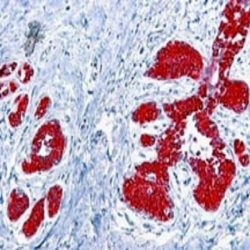
The mAb Anti-CD138 reacts with human CD138, syndecan-1, a 20-22 kDa protein that binds to interstitial extracellular matrix molecules. CD138 is predominantly expressed on epithelial cells and B lymphocytes at specific stages in their differentiation: precursor B cells in the bone marrow and antibody-secreting cells, including plasma cells but not mature peripheral B cell. The simultaneous assessment of the expression of CD38 and CD138 represents the best combination of markers for the identification of plasma cells.
REAGENT COMPOSITION
The purified mAb CD138 conjugated with different fluorochromes (see table above) is supplied in phosphate-buffered saline (PBS) containing 1% (m/v) BSA and 0,09% (m/v) sodium azide.
- Clone: B-A38.
- Isotype: Mouse / IgG1.
- Reagent is considered non-sterile.
-
 آنتی بادیهای فلوسایتومتری
آنتی بادی مونوکلونال فلوسایتومتری CD138-PECy5، کلون B-A38
Name: Flow Cytometry Antibody CD138-PECy5, Clone B-A38
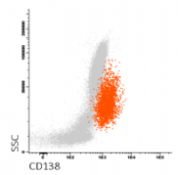
- Antibody CD138-PECy5 is a monoclonal antibody (mAb) labelled with R-phycoerythrin-cyanine 5 (PECy5) designed for flow cytometry (FC) use as a direct immunofluorescence reagent in the identification and enumeration of CD138 antigenexpressing cells.
SUMMARY AND EXPLANATION
Human lymphocytes may be classified in three main populations according to their biological function and their cell surface antigen expression: T lymphocytes, B lymphocytes and natural killer cells (NK). B lymphocytes are the producers of antibodies and mediate humoral immunity particularly effective against toxins, whole bacteria, and free viruses. The CYT-138C4 mAb reacts with CD138, syndecan-1, a 20-22 kDa protein that binds to interstitial extracellular matrix molecules. CD138 is predominantly expressed on epithelial cells and B lymphocytes at specific stages in their differentiation: precursor B cells in the bone marrow and antibody-secreting cells, including plasma cells but not mature peripheral B cell.
REAGENT COMPOSITION
The purified monoclonal CD138 Antibody conjugated with R-phycoerythrin-cyanine 5 (PECy5) is supplied in phosphatebuffered saline (PBS) containing 0,09% sodium azide.
- Clone: B-A38.
- Isotype: IgG1.
- Amount per 1 mL vial: 200 tests (5 µL mAb per determination).
- Reagent is considered non-sterile.
-
آنتی بادیهای فلوسایتومتری
آنتی بادی مونوکلونال فلوسایتومتری CD180-APC-C750TM ، کلون G28-8
Name: Flow Cytometry Antibody CD180-APC-C750, Clone G28-8
- Antibody CYT-180AC750 is a monoclonal antibody (mAb) labelled with tandem allophycocyanin-C750 (APC-C750TM) designed for use as a direct immunofluorescence reagent in the identification and enumeration of cells which express the CD180 antigen by flow cytometry. This reagent must be used by flow cytometry qualified personnel.
SUMMARY AND EXPLANATION
CD180 (RP105), belongs to the toll-like receptor (TLR) family and is expressed on most B cells, monocytes/macrophages, and dendritic cells. It is a 105 kDa protein consisting of extracellular leucine-rich repeats and a short cytoplasmic tail. MD-1, a secretory protein, is associated with CD180 at the cell surface and, in conjunction with TLR4, recognizes bacterial lipopolysaccharide. Ligation of CD180 by monoclonal antibodies leads to B cell activation.
REAGENT COMPOSITION
Purified monoclonal CD180 antibody conjugated with tandem allophycocyanin-C750 (APC-C750TM), supplied in phosphate buffered saline with 0,09% sodium azide.
- Clone: G28-8.
- Isotype: Mouse / IgG1.
- Amount per vial: 50 tests. 3 µl mAb per determination.
- Reagents are not considered sterile.
-
آنتی بادیهای فلوسایتومتری
آنتی بادی مونوکلونال فلوسایتومتری CD185-APC ، کلون MU5UBEE
Name: Flow Cytometry Antibody CD185-APC, Clone MU5UBEE
- Antibody CD185-APC is a monoclonal antibody (mAb) labelled with allophycocyanin (APC) designed for flow cytometry (FC) use as a direct immunofluorescence reagent in the identification and enumeration of CD185 antigen-expressing cells.
SUMMARY AND EXPLANATION
FC is a powerful tool in analytical and quantitative characterization of cells which provides rapid and multiparametric analysis of heterogeneous cell populations on a cell-by-cell basis. Flow cytometry is performed on cell suspension after incubating it with fluorescentlabelled antibodies directed against specific cellular proteins. Positive cells relative fluorescence intensity indicates the amount of antibody bonded to specific cell sites providing information about antigen expression. CD185, also known as C-X-C chemokine receptor 5 (CXCR5) and Burkitt lymphoma receptor 1 (BLR1), is a 7 transmembrane domain protein mainly expressed on B cells and CD4+ T cells (1, 2). CD185 has been selected by EuroFlow consortium in its B-CLPD (B-cell chronic lymphoproliferative diseases) panel.
REAGENT COMPOSITION
The purified monoclonal CD185 antibody conjugated with allophycocyanin (APC) is supplied in phosphate-buffered saline (PBS) containing 0.1% sodium azide.
- Clone: MU5UBEE.
- Isotype: IgG2b.
- Amount per vial: 100 tests (5 µl mAb per determination).
- Reagent is considered non-sterile.
-
 آنتی بادیهای فلوسایتومتری
آنتی بادی مونوکلونال فلوسایتومتری CD200-APC،کلون OX-104
Name: Flow Cytometry Antibody CD200-APC, Clone OX-104
- Antibody CD200-APC is a monoclonal antibody (mAb) labelled with allophycocyanin (APC) designed for flow cytometry (FC) use as a direct immunofluorescence reagent in the identification and enumeration of CD200 antigen-expressing cells. This reagent must be used by flow cytometry qualified personal.
SUMMARY AND EXPLANATION
CD200, also known as OX2, is expressed on thymocytes, neurons, endothelium, follicular dendritic cells in all lymphoid organs, a subset of CD34+ progenitor cells, and at low levels on some smooth muscle and B lymphocytes.CD200 has been selected by EuroFlow consortium in B-CPLD (B-cell chronic lymphoproliferative diseases) antibody panel to distinguish MCL (mantle cell lymphoma) cases from other B-CLPD entities.
REAGENT COMPOSITION
The purified monoclonal CD200 antibody conjugated with allophycocyanin (APC) is supplied in phosphate-buffered saline (PBS) containing 0,09 % sodium azide.
- Clone: OX-104.
- Isotype: IgG1.
- Amount per vial: 100 tests (5 ml mAb per determination).
- Reagent is considered non-sterile.
-
آنتی بادیهای فلوسایتومتری
آنتی بادی مونوکلونال فلوسایتومتری CD300e-APC ، کلون UP-H2
Name: Flow Cytometry Antibody CD300e-APC, Clone UP-H2
- Antibody CD300e-APC is a monoclonal antibody (mAb) labelled with allophycocyanin (APC) designed for flow cytometry (FC) use as a direct immunofluorescence reagent in the identification and enumeration of CD300e antigen-expressing cells.
SUMMARY AND EXPLANATION
FC is a powerful tool in analytical and quantitative characterization of cells which provides rapid and multiparametric analysis of heterogeneous cell populations on a cell-by-cell basis. Flow cytometry is performed on cell suspension after incubating it with fluorescentlabelled antibodies directed against specific cellular proteins. Positive cells relative fluorescence intensity indicates the amount of antibody bonded to specific cell sites providing information about antigen expression. IREM2, also known as CD300e, is a 34 kDa integral membrane glycoprotein which is a member of the CMFRF35 subfamily. IREM2 is expressed on monocytes and myeloid dendritic cell lineage. During normal monocytic development, CD300e is expressed in the last stages of maturation. CD300e has been selected by EuroFlow consortium in AML (acute myeloid leukemia) antibody panel.
REAGENT COMPOSITION
The purified monoclonal CD300e antibody conjugated with allophycocyanin (APC) is supplied in phosphate-buffered saline (PBS) containing 0.1% sodium azide.
- Clone: UP-H2.
- Isotype: IgG1.
- Amount per vial: 100 tests (10ul/test).
- Reagent is considered non-sterile.
-
 آنتی بادیهای فلوسایتومتری
آنتی بادی مونوکلونال فلوسایتومتری CyCD79a-FITC ، کلون HM57
Name: Flow Cytometry Antibody CyCD79a-FITC, Clone HM57
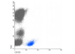
- Antibody CD79a-FITC is designed for use as a direct immunofluorescence reagent in the identification and enumeration of cells expressing the CD79α antigen by flow cytometry. In normal cells, CD79α expression is restricted to B cell lineage. CD79α is found in the cytoplasm of pro B-cells before Ig heavy-chain rearrangement and is extinguished in terminally differentiated plasma cells. CD79α is expressed in hairy cell leukaemia and in the majority of all low-grade B-cell leukaemias and lymphomas, such as lymphoplasmacytic, follicular, mantle cell, marginal zone and Burkitt’s lymphomas. Generally, the CD79α antigen is not found in acute myeloid leukaemia, but the FAB M3 class is a significant exception with more than 50% of cases displaying the antigen in the cytoplasm of malignant cells. Approximately 45% of T- cell ALL cases co-express CD3 and CD79α, and this co-expression phenotype may be clinically relevant. CYT-79aF4 should be used with a cell permeabilization kit which gives antibodies access to intracellular structures and leaves the morphological scatter characteristics of cells intact. This reagent must be used by personal qualified for flow cytometry.
REAGENT COMPOSITION
Purified monoclonal CD79a antibody (mAb) conjugated with Fluorescein isothiocyanate (FITC), supplied in phosphate buffered saline with ≤0,09% (m/v) sodium azide.
- Clone: HM57
- Isotype: Mouse IgG1
- Amount per 0,4 ml vial: 100 tests (4μl Ab to 106 cells)
-
 آنتی بادیهای فلوسایتومتری
آنتی بادی مونوکلونال فلوسایتومتری cyCD79a-PE ، کلون HM57
Name: Flow Cytometry Antibody cyCD79a-PE, Clone HM57
- Antibody cyCD79a-PE is a monoclonal antibody (mAb) labelled with R-phycoerythrin (PE) designed for use as a direct immunofluorescence reagent in the identification and enumeration of cells which express the CD79a antigen by flow cytometry (FC). CD79a is expressed on B cells at various stages of differentiation, from pre-B cell to plasma cell stage. This reagent must be used by flow cytometry qualified personal.
SUMMARY AND EXPLANATION
Human lymphocytes may be classified in three main populations according to their biological function and their cell surface antigen expression: T lymphocytes, B lymphocytes and natural killer cells (NK). B lymphocytes are the producers of antibodies and mediate humoral immunity particularly effective against toxins, whole bacteria, and free viruses. In normal cells CD79α expression is restricted to B cell lineage. cyCD79α is found in the cytoplasm of pro B- cells before Ig heavy-chain rearrangement and is extinguished in terminally differentiated plasma cells. CD79α is expressed in hairy cell leukaemia and in the majority of all low-grade B-cell leukaemias and lymphomas, such as lymphoplasmacytic, follicular, mantle cell, marginal zone and Burkitt’s lymphomas. Generally, the CD79α antigen is not found in acute myeloid leukaemia, but the FAB M3 class is a significant exception with more than 50% of cases displaying the antigen in the cytoplasm of malignant cells. Approximately 45% of T- cell ALL cases co-express CD3 and CD79α, and this co-expression phenotype may be clinically relevant.
REAGENT COMPOSITION
Purified monoclonal cyCD79a Antibody conjugated with R-phycoerythrin (PE), supplied in phosphate buffered saline with 0,09% sodium azide.
- Clone: HM57.
- Isotype: Mouse / IgG1.
- Amount per 1 ml vial: 100 tests (10 ml/ test).
- Reagents are not considered sterile.
-
آنتی بادیهای فلوسایتومتری
آنتی بادی مونوکلونال فلوسایتومتری IgA1-APC ، کلون SAA1
Name: Flow Cytometry Antibody IgA1-APC, Clone SAA1
IgA1-APC is designed for flow cytometry use as a direct immunofluorescence reagent in the identification and enumeration of IgA1 expressing cells. IgA1-APC reacts with the Fc portion of the heavy chain of human IgA1.
In humans, 85–90% of IgA antibodies are found in body secretions such us saliva, tears, breast milk; gastrointestinal mucous membranes, urogenital and respiratory tracts and prostatic fluid, among others. The remaining fraction of 10–15% of IgA antibodies is found in blood, produced by B cells in the lymph nodes, bone marrow, and spleen. Two subclasses of human serum IgA exist. IgA1 tends to be monomeric and is present at a fivefold higher concentration than IgA2. Soluble IgA2 tends to be present more often in polymeric forms.
CYT-IGA1AP should be used with BulkLysisTM (CYT-BL) buffer that allows you to stain a great number of cells and analyze low frequency subsets, as IgA1+ memory B cells and plasma cells. If another lysing protocol is used, two pre-staining washing steps using PBS + 1% (m/v) BSA + 0,09% (m/v) sodium azide must be performed. This reagent must be used by flow cytometry qualified personnel.
REAGENT COMPOSITION
Purified monoclonal anti-IgA1 antibody (mAb) conjugated with allophycocyanine (APC), supplied in phosphate buffered saline with 0,09% (m/v) sodium azide and BSA 1% (m/v).
Clone: SAA1.
Isotype: Mouse IgG1.
Amount per vial: 25 tests using 3µL of mAb to 106 cells.
-
آنتی بادیهای فلوسایتومتری
آنتی بادی مونوکلونال فلوسایتومتری IgA1-FITC ، کلون SAA1
Name: Flow Cytometry Antibody IgA1-FITC, Clone SAA1
- Antibody IgA1-FITC is designed for flow cytometry use as a direct immunofluorescence reagent in the identification and enumeration of IgA1 expressing cells. IgA1-FITC reacts with the Fc portion of the heavy chain of human IgA1.
In humans, 85–90% of IgA antibodies are found in body secretions such us saliva, tears, breast milk, gastrointestinal mucous membranes, urogenital and respiratory tracts, and prostatic fluid, among others. The remaining fraction of 10–15% of IgA antibodies is found in blood, produced by B cells in the lymph nodes, bone marrow, and spleen. Two subclasses of human serum IgA exist. IgA1 tends to be monomeric and is present at a fivefold higher concentration than IgA2. Soluble IgA2 tends to be present more often in polymeric forms.
CYT-IGA1F should be used with BulkLysisTM (CYT-BL) buffer that allows you to stain a great number of cells and analyze low-frequency subsets, as IgA1+ memory B cells and plasma cells. If another lysing protocol is used, two pre-staining washing steps using PBS + 1% (m/v) BSA + 0,09% (m/v) sodium azide must be performed.
This reagent must be used by flow cytometry qualified personnel.
REAGENT COMPOSITION
Purified monoclonal anti-IgA1 antibody (mAb) conjugated with fluorescein isothiocyanate (FITC), supplied in phosphate buffered saline with 0,09% (m/v) sodium azide and BSA 1% (m/v).
- Clone: SAA1.
- Isotype: Mouse IgG1.
- Amount per vial: 25 tests using 3µL of mAb to 106 cells.
-
آنتی بادیهای فلوسایتومتری
آنتی بادی مونوکلونال فلوسایتومتری IgA1-PE ، کلون SAA1
Name: Flow Cytometry Antibody IgA1-PE, Clone SAA1
- IgA1-PE is designed for flow cytometry use as a direct immunofluorescence reagent in the identification and enumeration of IgA1 expressing cells. IgA1-PE reacts with the Fc portion of the heavy chain of human IgA1.
In humans, 85–90% of IgA antibodies are found in body secretions such us saliva, tears, breast milk, gastrointestinal mucous membranes, urogenital and respiratory tracts and prostatic fluid, among others. The remaining fraction of 10–15% of IgA antibodies is found in blood, produced by B cells in the lymph nodes, bone marrow, and spleen. Two subclasses of human serum IgA exist. IgA1 tends to be monomeric and is present at a fivefold higher concentration than IgA2. Soluble IgA2 tends to be present more often in polymeric forms.
CYT-IGA1PE should be used with BulkLysisTM (CYT-BL) buffer that allows you to stain a great number of cells and analyze low frequency subsets, as IgA1+ memory B cells and plasma cells. If another lysing protocol is used, two pre-staining washing steps using PBS + 1% (m/v) BSA + 0,09% (m/v) sodium azide must be performed. This reagent must be used by flow cytometry qualified personnel.
REAGENT COMPOSITION
Purified monoclonal anti-IgA1 antibody (mAb) conjugated with R-phycoerythrin (PE), supplied in phosphate buffered saline with 0,09% (m/v) sodium azide and BSA 1% (m/v).
Clone: SAA1.
Isotype: Mouse IgG1.
Amount per vial: 25 tests using 3µL of mAb to 106 cells.
-
آنتی بادیهای فلوسایتومتری
آنتی بادی مونوکلونال IgA1-PerCP-Cyanine5.5 ، کلون SAA1
Name: Flow Cytometry Antibody IgA1-PerCP-Cyanine5.5, Clone SAA1
Antibody IgA1-PerCP-Cyanine5.5 is designed for flow cytometry use as a direct immunofluorescence reagent in the identification and enumeration of IgA1 expressing cells. IgA1-PerCP-Cyanine5.5 reacts with the Fc portion of the heavy chain of human IgA2.
In humans, 85–90% of IgA antibodies are found in body secretions such us saliva, tears, breast milk; gastrointestinal mucous membranes, urogenital and respiratory tracts and prostatic fluid, among others. The remaining fraction of 10–15% of IgA antibodies is found in blood, produced by B cells in the lymph nodes, bone marrow, and spleen. Two subclasses of human serum IgA exist. IgA1 tends to be monomeric and is present at a fivefold higher concentration than IgA2. Soluble IgA2 tends to be present more often in polymeric forms.
CYT-IGA1C should be used with BulkLysisTM (CYT-BL) buffer that allows you to stain a great number of cells and analyze low frequency subsets, as IgA1+ memory B cells and plasma cells. If another lysing protocol is used, two pre-staining washing steps using PBS + 1% (m/v) BSA + 0,09% (m/v) sodium azide must be performed.
REAGENT COMPOSITION
Purified monoclonal anti-IgA1 antibody (mAb) conjugated with Peridin chlorophyll protein-Cyanine 5.5 (PerCP-Cyanine5.5), supplied in phosphate buffered saline with 0,09% (m/v) sodium azide and BSA 1% (m/v).
- Clone: SAA1.
- Isotype: Mouse IgG1.
- Amount per vial: 25 tests using 3µL of mAb to 106 cells.
-
آنتی بادیهای فلوسایتومتری
آنتی بادی مونوکلونال فلوسایتومتری IgA2- PerCP-Cyanine5.5 ، کلون SAA2
Name: Flow Cytometry Antibody IgA2-PerCP-Cyanine5.5, Clone SAA2
Antibody IgA2-PerCP-Cyanine5.5 is designed for flow cytometry use as a direct immunofluorescence reagent in the identification and enumeration of IgA2 expressing cells. IgA2-PerCP-Cyanine5.5 reacts with the Fc portion of the heavy chain of human IgA2.
In humans, 85–90% of IgA antibodies are found in body secretions such us saliva, tears, breast milk; gastrointestinal mucous membranes, urogenital and respiratory tracts and prostatic fluid, among others. The remaining fraction of 10–15% of IgA antibodies is found in blood, produced by B cells in the lymph nodes, bone marrow, and spleen. Two subclasses of human serum IgA exist. IgA1 tends to be monomeric and is present at a fivefold higher concentration than IgA2. Soluble IgA2 tends to be present more often in polymeric forms.
CYT-IGA2C2 should be used with BulkLysisTM (CYT-BL) buffer that allows you to stain a great number of cells and analyze low frequency subsets, as IgA2+ memory B cells and plasma cells. If another lysing protocol is used, two pre-staining washing steps using PBS+1% (m/v) BSA + 0,09% (m/v) sodium azide must be performed.
REAGENT COMPOSITION
Purified monoclonal anti-IgA2 antibody (mAb) conjugated with Peridin chlorophyll protein-Cyanine 5.5 (PerCP-Cyanine5.5), supplied in phosphate buffered saline with 0,09% (m/v) sodium azide and BSA 1% (m/v).
- Clone: SAA2.
- Isotype: Mouse IgG1.
- Amount per vial: 25 tests using 3µL of mAb to 106 cells
-
آنتی بادیهای فلوسایتومتری
آنتی بادی مونوکلونال فلوسایتومتری IgA2-PE ، کلون SAA2
Name: Flow Cytometry Antibody IgA2-PE, Clone SAA2
- Antibody IgA2-PE is designed for flow cytometry use as a direct immunofluorescence reagent in the identification and enumeration of IgA2 expressing cells. IgA2-PE reacts with the Fc portion of the heavy chain of human IgA2.
In humans, 85–90% of IgA antibodies are found in body secretions such us saliva, tears, breast milk; gastrointestinal mucous membranes, urogenital and respiratory tracts and prostatic fluid, among others. The remaining fraction of 10–15% of IgA antibodies is found in blood, produced by B cells in the lymph nodes, bone marrow, and spleen. Two subclasses of human serum IgA exist. IgA1 tends to be monomeric and is present at a fivefold higher concentration than IgA2. Soluble IgA2 tends to be present more often in polymeric forms.
CYT-IGA2PE should be used with BulkLysisTM (CYT-BL) buffer that allows you to stain a great number of cells and analyze low frequency subsets, as IgA2+ memory B cells and plasma cells. If another lysing protocol is used, two pre-staining washing steps using PBS + 1% (m/v) BSA + 0,09% (m/v) sodium azide must be performed. This reagent must be used by flow cytometry qualified personnel.
REAGENT COMPOSITION
Purified monoclonal anti-IgA2 antibody (mAb) conjugated with R-phycoerythrin (PE), supplied in phosphate buffered saline with 0,09% (m/v) sodium azide and BSA 1% (m/v).
Clone: SAA2.
Isotype: Mouse IgG1.
Amount per vial: 25 tests using 3µL of mAb to 106 cells
-
آنتی بادیهای فلوسایتومتری
آنتی بادی مونوکلونال فلوسایتومتری IgD-FITC ، کلون IADB6
Name: Flow Cytometry Antibody IgD-FITC, Clone IADB6.
- Antibody IgD-FITC is designed for flow cytometry use as a direct immunofluorescence reagent in the identification and enumeration of IgD expressing cells. IgD-FITC reacts with the delta chain of human IgD. IgD is barely detectable in the blood (0,001% of total serum Ig) and is rarely secreted. It is observed only on the surface of immature, peripheral B cells that already express IgM. The expression of IgD is transient; upon activation of the B cell by the binding of antigen, the surface expression of mIgD, but not mIgM, is abrogated.
CYT-IGDF should be used with BulkLysisTM (CYT-BL) buffer that allows you to stain a great number of cells and analyze low-frequency subsets, as IgD+ memory B cells and plasma cells. If another lysing protocol is used, two pre-staining washing steps using PBS + 1% (m/v) BSA + 0,09% (m/v) sodium azide must be performed.
REAGENT COMPOSITION
Purified monoclonal anti-IgD antibody (mAb) conjugated with fluorescein isothiocyante (FITC), supplied in phosphate buffered saline with 0,09% (m/v) sodium azide and BSA 1% (m/v).
- Clone: IADB6
- Isotype: Mouse IgG2a.
- Amount per vial: 25 tests using 3µL of mAb to 106 cells.
-
آنتی بادیهای فلوسایتومتری
آنتی بادی مونوکلونال فلوسایتومتری IgD-PerCP-Cyanine5.5 ،کلون IADB6
Name: Flow Cytometry Antibody IgD-PerCP-Cyanine5.5 , Clone IADB6.
- Antibody IgD-PerCP-Cyanine5.5 is designed for flow cytometry use as a direct immunofluorescence reagent in the identification and enumeration of IgD expressing cells. IgD-PerCP-Cyanine5.5 reacts with the delta chain of human IgD. IgD is barely detectable in the blood (0,001% of total serum Ig) and is rarely secreted. It is observed mostly on the surface of immature, peripheral B cells that already express IgM. The expression of IgD is transient; upon activation of the B cell by the binding of antigen, the surface expression of mIgD, but not mIgM, is abrogated.
CYT-IGDC should be used with BulkLysisTM (CYT-BL) buffer that allows you to stain a great number of cells and analyze low-frequency subsets, as IgD+ memory B cells and plasma cells. If another lysing protocol is used, two pre-staining washing steps using PBS + 1% (m/v) BSA + 0,09% (m/v) sodium azide must be performed.
REAGENT COMPOSITION
Purified monoclonal anti-IgD antibody (mAb) conjugated with Peridin chlorophyll protein-Cyanine 5.5 (PerCP-Cyanine5.5), supplied in phosphate buffered saline with 0,09% (m/v) sodium azide and BSA 1% (m/v).
- Clone: IADB6.
- Isotype: Mouse IgG2a.
- Amount per vial: 25 tests using 3µL of mAb to 106 cells
-
 آنتی بادیهای فلوسایتومتری
آنتی بادی مونوکلونال فلوسایتومتری CD38-multi-epitope-FITC ، کلون multi -epitope
Name: Flow Cytometry Antibody CD38-multi-epitope-FITC, Clone multi epitope
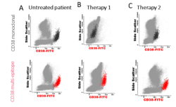
Antibody CYT-38F2 is designed for Flow Cytometry (FC) use for the identification and enumeration of human CD38 antigen-expressing cells even when cells are hampered by another monoclonal antibody. After an extensive selection of clones against different parts of the CD38 molecule, Cytognos has developed a multi-epitope reagent that allows the identification by FC of CD38 in plasma cells even if the patient is under treatment with Anti-CD38.
CD38, also known as ADP-ribosyl cyclase, is a type II transmembrane glycoprotein able to transform NADP+ into cyclic ADP-ribose, NAADP+ and ADP-ribose. CD38 is expressed at variable levels on the majority of hematopoietic cells (during early differentiation and activation) and in some non-hematopoietic tissues. It is expressed at high levels on plasma cells (CD38+++) and, due to its strong reactivity, CD38 is considered a “specific plasma cell marker. but it is also present on thymocytes, activated T cells, NKs monocytes, and basophils . CD38 has been reported to play a role as regulator (positive and negative) of cell activation and proliferation, and to be involved in adhesion between lymphocytes and endothelial cells. CD38 antibody is used in FC for diagnosis and monitoring of plasma cell disorders (e.g. multiple myeloma, MGUS, plasma cell leukemia, etc.). The simultaneous assessment of the expression of CD38 and CD138 represents the best combination of markers for the identification of plasma cells. Anti-CD38 monoclonal antibodies continue to demonstrate promising results as new treatments for patients with multiple myeloma. Therapeutic anti-CD38 targets this molecule, highly expressed on the surface of multiple myeloma cells, hampering its detection by FC. Cytognos has developed an anti-human CD38 multiepitope reagent containing a mixture of antibodies that allows the identification by FC of CD38 molecule on plasma cells, even in patients under anti-CD38 therapy.
Following is shown a comparative evaluation by FC of CD38-FITC monoclonal (dark grey dots) and CD38 multi-epitope-FITC (CYT38F2; red dots). A) Bone marrow samples from untreated patients. B) and C) patients treated with two different therapies against CD38.
REAGENT COMPOSITION
Purified pool of multi-epitope anti-CD38 antibodies labelled with fluorescein isothiocyanate (FITC) and supplied in phosphate-buffered saline (PBS) containing 1% (m/v) BSA and 0.09% (m/v) sodium azide. Amount per vial: 0,25 ml (50 tests using 5 µl Ab per 106 cells)
-
آنتی بادیهای فلوسایتومتری
آنتی بادی مونوکلونال فلوسایتومتری CD81-APC-C750™ (RUO) ، کلون M38
Name: Flow Cytometry Antibody CD81-APC-C750™ (RUO) Clone M38
- Anti-human CD81-APC-C750TM is a monoclonal antibody (mAb) labelled with the tandem allophycocyanine-C750 (APCC750TM) designed for use as a direct immunofluorescence reagent in the identification and enumeration of cells which express the CD81 antigen by flow cytometry (FC). This reagent must be used by flow cytometry qualified personnel.
SUMMARY AND EXPLANATION
The CD81 (TAPA-1) antigen is expressed on normal lymphocytes, monocytes and eosinophils, but not on platelets and neutrophils. This antigen may be present in some cases as multimolecular complexes, in association with other members of the TM4 superfamily (CD37, CD53), or on the surface of B cells, in association with CD19 and/or CD21 and/or MHC class II antigens. Most B lymphocytes, at all stages of cellular differentiation, express CD81 at relatively high levels. Because each flow cytometer has different operating characteristics each laboratory must determine its optimal operating procedure.
REAGENT COMPOSITION
Purified monoclonal CD81 antibody conjugated with the tandem allophycocyanine-C750 (APC-C750TM), supplied in phosphate-buffered saline (PBS) containing 1% (m/v) BSA and 0.09% (m/v) sodium azide.
- Clone: M38.
- Isotype: Mouse / IgG1.
- Amount per 0,15 mL vial: 50 tests (3 µL mAb per determination)
- Reagents are not considered sterile.
-
 آنتی بادیهای فلوسایتومتری
آنتی بادی مونوکلونال فلوسایتومتری CD103-FITC ، کلون B-ly7
Name: Flow Cytometry Antibody CD103-FITC Clone B-ly7
- Antibody CD103-FITC is designed for flow cytometry use as a direct immunofluorescence reagent in the identification and enumeration of CD103 antigen-expressing cells.
CD103 is normally expressed at very high levels on intra-epithelial lymphocytes in non-lymphoid tissues, such as skin, lung, and gastrointestinal organs that express E-cadherin.
In contrast, CD103 is poorly expressed by T cells in blood, spleen, and lymph nodes. Since its discovery, CD103 has been widely used for hairy cell leukaemia (HCL) diagnosis and despite rare exceptions CD103 has been detected together with CD25 in virtually all HCL studies using flow cytometry. CD103 is not ordinarily detected in other types of Lymphoproliferative disorders except for rare cases. CD103 can also be used to define a specialized subset of CD8+ cytolytic T lymphocytes. This reagent must be used by flow cytometry qualified personal.
REAGENT COMPOSITION
The purified monoclonal CD103 antibody (mAb) conjugated with fluorescein isothiocyanate (FITC) is supplied in phosphate-buffered saline (PBS) containing ≤0.1% sodium Azid.
- Clone: B-ly7.
- Isotype: IgG1.
- Amount per 1 ml vial: 200 tests (5 µl mAb per determination).
- Reagent is considered non-sterile.
برای تماس کلیک کنید
سما تشخیص آریا ، عرضه کننده متنوع ترین و کامل ترین محصولات کیت های آزمایشگاهی