-
 Quartett
آنتی بادی KI67 کلون QR015 برند Quartett
Ki-67 is a nuclear protein expressed in all proliferating cells, which are in active phases of cell cycle (late G1, S, G2, mitosis). Ki-67 is not detected in resting cells (G0 phase). Thus, the antibody is a general proliferation marker, especially used to assess the proliferative activity of a tumor. Different cancer types express Ki-67, including breast, prostate, lung, and colon.
-
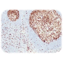
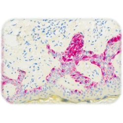
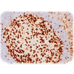
 Quartett
آنتی بادی Kappa Immunoglobulin Light Chain کلون QR051 برند Quartett
B lymphocytes produce immunoglobulins consisting of two identical heavy chains and either two identical kappa light chains or lambda light chains. Normal lymphoid tissues therefore contain a mixture of B cells that express kappa and lambda light chains in a ratio of 2:1. This ratio is lost in tumors of B cell origin as they arise from one transformed cell, and thus only one type of light chain is expressed. The monoclonality of malignant lymphoma is used as diagnostic marker in detection with Kappa Light Chain antibody. This antibody labels kappa light chains of human immunoglobulins expressed by B lymphocytes and plasma cells. It may be helpful in detection of leukemia, plasmacytoma and various non-Hodgkin lymphoma. Other cells may also express kappa light chains resulting from non-specific immunoglobulin uptake. Kappa light chain antibody may be used in a panel with Lambda light chain.
-
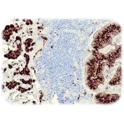
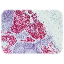
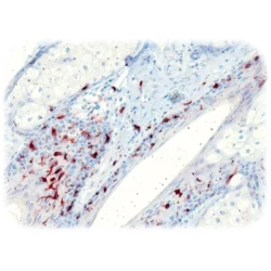 Quartett
آنتی بادی CD207(Langerin) کلون QR065 برند Quartett
Calcium-dependent (C-type) lectins are a family of lectins which share structural homology in their high-affinity carbohydrate-binding domain. Proteins of the CLEC superfamily function in a variety of biological processes, including cell adhesion, cell-cell signaling, glycoprotein turnover, apoptosis, inflammation, and immune response to pathogens. CLEC4K/Langerin is a type II membrane associated receptor expressed exclusively by Langerhans cells, in astrocytoma, malignant ependymoma, but not in normal brain tissues. It recognizes mannose residues, induces membrane superimposition and zippering leading to formation of Birbeck granules. Defects in CLEC4K cause Birbeck granule deficiency, a condition characterized by the absence of Birbeck granules in epidermal Langerhans cells.
-
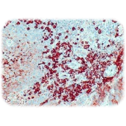
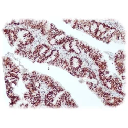
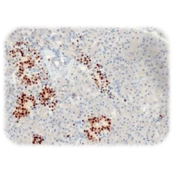
 Quartett
آنتی بادی Lambda Immunoglobulin Light Chain کلون QR052 برند Quartett
B lymphocytes produce immunoglobulins consisting of two identical heavy chains and either two identical kappa light chains or lambda light chains. Normal lymphoid tissues therefore contain a mixture of B cells that express kappa and lambda light chains in a ratio of 2:1. This ratio is lost in tumors of B cell origin as they arise from one transformed cell, and thus only one type of light chain is expressed. The monoclonality of malignant lymphoma is used as diagnostic marker in detection with lambda light chain antibody.
This antibody labels lambda light chains of human immunoglobulins expressed by B lymphocytes and plasma cells. It may be helpful in detection of leukemia, plasmacytoma and various non-Hodgkin lymphoma. Other cells may also express lambda light chains resulting from non-specific immunoglobulin uptake. Lambda light chain antibody may be used in a panel with kappa light chain. -
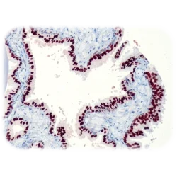
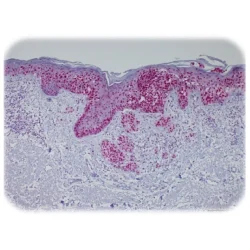
 Quartett
آنتی بادی Melanoma کلون HMB-45 برند Quartett
Melanoma (HMB-45) reacts against an antigen present in melanocytic tumors and is absolute specific for melanoma. The antibody stains fetal and neonatal melanocytes, junctional and blue nevus cells, and malignant melanocytes. Intradermal nevi, normal adult melanocytes, and non-melanocytic cells are negative. It does not stain tumor cells of epithelial, lymphoid, glial, or mesenchymal origin.
-
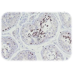
 Quartett
آنتی بادی MSH6 کلون QR011 برند Quartett
DNA mismatch repair (MMR) system consists of four major proteins called MLH1, MSH2, MSH6, and PMS2. These proteins work two by two, MLH1 with PMS2 and MSH2 with MSH6. Loss of function of one of the four proteins leads to inactivation of the MMR system, resulting in a loss of fidelity of the replication and an accumulation of mutations thereby leading to microsatellite instability (MSI). MSI is associated with hereditary nonpolyposis colorectal cancer (HNPCC, Lynch syndrome), which is characterized by the development of colorectal cancer, endometrial cancer and various other tumors at early age.
Loss of MSH6 function due to gene mutation or epigenetic changes is characterized by absence of nuclear expression in neoplastic cells, whereas intact nuclear MSH6 expression indicates normal MSH6 function and no gene mutations. MSH6 is normally expressed in most cases of sporadic colorectal cancer, loss of MSH6 expression is found in 2-16%.
Anti-MSH6 is useful in detection of MSI, especially in a panel with MSH2 (QR010), PMS2 (QR009) and MLH1 (QM003). -
 Quartett
آنتی بادی NUT1 کلون QR043 برند Quartett
NUT carcinoma (NC, formerly NUT midline carcinoma) is a rare, aggressive subtype of squamous cell carcinoma defined by a chromosomal rearrangement of the NUT gene (also known as NUTM1, nuclear protein in testis). It usually arises in the midline of the body including the thorax, mediastinum, lung (thoracic regions ~50 %) and head and neck area (~40 %), but has also been diagnosed arising outside the midline including salivary gland, pancreas, bladder, kidney, adrenal gland as well as various soft tissue and bone locations. NC is a nearly uniformly lethal cance with a reproducible 6.5 month median overall survival. Although NC can occur at any age, it affects primarily adolescents and young adults with median age of 24. In the majority of cases (~75 %), NUT is fused to BRD4. This results in a chimeric powerful BRD4 NUT oncoprotein. Variant NUT fusion partners, including BRD3, NSD3, ZNF532, and ZNF592, encode BRD4 interacting proteins that serve to link NUT with BRD4. Diagnosis of NC can be established by positive NUT nuclear immunohistochemical staining.
-
 Quartett
آنتی بادی Mammaglobin A & B کلون QR080 برند Quartett
Mammaglobin is a 10 kDa glycoprotein that is associated to breast. A correlation between increased expression of mammaglobin gene and breast cancer has been reported.
-
 Quartett
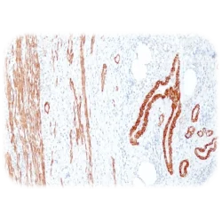
آنتی بادی Smooth Muscle Myosin, heavy chain کلون QR064 برند Quartett
Myosin heavy chain 11 (MYH11) is a smooth muscle myosin and a subunit of a hexameric protein that consists of two heavy chain subunits and two light chain subunits. It functions as a major contractile protein, converting chemical into mechanical energy through the ATP hydrolysis. MYH has been a useful marker for myoepithelial cell as well as smooth muscle cell differentiation.
-
 Quartett
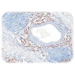
آنتی بادی MPO(Myeloperoxidase) کلون QR101 برند Quartett
Myeloperoxidase is a peroxidase enzyme. This antibody detects granulocytes and monocytes in blood and precursors of granulocytes in the bone marrow. In normal tissues and in a variety of myeloproliferative disorders myeloid cells of both neutrophilic and eosinophilic types, at all stages of maturation, exhibit strong cytoplasmic reactivity for MPO. Erythroid precursors, megakaryocytes, lymphoid cells, mast cells, and plasma cells are nonreactive. MPO is not observed in the neoplastic cells of a wide variety of epithelial tumors and sarcomas. MPO is useful in differentiating between myeloid and lymphoid leukemias.
-
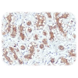
 Quartett
آنتی بادی MSH2 کلون QR010 برند Quartett
DNA mismatch repair (MMR) system consists of four major proteins called MLH1, MSH2, MSH6, and PMS2. These proteins work two by two, MLH1 with PMS2 and MSH2 with MSH6. Loss of function of one of the four proteins leads to inactivation of the MMR system, resulting in a loss of fidelity of the replication and an accumulation of mutations thereby leading to microsatellite instability (MSI). MSI is associated with hereditary nonpolyposis colorectal cancer (HNPCC, Lynch syndrome), which is characterized by the development of colorectal cancer, endometrial cancer and various other tumors at early age.
Loss of MSH2 function due to gene mutation or epigenetic changes is characterized by absence of nuclear expression in neoplastic cells, whereas intact nuclear MSH2 expression indicates normal MSH2 function and no gene mutations. MSH2 is normally expressed in most cases of sporadic colorectal cancer, loss of MSH2 expression is found in 13-40%.
Anti-MSH2 is useful in detection of MSI, especially in a panel with MSH6 (QR011), PMS2 (QR009) and MLH1 (QM003).
-
 Quartett
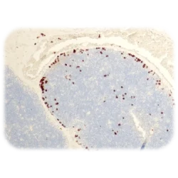
آنتی بادی MUM1(IRF4) کلون QR075 برند Quartett
MUM1 is a transcriptional activator that binds to the interferon-stimulated response element (ISRE) of the MHC class I promoter as well as immunoglobulin lambda light chain enhancer, together with PU.1. It is expressed in the nuclei and cytoplasm of plasma cells, a subset of B-cells in the light zone of the germinal center, activated T-cells and a wide spectrum of related hematolymphoid neoplasms derived from these cells. This antibody is used to detect lymphoid malignancies and HodgkinÕs and Reed Sternberg cells of classic HodgkinÕs disease in combination with anti-CD30.
-
 Quartett
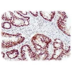
آنتی بادی Napsin A کلون QR058 برند Quartett
Napsin A is a pepsin-like aspartate protease with a molecular weight of about 38 kDa. Napsin A is closely related to Napsin B. Napsin is expressed as a single-chain protein in type II pneumocytes and in adenocarcinomas of the lung and kidney. The high specificity of Napsin A in lung adenocarcinomas is useful in distinguishing such primary adenocarcinomas from adenocarcinomas of unknown origin.
-
 Quartett
آنتی بادی NKX2.2 کلون QR077 برند Quartett
The homeodomain protein NK2 homeobox 2 (NKX2.2) is a transcription factor that plays a critical role in the control of cell fate specification and differentiation in many tissues. In the developing central nervous system, this developmentally important transcription factor functions as a transcriptional repressor that governs oligodendrocyte differentiation and myelin gene expression, but the roles of various NKX2.2 structural domains in this process are unclear.
-
 Quartett
آنتی بادی NKX3.1 کلون QR055 برند Quartett
NKX3.1 is a transcription factor that acts as a putative prostate tumor suppressor. It is largely expressed in a prostate and androgen-regulated manner. NKX3.1 is expressed in normal prostate epithelium. Loss of the protein is a common finding in human prostate cancer and intraepithelial prostate neoplasia. The antibody NKX3.1 is a highly sensitive and specific marker for prostate adenocarcinoma and is useful in a panel with other prostate markers such as PSA, PSAP or Prostein, particularly in the case of low-differentiation tumors in which PSA, PSAP and / or Prostein are poorly expressed or can be lost. Primary and metastatic prostate carcinoma show a lower NKX3.1 staining intensity than the normal prostate.
-
 Quartett
آنتی بادی Olig 2 Protein کلون QR071 برند Quartett
Olig2 is a transcription factor involved in oligodendroglial specification. It is expressed in most glial tumors, such as oligodendrogliomas and astrocytomas, but has been not found in the non-glial tumors including neuroepithelial tumors, ependymomas, sub ependymomas, medulloblastomas, and nonneuroepithelial tumors, such as CNS lymphomas, meningiomas, schwannomas, atypical teratoid/rhabdoid tumor, and hemangioblastomas. Although more than half of glioblastomas are positive for Olig2, expression is very weak in terms of both percentage of labeled cells and intensity. Compared to the strong staining seen in glioma samples, a weak expression is observed in non-tumoral brain tissue.
-
 Quartett
آنتی بادی PRAME 1 کلون QR005 برند Quartett
PRAME is a tumor-associated antigen that is preferentially expressed on the nuclei of neoplastic melanocytes. In normal tissue, expression of this cancer testis antigen is largely restricted to testis.
Anti-PRAME can be used for differential diagnosis of melanoma with diffuse positive nuclear staining versus nevi which are negative or only focally positive for PRAME. Therefore, it can be a helpful additional examination for situations in which antibodies against Hmb-45, Melan A and SOX10 do not provide sufficient information to distinguish between benign and malignant melanocytic lesions.
Clear cell sarcoma is reported to give consistently negative staining in contrast to malignant melanomas and can therefore be used for differential diagnosis between clear cell sarcoma (negative) vs. malignant melanoma (positive). Most synovial sarcomas and myxoid liposarcomas are diffusely positive for PRAME. Literature using other clones documented that PRAME is expressed not only by melanoma but also by various non melanoma neoplasms including non-small cell lung cancer (NSCLC), breast carcinoma, renal carcinoma, ovarian carcinoma, and leukemia. -
 Quartett
آنتی بادی PD1 کلون QR002 برند Quartett
Programmed Death 1 (PD-1; CD279) is a transmembrane protein functioning as surface receptor for its ligands PD L1 and PD-L2. PD-1 is expressed mainly on activated T cells, B-cells and myeloid cells.
PD-1 is an immune checkpoint and plays a crucial role in down regulation of the immune system through inhibition of T-cell activation, which in turn reduces autoimmunity. Some tumor cells use this mechanism to prevent apoptosis by inhibiting T-cells directed to them. PD-1 is a useful marker for angioimmunoblastic lymphoma. Furthermore, increased expression of PD-1 has been found to be associated with poor prognosis in hepatocellular carcinoma and renal cell carcinoma. Treatments targeting PD-1 have shown encouraging results in breast cancer, non-small cell lung cancer (NSCLC) and melanoma. -
 Quartett
آنتی بادی PDL1(CD274) کلون QR001 برند Quartett
Programmed Death Ligand 1 (PD-L1), also known as CD274 and B7-H1, is a transmembrane protein expressed on the surface of resting T-cells. Binding to its receptor PD-1, a T-cell immune checkpoint, T-cell activation is inhibited and autoimmune reaction is stopped. Some tumor cells use this mechanism to prevent apoptosis and obtain resistance against CD8+ T-cell mediated cell lysis. Blockade of the PD-1/PD-L1 pathway has now shown useful in therapy of multiple cancer types, causing durable tumor regressions in a substantial proportion of otherwise treatment refractory cases of melanoma, and carcinomas of e.g., lung, kidney, andurinary tract. Anti-PD-L1 is suitable to detect non-small cell lung cancer (NSCLC), gastric carcinoma or melanoma. According to literature PD-L1 is known to be expressed in 36-72% of NSCLC, 33-83% of melanoma and 15-69% of gastric cancer.
-
 Quartett
آنتی بادی P40 کلون QR020 برند Quartett
p40, also known as deltaNp63, is an isoform of TP63 that lacks the transactivation domain. p40 is expressed in the nucleus of the normal breast myoepithelium and in the epidermal cells of the skin. In tumor tissues p40 staining is observed in lung and cervical squamous cell carcinoma. p40 antibody recognizes the shortest variant of human p53 and can be a valuable marker in cases where p63 has been used traditionally. The transcription factor p63 was identified as a p53 homolog. p63 is currently the most commonly used marker for squamous cell carcinoma of the lung (SqCC). In a recent study, the p40 staining was equivalent to the sensitivity to SqCC of p63 but was significantly better specific than p63. This report strongly supports the routine use of p40 as an alternative to p63 in the diagnosis of pulmonary squamous cell carcinoma. p40 can prove to be an important antibody in the differential diagnosis of lung adenocarcinoma against lung SqCC.
برای تماس کلیک کنید
سما تشخیص آریا ، عرضه کننده متنوع ترین و کامل ترین محصولات کیت های آزمایشگاهی
