حجم موجود: 3 / 7 / 12 میلیلیتر
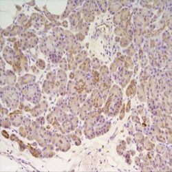
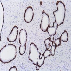
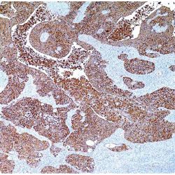
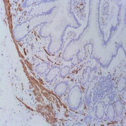
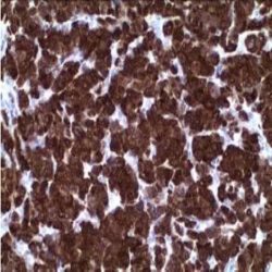
مورد استفاده: ایمونوهیستوشیمی روی بافت پارافین. بر روی بافتهای فروزن یا وسترن بلات تست نشده است.
کلون: 211F1.1
ایزوتایپ: Mouse IgG2ak
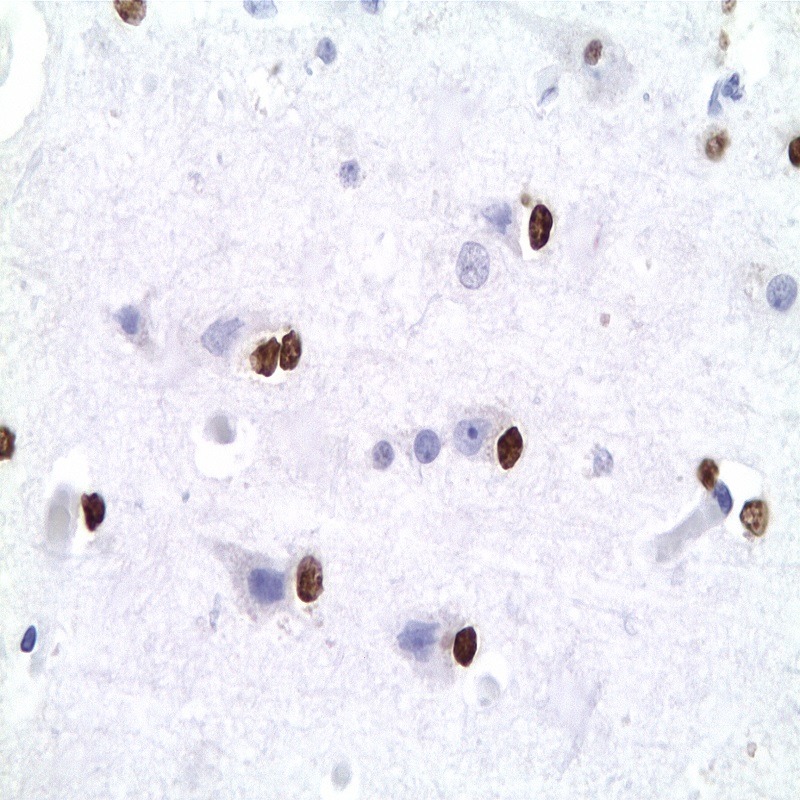
کنترل مثبت IHC: غشاء مغزی
قابلیت دیده شدن: هسته ای
روش پیشنهادی IHC:
- برش تهیه شده با ضخامت μm4 باید روی لام شارژ شده در طول شب و در دمای 60 درجه سانتی گراد خشک شود.
- دپارافینه کردن، رهیدراته کردن و استفاده از حرارت جهت بازیابی اپی توپ : بافت را با استفاده از بافر EDTA pH 8 برای بیست دقیقه در دمای 95 درجه سانتی گراد بجوشانید. سپس 5-3 بار با استفاده از آب مقطر یا دیونیزه شده شسته و سپس بیست دقیقه در دمای اتاق خنک کنید.
- بلاک پروکسیدازهای درونی : بلاک با استفاده از محلول پروکسیداز به مدت ده دقیقه
- آنتی بادی اولیه: برای 10 دقیقه در انکوبه کنید پروتکل ممکن است بسته به تهیه نمونه و کاربرد خاص متفاوت باشد. شرایط استاندارد باتوجه به آزمایشگاه و کاربر متفاوت می باشد.
- برای رنگ آمیزی ، می توانید از کیت Master Polymer Plus Detection یا کیتهای دیتکشن سایر کمپانی ها استفاده کنید.
- رنگ میزی نهایی با هماتوکسیلین و فیکس نهایی اسلاید.
شرایط نگهداری و پایداری : حداکثر 18 ماه؛ در دمای 2-8 درجه سانتیگراد نگهداری شود. فریز نشود.
دیدگاهها
هیچ دیدگاهی برای این محصول نوشته نشده است.