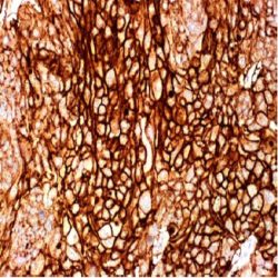
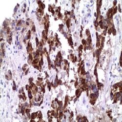
Name: SOX-11 Antibody clone MD-58 also known as MRQ-58
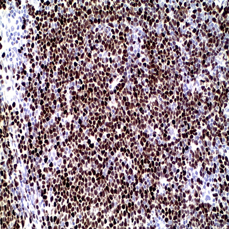
Description and applications:Mantle cell lymphoma (MCL) accounts for 5% to 10% of mature Bcell neoplasms and is an aggressive disease genetically characterized by overexpression of cyclin D1 (CCND1) due to the specific translocation t(11;14)(q13;q32). It is necessary that MCL be distinguished from potential morphologic mimics, including chronic lymphocytic leukemia/ small lymphocytic lymphoma (CLL/SLL), follicular lymphoma (FL), and marginal zone lymphoma (MZL) based on immunohistochemical (IHC) staining for CD5, CD23, and CD10. MCL and CLL both express CD5 but MCL, in contrast to CLL, generally lacks CD23 expression by IHC. FL lacks expression of both CD5 and CD23 but most often expresses CD10, whereas MZL is typically negative for all 3 antigens. Cyclin D1 overexpression is thus the hallmark of MCL even though approximately 5%-10% of MCLs lack Cyclin D1 expression and may be misdiagnosed by overreliance on CyclinD1 IHC. The recognition of cyclin D1-negative MCL is difficult because it may resemble other small B-cell lymphomas morphologically and phenotypically. Although the clinical information on cyclin D1negative MCL is limited, published data indicate that the behaviour of the variant is as aggressive as that of conventional MCL. On the other hand, patients with small B-cell lymphomas mimicking MCL have a significantly better outcome than those with true MCL. It is, therefore, important to find reliable biomarkers that may allow the identification of cyclin D1-negative MCL in clinical practice. SOX-11, the SRY (sex-determining region Y)-box11 gene, a transcription factor, normally is expressed in the developing human central nervous system, medulloblastoma, and glioma. In a series study, SOX- 11 expression was investigated in 54 cyclin D1positive MCL and 209 other lymphoid neoplasms. Interestingly, nearly all MCL were strongly positive for anti-SOX-11 (50/54, 93%), with a nuclear pattern. The staining was intense and relatively homogeneous in most of the cells. Compared to anti-cyclin D1 staining, anti-SOX-11 reactivity was stronger and more homogeneous. Five T-cell and B-cell lymphoblastic leukemia/lymphomas showed strong SOX-11 nuclear expression. One case of classic Hodgkin lymphoma, two of eight BL and two of three T-cell prolymphocytic leukemias were also positive. SOX-11 protein expression was examined by immunohistochemistry in the 12 cyclin D1-negative MCL, and all of them showed strong nuclear positive staining similar to that occurring in conventional cyclin D1-positive MCL. The expression of SOX-11 in reactive tonsil, lymph node and spleen specimens was studied. No nuclear expression was observed in any lymphocyte compartment. Only cytoplasmic staining was seen in cells from reactive germinal centers. Therefore, the authors confirmed prior reports that SOX-11 nuclear expression was a specific marker for MCL, including cyclin D1-negative MCL with typical morphology. Their study indicates that SOX-11 IHC is of value in further defining pathologic features of CD5+ DLBCL. Routine use of anti-SOX-11 in cases of suspected CD5+ DLBCL might help identify additional cases of cyclin D1-negative blastoid MCL. In summary, nuclear protein expression of SOX-11 is highly associated with both cyclin D1-positve and negative MCL. The detection of this transcription factor is a useful biomarker for identifying true cyclin D1- negative MCL. SOX-11 IHC is of value in further defining pathologic features of CD5+ DLBCL. Routine use of anti-SOX-11 in cases of suspected CD5+ DLBCL might help identify additional cases of cyclin D1- negative blastoid MCL. SOX-11 can also be detected in some BL, LBL and T-PLL, although the different morphological and phenotypic features of these malignancies allow easy recognition of the cases of cyclin D1-negative MCL
Composition:Anti-human SOX-11 mouse monoclonal antibody purified from serum and prepared in 10mM PBS, pH 7.4, with 0.2% BSA and 0.09% sodium azide
Intended use: Immunohistochemistry (IHC) on paraffin embedded tissues. Not tested on frozen tissues or Western-Blotting
دیدگاهها
هیچ دیدگاهی برای این محصول نوشته نشده است.