-
 آنتی بادیهای فلوسایتومتری
آنتی بادی مونوکلونال فلوسایتومتری CD81-APC-C750™ (RUO) ، کلون M38
Name: Flow Cytometry Antibody CD81-APC-C750™ (RUO) Clone M38
- Anti-human CD81-APC-C750TM is a monoclonal antibody (mAb) labelled with the tandem allophycocyanine-C750 (APCC750TM) designed for use as a direct immunofluorescence reagent in the identification and enumeration of cells which express the CD81 antigen by flow cytometry (FC). This reagent must be used by flow cytometry qualified personnel.
SUMMARY AND EXPLANATION
The CD81 (TAPA-1) antigen is expressed on normal lymphocytes, monocytes and eosinophils, but not on platelets and neutrophils. This antigen may be present in some cases as multimolecular complexes, in association with other members of the TM4 superfamily (CD37, CD53), or on the surface of B cells, in association with CD19 and/or CD21 and/or MHC class II antigens. Most B lymphocytes, at all stages of cellular differentiation, express CD81 at relatively high levels. Because each flow cytometer has different operating characteristics each laboratory must determine its optimal operating procedure.
REAGENT COMPOSITION
Purified monoclonal CD81 antibody conjugated with the tandem allophycocyanine-C750 (APC-C750TM), supplied in phosphate-buffered saline (PBS) containing 1% (m/v) BSA and 0.09% (m/v) sodium azide.
- Clone: M38.
- Isotype: Mouse / IgG1.
- Amount per 0,15 mL vial: 50 tests (3 µL mAb per determination)
- Reagents are not considered sterile.
-
 آنتی بادیهای فلوسایتومتری
آنتی بادی مونوکلونال فلوسایتومتری CD103-FITC ، کلون B-ly7
Name: Flow Cytometry Antibody CD103-FITC Clone B-ly7
- Antibody CD103-FITC is designed for flow cytometry use as a direct immunofluorescence reagent in the identification and enumeration of CD103 antigen-expressing cells.
CD103 is normally expressed at very high levels on intra-epithelial lymphocytes in non-lymphoid tissues, such as skin, lung, and gastrointestinal organs that express E-cadherin.
In contrast, CD103 is poorly expressed by T cells in blood, spleen, and lymph nodes. Since its discovery, CD103 has been widely used for hairy cell leukaemia (HCL) diagnosis and despite rare exceptions CD103 has been detected together with CD25 in virtually all HCL studies using flow cytometry. CD103 is not ordinarily detected in other types of Lymphoproliferative disorders except for rare cases. CD103 can also be used to define a specialized subset of CD8+ cytolytic T lymphocytes. This reagent must be used by flow cytometry qualified personal.
REAGENT COMPOSITION
The purified monoclonal CD103 antibody (mAb) conjugated with fluorescein isothiocyanate (FITC) is supplied in phosphate-buffered saline (PBS) containing ≤0.1% sodium Azid.
- Clone: B-ly7.
- Isotype: IgG1.
- Amount per 1 ml vial: 200 tests (5 µl mAb per determination).
- Reagent is considered non-sterile.
-
 آنتی بادیهای فلوسایتومتری
آنتی بادی مونوکلونال فلوسایتومتری CD117-PE ، کلون 104D2
Name: Flow Cytometry Antibody CD117-APC, Clone 104D2
Antibody CD117 is a mAb designed for flow cytometry use as a direct immunofluorescence reagent in the identification and enumeration of CD117 antigen-expressing cells. CD117 is a specific marker to detect leukemic cells committed to the myeloid lineage and therefore it represents a useful reagent in the characterization of acute leukaemias. C-kit expression would be particularly relevant for the diagnosis of biphenotypic acute leukaemias as well since in these cases its association with myeloid lineage is greater than that of the CD13 and CD33 antigens.
CD117 reacts specifically with human c-kit gene product (SCF receptor). The c-kit proto-oncogen (CD117) has been shown to be present in several cell types including normal and neoplastic haemopoietic cells. The majority of CD117+ bone marrow (BM) cells (50-70%), which mainly correspond to myeloid precursors, coexpress the progenitor associated CD34 antigen. The c-kit+/CD34- cells mainly consist of immature cells, mast cells and CD34- erythroid precursors. In acute leukaemias CD117 expression was initially associated with AML. Nevertheless, at present it is well established that CD117 expression may also be found in a relatively important proportion of T-ALL while it is usually absent in B-lineage ALL. The analysis of CD117 could be relevant for the investigation of minimal residual disease (MRD) since in combination with other antigens may be useful for the identification of leukaemia associated phenotypes in patients who achieved morphological complete remission. The combination of CD117 and other myeloid-associated antigens such as CD11b and CD15 may be of great help to monitoring MRD in AML patients although it defines a subpopulation of myeloid cells which are either absent or present at very low frequencies in normal human BM . Additionally, some acute leukaemias express CD117 over-expression which is not detected in normal cells. Moreover, different studies have shown that in around one-third of both multiple myeloma cases and patients with monoclonal gammopathy of undetermined significance myelomatous plasma cells display reactivity for CD117 .BM mast cells are clearly identifiable on the basis of their light scatter properties and strong CD117 expression. BM mast cells studies result interesting for the diagnosis of adult indolent systemic mast cell disease. and mast cell leukaemia.
REAGENT COMPOSITION
The purified monoclonal CD117 antibody (mAb) conjugated with one fluorochrome (see table above) and supplied in phosphate buffered saline with <0,1% (m/v) sodium azide.
- Clone: 104D2
- Isotype: IgG1
- Amount per 1 ml vial: 100 tests (10 µl mAb per determination)
- Reagent is considered non-sterile.
-
 آنتی بادیهای فلوسایتومتری
آنتی بادی مونوکلونال فلوسایتومتری CD117-PE ، کلون 104D2
Name: Flow Cytometry Antibody CD117-PE Clone 104D2
Antibody CD117 is a mAb designed for flow cytometry use as a direct immunofluorescence reagent in the identification and enumeration of CD117 antigen-expressing cells. CD117 is a specific marker to detect leukemic cells committed to the myeloid lineage and therefore it represents a useful reagent in the characterization of acute leukaemias. C-kit expression would be particularly relevant for the diagnosis of biphenotypic acute leukaemias as well since in these cases its association with myeloid lineage is greater than that of the CD13 and CD33 antigens.
CD117 reacts specifically with human c-kit gene product (SCF receptor). The c-kit proto-oncogen (CD117) has been shown to be present in several cell types including normal and neoplastic haemopoietic cells. The majority of CD117+ bone marrow (BM) cells (50-70%), which mainly correspond to myeloid precursors, coexpress the progenitor associated CD34 antigen. The c-kit+/CD34- cells mainly consist of immature cells, mast cells and CD34- erythroid precursors. In acute leukaemias CD117 expression was initially associated with AML. Nevertheless, at present it is well established that CD117 expression may also be found in a relatively important proportion of T-ALL while it is usually absent in B-lineage ALL. The analysis of CD117 could be relevant for the investigation of minimal residual disease (MRD) since in combination with other antigens may be useful for the identification of leukaemia associated phenotypes in patients who achieved morphological complete remission. The combination of CD117 and other myeloid-associated antigens such as CD11b and CD15 may be of great help to monitoring MRD in AML patients although it defines a subpopulation of myeloid cells which are either absent or present at very low frequencies in normal human BM . Additionally, some acute leukaemias express CD117 over-expression which is not detected in normal cells. Moreover, different studies have shown that in around one-third of both multiple myeloma cases and patients with monoclonal gammopathy of undetermined significance myelomatous plasma cells display reactivity for CD117 .BM mast cells are clearly identifiable on the basis of their light scatter properties and strong CD117 expression. BM mast cells studies result interesting for the diagnosis of adult indolent systemic mast cell disease. and mast cell leukaemia.
REAGENT COMPOSITION
The purified monoclonal CD117 antibody (mAb) conjugated with one fluorochrome (see table above) and supplied in phosphate buffered saline with <0,1% (m/v) sodium azide.
- Clone: 104D2
- Isotype: IgG1
- Amount per 1 ml vial: 100 tests (10 µl mAb per determination)
- Reagent is considered non-sterile.
-
آنتی بادیهای فلوسایتومتری
آنتی بادی مونوکلونال فلوسایتومتری IgG1-FITC ، کلون SAG1
Name: Flow Cytometry Antibody IgG1-FITC Clone SAG1
Antibody IgG1-FITC is designed for flow cytometry use as a direct immunofluorescence reagent in the identification and enumeration of IgG1 expressing cells. IgG1-FITC reacts with the Fc region of human IgG1.
IgG is the most abundant immunoglobulin in human serum (approximately 80% of total). Soluble IgG (sIgG) antibodies are distributed equally between the intra- and extravascular pools.
There are four different IgG subclasses responsible of protection against microorganisms: IgG1 (67% overall), IgG2 (22% overall), IgG3 (7% overall) and IgG4 (4% overall). These molecules have different biological properties as distinct ability to fix complement proteins and interact with phagocytic cell receptors. The IgG1 and IgG3 subclasses are particularly good opsonins and mediators of antibodydependent cell-mediated cytotoxicity (ADCC).
sIgG antibodies are important activators of complement but not as efficient as sIgM antibodies. IgG3 is the most efficient complementfixing IgG subclass, while IgG1 is somewhat less efficient, and IgG2 is even less so. IgG4 is unable to bind C1q and so cannot fix complement at all in the classical pathway.
Only IgG antibodies can cross the mammalian placenta, and maternal IgG1, IgG3, and IgG4 molecules have been found to be crucial for conferring the passive immunity that protects the developing human fetus and newborn in the first few months of life. IgG2 crosses the placenta with much lower efficiency.
CYT-IGG1F should be used with BulkLysisTM (CYT-BL) buffer that allows you to stain a great number of cells and analyze lowfrequency subsets, as IgG1+ memory B cells and plasma cells. If another lysing protocol is used, two pre-staining washing steps using PBS+1% (m/v) BSA + 0,09% (m/v) sodium azide must be performed.
This reagent must be used by flow cytometry qualified personnel.
REAGENT COMPOSITION
Purified monoclonal anti-IgG1 antibody (mAb) conjugated with fluorescein isothiocyanate (FITC), supplied in phosphate buffered saline with 0,09% (m/v) sodium azide and BSA 1% (m/v).
- Clone: SAG1.
- Isotype: Mouse IgG2b.
- Amount per vial: 25 tests using 3µL of mAb to 106 cells
-
آنتی بادیهای فلوسایتومتری
آنتی بادی مونوکلونال فلوسایتومتری IgG1-PE ، کلون SAG1
Name: Flow Cytometry Antibody IgG1-PE, Clone SAG1
Antibody IgG1-PE is designed for flow cytometry use as a direct immunofluorescence reagent in the identification and enumeration of IgG1 expressing cells. IgG1-PE reacts with the Fc region of human IgG1.
IgG is the most abundant immunoglobulin in human serum (approximately 80% of total). Soluble IgG (sIgG) antibodies are distributed equally between the intra- and extravascular pools.
There are four different IgG subclasses responsible of protection against microorganisms: IgG1 (67% overall), IgG2 (22% overall), IgG3 (7% overall) and IgG4 (4% overall). These molecules have different biological properties as distinct ability to fix complement proteins and interact with phagocytic cell receptors. The IgG1 and IgG3 subclasses are particularly good opsonins and mediators of antibody dependent cell-mediated cytotoxicity (ADCC) (1).
sIgG antibodies are important activators of complement but not as efficient as sIgM antibodies. IgG3 is the most efficient complement fixing IgG subclass, while IgG1 is somewhat less efficient, and IgG2 is even less so. IgG4 is unable to bind C1q and so cannot fix complement at all in the classical pathway (1).
Only IgG antibodies can cross the mammalian placenta, and maternal IgG1, IgG3, and IgG4 molecules have been found to be crucial for conferring the passive immunity that protects the developing human fetus and newborn in the first few months of life. IgG2 crosses the placenta with much lower efficiency (1).
CYT-IGG1PE should be used with BulkLysisTM (CYT-BL) buffer that allows you to stain a great number of cells and analyze low frequency subsets, as IgG1+ memory B cells and plasma cells. If another lysing protocol is used, two pre-staining washing steps using PBS + 1% (m/v) BSA + 0,09% (m/v) sodium azide must be performed.
This reagent must be used by flow cytometry qualified personnel.
REAGENT COMPOSITION
Purified monoclonal anti-IgG1 antibody (mAb) conjugated with R-phycoerythrin (PE), supplied in phosphate buffered saline with 0,09% (m/v) sodium azide and BSA 1% (m/v).
- Clone: SAG1.
- Isotype: Mouse IgG2b.
- Amount per vial: 25 tests using 3µL of mAb to 106 cells.
-
آنتی بادیهای فلوسایتومتری
آنتی بادی مونوکلونال فلوسایتومتری IgG2-FITC ، کلون SAG2
Name: Flow Cytometry Antibody IgG2-FITC, Clone SAG2
Antibody IgG2-FITC is designed for flow cytometry use as a direct immunofluorescence reagent in the identification and enumeration of IgG2 expressing cells. IgG2-FITC reacts with the Fc region of human IgG2.
IgG is the most abundant immunoglobulin in human serum (approximately 80% of total). Soluble IgG (sIgG) antibodies are distributed equally between the intra- and extravascular pools.
There are four different IgG subclasses responsible of protection against microorganisms: IgG1 (67% overall), IgG2 (22% overall), IgG3 (7% overall) and IgG4 (4% overall). These molecules have different biological properties as distinct ability to fix complement proteins and interact with phagocytic cell receptors. The IgG1 and IgG3 subclasses are particularly good opsonins and mediators of antibodydependent cell-mediated cytotoxicity (ADCC).
sIgG antibodies are important activators of complement but not as efficient as sIgM antibodies. IgG3 is the most efficient complement fixing IgG subclass, while IgG1 is somewhat less efficient, and IgG2 is even less so. IgG4 is unable to bind C1q and so cannot fix complement at all in the classical pathway.
Only IgG antibodies can cross the mammalian placenta, and maternal IgG1, IgG3, and IgG4 molecules have been found to be crucial for conferring the passive immunity that protects the developing human fetus and newborn in the first few months of life. IgG2 crosses the placenta with much lower efficiency.
CYT-IGG2F should be used with BulkLysisTM (CYT-BL) buffer that allows you to stain a great number of cells and analyze lowfrequency subsets, as IgG2+ memory B cells and plasma cells. If another lysing protocol is used, two pre-staining washing steps using PBS + 1% (m/v) BSA + 0,09% (m/v) sodium azide must be performed. This reagent must be used by flow cytometry qualified personnel.
REAGENT COMPOSITION
Purified monoclonal anti-IgG2 antibody (mAb) conjugated with fluorescein isothiocyanate (FITC), supplied in phosphate buffered saline with 0,09% (m/v) sodium azide and BSA 1% (m/v).
- Clone: SAG2.
- Isotype: Mouse IgG1.
- Amount per vial: 25 tests using 3µL of mAb to 106 cells.
-
آنتی بادیهای فلوسایتومتری
آنتی بادی مونوکلونال فلوسایتومتری IgG2-PE ، کلون SAG2
Name: Flow Cytometry Antibody IgG2-PE, Clone SAG2
Antibody IgG2-PE is designed for flow cytometry use as a direct immunofluorescence reagent in the identification and enumeration of IgG2 expressing cells. IgG2-PE reacts with the Fc region of human IgG2.
IgG is the most abundant immunoglobulin in human serum (approximately 80% of total). Soluble IgG (sIgG) antibodies are distributed equally between the intra- and extravascular pools.
There are four different IgG subclasses responsible of protection against microorganisms: IgG1 (67% overall), IgG2 (22% overall), IgG3 (7% overall) and IgG4 (4% overall). These molecules have different biological properties as distinct ability to fix complement proteins and interact with phagocytic cell receptors. The IgG1 and IgG3 subclasses are particularly good opsonins and mediators of antibody dependent cell-mediated cytotoxicity (ADCC).
sIgG antibodies are important activators of complement but not as efficient as sIgM antibodies. IgG3 is the most efficient complement fixing IgG subclass, while IgG1 is somewhat less efficient, and IgG2 is even less so. IgG4 is unable to bind C1q and so cannot fix complement at all in the classical pathway.
Only IgG antibodies can cross the mammalian placenta, and maternal IgG1, IgG3, and IgG4 molecules have been found to be crucial for conferring the passive immunity that protects the developing human fetus and newborn in the first few months of life. IgG2 crosses the placenta with much lower efficiency.
CYT-IGG2PE should be used with BulklysisTM (CYT-BL) buffer that allows you to stain a great number of cells and analyze low frequency subsets, as IgG2+ memory B cells and plasma cells. If another lysing protocol is used, two pre-staining washing steps using PBS + 1% (m/v) BSA + 0,09% (m/v) sodium azide must be performed.
This reagent must be used by flow cytometry qualified personnel
REAGENT COMPOSITION
Purified monoclonal anti-IgG2 antibody (mAb) conjugated with R-phycoerythrin (PE), supplied in phosphate buffered saline with 0,09% (m/v) sodium azide and BSA 1% (m/v).
- Clone: SAG2.
- Isotype: Mouse IgG1.
- Amount per vial: 25 tests using 3µL of mAb to 106 cells.
-
آنتی بادیهای فلوسایتومتری
آنتی بادی مونوکلونال فلوسایتومتری IgG3-FITC ، کلون SAG3
Name: Flow Cytometry Antibody IgG3-FITC, Clone SAG3
Antibody IgG3-FITC is designed for flow cytometry use as a direct immunofluorescence reagent in the identification and enumeration of IgG3 expressing cells. IgG3-FITC reacts with the hinge region of the heavy chain of human IgG3.
IgG is the most abundant immunoglobulin in human serum (approximately 80% of total). Soluble IgG (sIgG) antibodies are distributed equally between the intra- and extravascular pools.
There are four different IgG subclasses responsible of protection against microorganisms: IgG1 (67% overall), IgG2 (22% overall), IgG3 (7% overall) and IgG4 (4% overall). These molecules have different biological properties as distinct ability to fix complement proteins and interact with phagocytic cell receptors. The IgG1 and IgG3 subclasses are particularly good opsonins and mediators of antibody dependent cell-mediated cytotoxicity (ADCC) (1).
sIgG antibodies are important activators of complement but not as efficient as sIgM antibodies. IgG3 is the most efficient complement fixing IgG subclass, while IgG1 is somewhat less efficient, and IgG2 is even less so. IgG4 is unable to bind C1q and so cannot fix complement at all in the classical pathway.
Only IgG antibodies can cross the mammalian placenta, and maternal IgG1, IgG3, and IgG4 molecules have been found to be crucial for conferring the passive immunity that protects the developing human fetus and newborn in the first few months of life. IgG2 crosses the placenta with much lower efficiency.
CYT-IGG3F should be used with BulkLysisTM (CYT-BL) buffer that allows you to stain a great number of cells and analyze low frequency subsets, as IgG3+ memory B cells and plasma cells. If another lysing protocol is used, two pre-staining washing steps using PBS + 1% (m/v) BSA + 0,09% (m/v) sodium azide must be performed.
This reagent must be used by flow cytometry qualified personnel. This reagent must be used by flow cytometry qualified personnel
REAGENT COMPOSITION
Purified monoclonal anti-IgG3 antibody (mAb) conjugated with fluorescein isothiocyanate (FITC), supplied in phosphate buffered saline with 0,09% (m/v) sodium azide and BSA 1% (m/v).
- Clone: SAG3.
- Isotype: Mouse IgG1.
- Amount per vial: 25 tests using 3µL of mAb to 106 cells.
-
آنتی بادیهای فلوسایتومتری
آنتی بادی مونوکلونال فلوسایتومتری IgG4-APC ، کلون SAG4
Name: Flow Cytometry Antibody IgG4-APC, Clone SAG4
Antibody IgG4-APC is designed for flow cytometry use as a direct immunofluorescence reagent in the identification and enumeration of IgG4 expressing cells. IgG4-APC recognizes an epitope within Fc region (CH2 domain) of human IgG4.
IgG is the most abundant immunoglobulin in human serum (approximately 80% of total). Soluble IgG (sIgG) antibodies are distributed equally between the intra- and extravascular pools.
There are four different IgG subclasses responsible of protection against microorganisms: IgG1 (67% overall), IgG2 (22% overall), IgG3 (7% overall) and IgG4 (4% overall). These molecules have different biological properties as distinct ability to fix complement proteins and interact with phagocytic cell receptors. The IgG1 and IgG3 subclasses are particularly good opsonins and mediators of antibody dependent cell-mediated cytotoxicity (ADCC).
sIgG antibodies are important activators of complement but not as efficient as sIgM antibodies. IgG3 is the most efficient complement fixing IgG subclass, while IgG1 is somewhat less efficient, and IgG2 is even less so. IgG4 is unable to bind C1q and so cannot fix complement at all in the classical pathway.
Only IgG antibodies can cross the mammalian placenta, and maternal IgG1, IgG3, and IgG4 molecules have been found to be crucial for conferring the passive immunity that protects the developing human fetus and newborn in the first few months of life. IgG2 crosses the placenta with much lower efficiency.
CYT-IGG4AP should be used with BulkLysisTM (CYT-BL) buffer that allows you to stain a great number of cells and analyze low frequency subsets, as IgG4+ memory B cells and plasma cells. If another lysing protocol is used, two pre-staining washing steps using PBS + 1% (m/v) BSA + 0,09% (m/v) sodium azide must be performed.
This reagent must be used by flow cytometry qualified personnel.
REAGENT COMPOSITION
Purified monoclonal anti-IgG4 antibody (mAb) conjugated with allophycocyanin (APC), supplied in phosphate buffered saline with 0,09% (m/v) sodium azide and BSA 1% (m/v).
- Clone: SAG4.
- Isotype: Mouse IgG1.
- Amount per vial: 25 tests using 3µL of mAb to 106 cells
-
آنتی بادیهای فلوسایتومتری
آنتی بادی مونوکلونال فلوسایتومتری IgG4-FITC ، کلون SAG4
Name: Flow Cytometry Antibody IgG4-FITC, Clone SAG4
Antibody IgG4-FITC is designed for flow cytometry use as a direct immunofluorescence reagent in the identification and enumeration of IgG4 expressing cells. IgG4-FITC recognizes an epitope within Fc region (CH2 domain) of human IgG4.
IgG is the most abundant immunoglobulin in human serum (approximately 80% of total). Soluble IgG (sIgG) antibodies are distributed equally between the intra- and extravascular pools.
There are four different IgG subclasses responsible of protection against microorganisms: IgG1 (67% overall), IgG2 (22% overall), IgG3 (7% overall) and IgG4 (4% overall). These molecules have different biological properties as distinct ability to fix complement proteins and interact with phagocytic cell receptors. The IgG1 and IgG3 subclasses are particularly good opsonins and mediators of antibody dependent cell-mediated cytotoxicity (ADCC).
sIgG antibodies are important activators of complement but not as efficient as sIgM antibodies. IgG3 is the most efficient complement fixing IgG subclass, while IgG1 is somewhat less efficient, and IgG2 is even less so. IgG4 is unable to bind C1q and so cannot fix complement at all in the classical pathway.
Only IgG antibodies can cross the mammalian placenta, and maternal IgG1, IgG3, and IgG4 molecules have been found to be crucial for conferring the passive immunity that protects the developing human fetus and newborn in the first few months of life. IgG2 crosses the placenta with much lower efficiency.
CYT-IGG4F should be used with BulkLysisTM (CYT-BL) buffer that allows you to stain a great number of cells and analyze low frequency subsets, as IgG4+ memory B cells and plasma cells. If another lysing protocol is used, two pre-staining washing steps using PBS + 1% (m/v) BSA + 0,09% (m/v) sodium azide must be performed.
This reagent must be used by flow cytometry qualified personnel.
REAGENT COMPOSITION
Purified monoclonal anti-IgG4 antibody (mAb) conjugated with fluorescein isothiocyanate (FITC), supplied in phosphate buffered saline with 0,09% (m/v) sodium azide and BSA 1% (m/v).
- Clone: SAG4.
- Isotype: Mouse IgG1.
- Amount per vial: 25 tests using 3µL of mAb to 106 cells.
-
آنتی بادیهای فلوسایتومتری
آنتی بادی مونوکلونال فلوسایتومتری IgM-APC ، کلون SADA4
Name: Flow Cytometry Antibody IgM-APC, Clone SADA4
Antibody IgM-APC is designed for flow cytometry use as a direct immunofluorescence reagent in the identification and enumeration of IgM expressing cells.
Monomeric IgM is always the first form and isotype of Ig generated by naive B cells. Following its initial activation by antigen, the naive B cell proliferates and differentiates, and its progeny produce the pentameric secreted form of IgM. Thus, it is IgM antibodies that are expressed first in any primary immune response. IgM antibodies comprise only about 5–10% of normal serum Ig. Although the bulk of IgM is found in the blood, if vascular permeability has been increased in a local area by the release of vasoactive compounds during an inflammatory response, IgM antibodies can exit the blood and enter the tissues to reach sites of infection. Although the concentration of IgM antibodies in the external secretions is very low compared to that of IgA, secretory IgM antibodies do play an important role in mucosal humoral immunity.
CYT-IGMAP should be used with BulklysisTM (CYT-BL) buffer that allows you to stain a great number of cells and analyze low-frequency subsets, as IgM+ memory B cells and plasma cells. If another lysing protocol is used, two pre-staining washing steps using PBS+1% (m/v) BSA + ≤0.09% (m/v) sodium azide must be performed. This reagent must be used by flow cytometry qualified personal. This reagent must be used by flow cytometry qualified personnel
REAGENT COMPOSITION
Purified monoclonal anti-IgM antibody (mAb) conjugated with Allophycocyanin (APC), supplied in phosphate buffered saline with <0,1% (m/v) sodium azide.
- Clone: SA-DA4
- Isotype: Mouse IgG1
- Amount per vial: sufficient volume for 25 tests using 5µl per 106 cells
-
آنتی بادیهای فلوسایتومتری
آنتی بادی مونوکلونال فلوسایتومتری IgM-PerCP-Cyanine5.5 ، کلون MHM-88.
Name: Flow Cytometry Antibody IgM-PerCP-Cyanine5.5, Clone MHM-88.
Antibody IgM-PerCP-Cyanine5.5 is designed for flow cytometry use as a direct immunofluorescence reagent in the identification and enumeration of IgM expressing cells.
Monomeric IgM is always the first form and isotype of Ig generated by naive B cells. Following its initial activation by antigen, the naive B cell proliferates and differentiates, and its progeny produce the pentameric secreted form of IgM. Thus, it is IgM antibodies that are expressed first in any primary immune response. IgM antibodies comprise only about 5–10% of normal serum Ig.
Although the bulk of IgM is found in the blood, if vascular permeability has been increased in a local area by the release of vasoactive compounds during an inflammatory response, IgM antibodies can exit the blood and enter the tissues to reach sites of infection. Although the concentration of IgM antibodies in the external secretions is very low compared to that of IgA, secretory IgM antibodies do play an important role in mucosal humoral immunity.
CYT-IGMC2 should be used with BulkLysisTM (CYT-BL) buffer that allows you to stain a great number of cells and analyze low frequency subsets, as IgM+ memory B cells and plasma cells. If another lysing protocol is used, two pre-staining washing steps using PBS + 1% (m/v) BSA + 0,09% (m/v) sodium azide must be performed.
This reagent must be used by flow cytometry qualified personnel.
REAGENT COMPOSITION
Purified monoclonal anti-IgM antibody (mAb) conjugated with Peridin chlorophyll protein-Cyanine 5.5 (PerCP-Cyanine5.5), supplied in phosphate buffered saline with 0,09% (m/v) sodium azide and BSA 1% (m/v).
- Clone: MHM-88.
- Isotype: Mouse IgG1.
- Amount per vial: 25 tests using 3µL per 106 cells
-
 آنتی بادیهای فلوسایتومتری
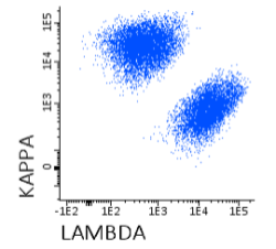
آنتی بادی مونوکلونال فلوسایتومتری KAPPA-PE ، کلون Polyclonal
Name: Flow Cytometry Antibody KAPPA-PE, Clone Polyclonal
- Antibody Kappa-PE is a polyclonal antibody (pAb) labelled with R-Phycoerythrin (PE) and designed for use as a direct immunofluorescence reagent in the identification and enumeration of cells that express human Kappa immunoglobulin Light Chains by flow cytometry. Antibodies to Kappa Light Chains are useful for the identification of clonal excess in B-cell lymphoproliferative disorders together with a panel of other antibodies. Anti-Kappa Light Chains reacts with free Kappa chains as well as Kappa chains in intact immunoglobulin molecules. This reagent must be used by flow cytometry qualified personal.
REAGENT COMPOSITION
Purified polyclonal antibody Anti-human Kappa Light Chains, Goat F(ab’)2, conjugated with R-Phycoerythrin (PE), supplied in phosphate buffered saline with <0,1% (m/v) sodium azide.
- Amount per 0,5 ml
- vial: 100 tests (5 µl pAb to 106 cells).
- Reagents are not considered sterile.
-
-
-
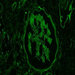 ایمونوفلورسانس
آنتی بادی Human Albumin (Polyclonal) -FITC
Anti-Human Albumin
DESCRIPTION AND APPLICATIONS: Albumin is a soluble, monomeric protein which comprises about one-half of the blood serum protein. Albumin functions primarily as a carrier protein for steroids, fatty acids, and thyroid hormones and plays a role in stabilizing extracellular fluid volume. Albumin is synthesized in the liver as preproalbumin which has an N-terminal peptide that is removed before the nascent protein is released from the rough endoplasmic reticulum. The product, proalbumin, is in turn cleaved in the Golgi vesicles to produce the secreted albumin. This antibody should be used in conjunction with a panel of antibodies to aid in the identification of albumin in target tissue (e.g., in the diagnosis of renal or dermal pathologies). The interpretation of the results should be always corroborated with those of external controls and integrated with the morphological and clinical aspectsfor the final diagnosis.
COMPOSITION Anti-human Rabbit Antibody to Albumin. Fluorescein Conjugated Polyclonal antibody purified from serum and prepared in buffer with carrier protein and preservative for stabilization
IMMUNOGEN: Albumin (Human Serum)
-
-
-
 ایمونوفلورسانس
آنتی بادی Human C1q (Polyclonal) -FITC
Anti-Human C1q
DESCRIPTION AND APPLICATIONS :C1q associates with the proenzymes C1r and C1s to yield C1, the first component of the serum complement system. The collagen-like regions of C1q interact with the Ca(2+)-dependent C1r(2)C1s(2) proenzyme complex, and efficient activation of C1 takes place on interaction of the globular heads of C1q with the Fc regions of IgG or IgM antibody present in immune complexes.
C1q is predominantly produced by macrophages but also by follicular dendritic cells, interdigitating cells and cells of the monocyte-macrophage lineage. C1q deficiency has a profound effect on host defence and clearance of immune complexes. Absence of C1q may cause autoimmunity by impairment of the clearance of apoptotic cells. Inherited C1q deficiency is also associated with the development of systemic lupus erythematosus (SLE).
This antibody should be used in conjunction with a panel of antibodies to aid in the identification of C1q fragment in target tissue (e.g., in the diagnosis of renal or dermal pathologies).
The interpretation of the results should be always corroborated with those of external controls and integrated with the morphological and clinical aspects for the final diagnosis
COMPOSITION: Anti-human Sheep Antibody to C1q. Fluorescein Conjugated Polyclonal antibody purified from serum and prepared in buffer with carrier protein and preservative for stabilization.
IMMUNOGEN: Human C1q, purified from plasma
برای تماس کلیک کنید
سما تشخیص آریا ، عرضه کننده متنوع ترین و کامل ترین محصولات کیت های آزمایشگاهی