Category: فلوسایتومتری
Showing 161–180 of 189 results
فیلتر ها-
آنتی بادیهای فلوسایتومتری
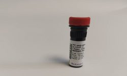
آنتی بادی فلوسایتومتری FITC anti-mouse CD49b (pan-NK cells) Antibody
Rated 0 out of 5FITC anti-mouse CD49b (pan-NK cells) Antibody
Company:BioLegend
Catalog Number : 108905
Size : 50 µg
Clone : DX5
Isotype : Rat IgM, κ
Reactivity : Mouse
Description :
DX5 antigen has been recently characterized as CD49b. It is a 150 kD integrin α chain also known as α integrin, VLA-2 α chain, and integrin α chain. CD49b non-covalently associates with CD29 (β integrin) to form the CD49b/CD29 complex known as VLA-2, a receptor for collagen and laminin. CD49b is expressed on platelets, the majority of NK cells, NKT cells, and a small subset of CD8+ T cells (this population can be significantly increased following viral infection). DX5 is used for the identification and isolation of NK cells, and is especially useful for identifying NK cells in mice lacking the NK1.1 antigen.
-
آنتی بادیهای فلوسایتومتری
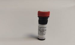
آنتی بادی فلوسایتومتری FITC anti-mouse/human CD11b Antibody
Rated 0 out of 5FITC anti-mouse/human CD11b Antibody
Company:BioLegend
Catalog Number : 101205
Size : 50 µg
Clone : M1/70
Isotype : Rat IgG2b, κ
Reactivity : Mouse, Human, Cross-Reactivity: Chimpanzee, Baboon, Cynomolgus, Rhesus, Rabbit (Lapine)
Description :
CD11b is a 170 kD glycoprotein also known as αM integrin, Mac-1 α subunit, Mol, CR3, and Ly-40. CD11b is a member of the integrin family, primarily expressed on granulocytes, monocytes/macrophages, dendritic cells, NK cells, and subsets of T and B cells. CD11b non-covalently associates with CD18 (β2 integrin) to form Mac-1. Mac-1 plays an important role in cell-cell interaction by binding its ligands ICAM-1 (CD54), ICAM-2 (CD102), ICAM-4 (CD242), iC3b, and fibrinogen.
-
آنتی بادیهای فلوسایتومتری
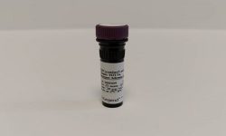
آنتی بادی فلوسایتومتری FITC F(ab’)2 Goat anti-human IgG Fcγ Antibody
Rated 0 out of 5FITC F(ab’)2 Goat anti-human IgG Fcγ Antibody
Company:BioLegend
Catalog Number: 398006
Size : 100 µg
Clone : Poly23980
Isotype : Goat Polyclonal Ig
Reactivity : Human
Description :
gG Fc is a homodimer that is composed of the constant region of the two heavy chains that form the IgG molecule. The Fc fragment mediates opsonization, antibody dependent cellular cytotoxicity (ADCC), and complement activation through binding to Fc receptors such as CD16, CD32, CD64, and the complement factor C1.
-
آنتی بادیهای فلوسایتومتری
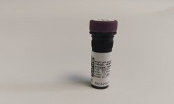
آنتی بادی فلوسایتومتری PE anti-human CD79a (Igα) Antibody
Rated 0 out of 5PE anti-human CD79a (Igα) Antibody
Company:BioLegend
Catalog Number : 333503
Size: 25 tests
Clone : HM47
Isotype : Mouse IgG1, κ
Reactivity: Human
Description:
CD79a is a 47 kD type I integral membrane protein, also known as mb-1 or Iga. It is a member of the Ig superfamily and disulphide-associated with CD79b (B29). The interaction of CD79a/CD79b heterodimer with B cell suface Ig forms B cell antigen complex. CD79a is expressed in B cells from early pre-B to plasma cell stage. It has been shown that CD79a is also weakly expressed in some precursors of T- and myeloid cells. CD79 mediates the transport of IgM to B cell surface and transduces signals initiated by BCR aggregation.
-
آنتی بادیهای فلوسایتومتری
آنتی بادی فلوسایتومتری PE anti-mouse CD366 (Tim-3) Antibody
Rated 0 out of 5PE anti-mouse CD366 (Tim-3) Antibody
Company:BioLegend
Catalog Number : 119703
Size : 50 µg
Clone : RMT3-23
Isotype : Rat IgG2a, κ
Reactivity : Mouse
Description :
CD366 (Tim-3) is a transmembrane protein also known as T cell immunoglobulin and mucin domain containing protein-3. Tim-3 is expressed at high levels on Th1 lymphocytes and CD11b macrophages. Tim-3 has also been shown to exist as a soluble protein. Cells expressing Tim-3 are present at high levels in the CNS of animals at the onset of experimental autoimmune encephalomyelitis (EAE), a disease mediated by lymphocytes secreting Th1-like cytokines. Tim-3 has been proposed to inhibit Th1-mediated immune responses and promote immunological tolerance.
-
آنتی بادیهای فلوسایتومتری
آنتی بادی فلوسایتومتری PE anti-mouse CD107a (LAMP-1) Antibody
Rated 0 out of 5 -
آنتی بادیهای فلوسایتومتری
آنتی بادی فلوسایتومتری PE anti-mouse Ly-6G/Ly-6C (Gr-1) Antibody
Rated 0 out of 5PE anti-mouse Ly-6G/Ly-6C (Gr-1) Antibody
Company:BioLegend
Catalog Number : 108407
Size : 50 µg
Clone : RB6-8C5
Isotype : Rat IgG2b, κ
Reactivity : Mouse
Description :
Gr-1 is a 21-25 kD protein also known as Ly-6G/Ly-6C. This myeloid differentiation antigen is a glycosylphosphatidylinositol (GPI)-linked protein expressed on granulocytes and macrophages. In bone marrow, the expression levels of Gr-1 directly correlate with granulocyte differentiation and maturation; Gr-1 is also transiently expressed on bone marrow cells in the monocyte lineage. Immature Myeloid Gr-1+ cells play a role in the development of antitumor immunity.
-
آنتی بادیهای فلوسایتومتری
آنتی بادی فلوسایتومتری PE anti-TCF1 (TCF7) Antibody
Rated 0 out of 5PE anti-TCF1 (TCF7) Antibody
Company:BioLegend
Catalog Number : 655208
Size : 100 tests
Clone : 7F11A10
Isotype : Mouse IgG1, κ
Reactivity : Human
Description :
TCF1 is the first identified member of the T-cell-specific transcription factor family. It plays an important role in T cell development and differentiation. TCF1 is inactivated by association with the transcriptional repressor TLE proteins. During Wnt signaling, the transcriptional coactivator CTNNB1 accumulates and, in turn, replaces the transcriptional repressor associated with TCF1. Interaction with CTNNB1 results in transactivation of TCF1 target genes. Deletion of TCF1 causes massive apoptosis of double positive thymocyte, suggesting that TCF1 is required for thymocyte survival during T cell development. In addition to its function in thymus, TCF1 promotes T cell differentiation to Th2 cells in the periphery through transcriptional activation of GATA3.
-
آنتی بادیهای فلوسایتومتری
آنتی بادی فلوسایتومتری PE/Cyanine5 anti-human CD3 Antibody
Rated 0 out of 5PE/Cyanine5 anti-human CD3 Antibody
Company:BioLegend
Clone: HIT3a
Isotype: Mouse IgG2a, κ
Cat Number: 300309
Reactivity: Human
Antibody Type: Monoclonal
Description:
CD3ε is a 20 kD chain of the CD3/T-cell receptor (TCR) complex which is composed of two CD3ε, one CD3γ, one CD3δ, one CD3ζ (CD247), and a T-cell receptor (α/β or γ/δ) heterodimer. It is found on all mature T lymphocytes, NK-T cells, and some thymocytes. CD3, also known as T3, is a member of the immunoglobulin superfamily that plays a role in antigen recognition, signal transduction, and T cell activation.
-
آنتی بادیهای فلوسایتومتری
آنتی بادی فلوسایتومتری PE/Cyanine5 anti-human CD19 Antibody
Rated 0 out of 5PE/Cyanine5 anti-human CD19 Antibody
Company:BioLegend
Catalog Numbeer: 302209
Size : 25 tests
Clone : HIB19
Isotype: Mouse IgG1, κ
Reactivity: Human, Chimpanzee, Rhesus
Description :
CD19 is a 95 kD type I transmembrane glycoprotein also known as B4. It is a member of the immunoglobulin superfamily expressed on B-cells (from pro-B to blastoid B cells, absent on plasma cells) and follicular dendritic cells. CD19 is involved in B cell development, activation, and differentiation. CD19 forms a complex with CD21 (CR2) and CD81 (TAPA-1), and functions as a BCR coreceptor.
-
آنتی بادیهای فلوسایتومتری
آنتی بادی فلوسایتومتری PerCP/Cyanine5.5 anti-mouse CD279 (PD-1) Antibody
Rated 0 out of 5PerCP/Cyanine5.5 anti-mouse CD279 (PD-1) Antibody
Company:BioLegend
Catalog Number : 135207
Size : 25 µg
Clone : 29F.1A12
Isotype : Rat IgG2a, κ
Reactivity : Mouse
Description :
CD279, also known as programmed death-1 (PD-1), is a 50-55 kD glycoprotein belonging to the CD28 family of the Ig superfamily. PD-1 is expressed on activated splenic T and B cells and thymocytes. It is induced on activated myeloid cells as well. PD-1 is involved in lymphocyte clonal selection and peripheral tolerance through binding its ligands, B7-H1 (PD-L1) and B7-DC (PD-L2). It has been reported that PD-1 and PD-L1 interactions are critical to positive selection and play a role in shaping the T cell repertoire. PD-L1 negative costimulation is essential for prolonged survival of intratesticular islet allografts.
-
محلول های فلوسایتومتری
محلول فلوسایتومتری – Annexin V Binding Buffer
Rated 0 out of 5Annexin V Binding Buffer
Company: BioLegend
Catalog : 422201
Size : 100 mL
:Description
Annexin V Binding Buffer has been formulated for flow cytometric labeling of apoptotic cells with Annexin V reagents. The buffer is provided as a ready-to-use solution.
-
محلول های فلوسایتومتری
محلول فلوسایتومتری – Cell Staining Buffer
Rated 0 out of 5Cell Staining Buffer
Company: BioLegend
Catalog Number : 420201
Size : 500 mL
Storage & Handling : Store between 2°C and 8°C
Description :
Cell Staining Buffer is an antibody diluent and cell wash buffer optimized for use in immunofluorescent staining of viable or fixed single cell suspensions. Cell Staining Buffer contains bovine calf serum as a protein carrier to reduce nonspecific binding of antibodies and fluorochrome reagents to target cells. It also contains a metabolic inhibitor, sodium azide (NaN ), to inhibit patching and capping of cell surface antigens. To prevent interference in biotin/avidin indirect staining protocols, Cell Staining Buffer is formulated without biotin.
-
محلول های فلوسایتومتری
محلول فلوسایتومتری – Flow Cytometry Antibody Diluent Buffer
Rated 0 out of 5Flow Cytometry Antibody Diluent Buffer
Company: BioLegend
Catalog : 425501
Size : 5 mL
:Description
This buffer is used to dilute antibodies intended to be used in flow cytometry. The components are compatible with our Cell Staining Buffer and it is useful to prepare concentrated antibodies or preparing staining cocktails.
-
محلول های فلوسایتومتری
محلول فلوسایتومتری – FluoroFix™ Buffer
Rated 0 out of 5FluoroFix™ Buffer
Company: BioLegend
Catalog Number : 422101
Size : 200 tests
Storage & Handling : The buffer solution should be stored between 2°C and 8°C
:Description
FluoroFix™ Buffer is a ready-to-use buffer, specially formulated for fixation of immunofluorescence stained cells, optimized to stabilize tandem dyes. It can be used as the final resuspension of the cell pellet in immunofluorescence staining procedures. FluoroFix™ Buffer is provided as 100 mL.This quantity is sufficient for 200 tests.
-
محلول های فلوسایتومتری
محلول فلوسایتومتری – Intracellular Staining Permeabilization Wash Buffer (10X)
Rated 0 out of 5Intracellular Staining Permeabilization Wash Buffer (10X)
Company: BioLegend
Catalog : 421002
Size : 100 mL
:Description
ntracellular Staining Permeabilization Wash Buffer is useful for intracellular staining procedures, e.g., in preparation of cells for staining intracellular cytokines or other proteins. Intracellular Staining ermeabilization Wash Buffer is used to permeabilize cells following fixation with Intracellular Staining Fixation Buffer (Cat. No. 420801). It is supplied as a 10X solution and should be diluted in deionized water prior to use. Intracellular Staining Permeabilization Wash Buffer has been formulated to have minimal effects on cells, reduce non-specific staining and enhance the signal to noise ratio. It can be used for antibody dilutions and cell washing during intracellular staining.
-
محلول های فلوسایتومتری
محلول های فلوسایتومتری – Propidium Iodide Solution
Rated 0 out of 5Propidium Iodide Solution
Company: BioLegend
Catalog : 421301
Size : 2 mL
Description :
Propidium iodide (PI) is a fluorescent dye that binds to DNA. When excited by 488nm laser light, it can be detected with in the PE/Texas Red® channel with a bandpass filter 610/10. It is commonly used in evaluation of cell viability or DNA content in cell cycle analysis by flow cytometry.
-
محلول های فلوسایتومتری
محلول های فلوسایتومتری – RBC Lysis Buffer (10X)
Rated 0 out of 5RBC Lysis Buffer (10X)
Company: BioLegend
Catalog : 420301
Size : 100 mL
:Description
Red Blood Cell (RBC) Lysis Buffer has been designed, formulated, and tested to ensure optimal lysis of RBCs in single cell suspensions with minimal effects on leukocytes. RBC Lysis Buffer is supplied as a 10X solution containing ammonium chloride, potassium carbonate, and EDTA, and should be diluted in deionized water prior to use. Nucleated RBCs are not effectively lysed with ammonium chloride.

