Category: FISH Probes
Showing 1–20 of 35 results
فیلتر ها-
پروبهای فیش
پروب فیش HER2/CEN17 FISH Probe
Rated 0 out of 5INTRODUCTION: HER2, also known as epidermal growth factor receptor 2, neu, ERBB2 or CD340 is a proto-oncogene located in the human chromosome 17, band 21 that code a transmembrane glycoprotein (p185) with tyrosine-kinase activity. Because of its interaction and similarity to EGFR and other members of the epidermal growth factor receptor family, HER2 takes part in the differentiation of some cell-lines during embryogenesis after the interaction between mesenchyme–epithelium-neuroectoderm. It also has an influence on the migration, differentiation and interaction between numerous cells.
The overexpression of HER2 has been proven in over 20% of adenocarcinomas of different locations, including lung, uterus, gastrointestinal and urinary tract, breast and skin appendages in different studies. In case of breast and stomach cancers, the overexpression of this oncoprotein, which is associated with a worse prognosis, is related to the gene amplification and it is subsidiary of responding to treatment anti HER2 with Trastuzumab (Herceptin), the humanized monoclonal antibody able to block this growth receptor and for which purpose, similar results have been obtained in malignant neoplasias, such as ovarian, stomach and salivary gland carcinomas.
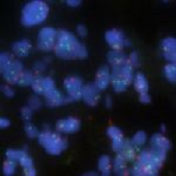
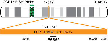
INTENDED USE: The HER2 FISH probe is designed to detect the number of copies of the gen ERBB2 (Her2/neu) in the 17q12 chromosome region. The probe CEN17 is designed to detect an alpha-satellite sequence of DNA located in the 17p11.1 region and allows identifying and counting the number of copies of chromosome 17.PROBE DESCRIPTION: The Her2 FISH Probe labeled with the CytoOrange fluorochrome covers a chromosomal region which includes the entire ERBB2 gene. The CCP17 FISH Probe labeled with the CytoGreen fluorochrome is covering the alpha satellite region of the centromere of the chromosome 17 and is designed as a control to determine the number of copies per cell of the chromosome 17.

-
پروبهای فیش
پروب فیش ALK Break Apart FISH Probe
Rated 0 out of 5INTRODUCTION:Rearrangements of the ALK gene (also known as CD246 or NBLST3), initially discovered in anaplastic large cell lymphoma, have also been found in many types of malignant tumors, including B and T-cell lymphomas, plasmacytomas, neuroblastomas, esophageal, breast, kidney, colon, thyroid and lungcarcinomas, among others.A significant percentage of cases of non-small cell lung cancer (3-13%) show abnormalities of the ALK gene and are likely to targeted treatments withspecific tyrosine kinases inhibitors that show significant benefits as compared to conventional chemotherapy.
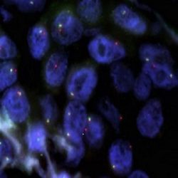
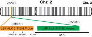
INTENDED USE: The ALK Break Apart FISH probe is designed to detect rearrangements in the ALK human gene located in the band of the chromosome 2p23.2. Aside from detecting breaks that can lead to translocation of parts of the gene, to its inversion or to its fusion with other genes, the probe can also be used to identify other aberrations of ALK such as deletions or amplifications.PROBE DESCRIPTION:The 5’ centromeric fragment of the ALK Break Apart FISH probe labeled with the fluorochrome CytoGreen covers the 5’ and central sequences of the ALK gene. The 3’ telomeric fragment of the ALK Break apart FISH probe labeled with CytoOrange covers the 3’ end and the contiguous neighboring region. Both probes are designed to detect sequences in both sides in a mutual cut-off point that is located within the ALK gene.

-
پروبهای فیش
پروب فیش ROS1 Break Apart FISH Probe
Rated 0 out of 5INTRODUCTION:
Rearrangements and abnormal expression of the ROS1 gene (also known as ROS, MCF3 or c-ros-1) have been described in approximately 1-2% of non-small cell lung carcinomas and other types of tumors. The detection of the ROS1 rearrangement in lung tumors allows applying a treatment with specific inhibitors in patients, which has shown a greater survival as compared to conventional chemotherapy.
INTENDED USE:
The ROS1 Break Apart FISH probe is designed to detect rearrangements in the ROS1 human gene located in the band of the chromosome 6q22.1. Aside from detecting breaks that can lead to translocation of parts of the gene, to its inversion or to its fusion with other genes, the probe can also be used to identify other aberrations of ROS1 such as deletions or amplifications.
PROBE DESCRIPTION:
The 5’ telomeric fragment of the ROS1 Break Apart FISH probe labeled with the fluorochrome CytoGreen covers the 5’ portion (beginning) of the ROS1 gene and some adjacent genomic sequences. The 3’ centromeric fragment of the ROS1 Break apart FISH probe labeled with CytoOrange covers the 3’ part (end) and the adjacent and inferior sequences of the gene.Both probes are flanking sequences in the ROS1 gene, in which variable cut-off points have been observed.

-
پروبهای فیش
پروب فیش CCND1 Break Apart FISH Probe
Rated 0 out of 5INTRODUCTION: Rearrangements and abnormal expression of the CCND1 gene (also known as BCL1, PRAD1, U21B31 or D11S287E) have been described in several types of hematological malignancies such as follicular mantle lymphoma [t(11;14) CCND1/IGH] or some cases of diffuse large B-cell lymphoma. Although up to 15-20% of the malignant myelomas usually show Cyclin D nuclear expression, these cases do not have CCND1 gene translocation. On the other hand, there are cases of cyclin D1-negative mantle cell lymphoma and only the detection of the CCND1 gene translocation after a hybridization study can establish a diagnosis.
INTENDED USE: The CCND1 Break Apart FISH probe is designed to detect rearrangements in the CCND1 human gene located in the band of the chromosome 11q13.3. Aside from detecting breaks that can lead to translocation of parts of the gene, to its inversion or to its fusion with other genes, the probe can also be used to identify other aberrations of CCND1 such as deletions or amplifications.
PROBE DESCRIPTION: The 3’ telomeric fragment of the CCMD1 Break Apart FISH probe labeled with the fluorochrome CytoOrange covers some genomic sequences adjacent to the 3’ portion (beginning) of the CCND1. The 5’ centromeric fragment of the CCND1 Break apart FISH probe labeled with CytoGreen covers the sequences adjacent to the gene’s 3’ end. Both probes are flanking sequences of the CCND1 gene, in which variable cut-off points have been observed.

-
پروبهای فیش
پروب فیش BCL2 Break Apart FISH Probe
Rated 0 out of 5INTRODUCTION:Rearrangements and abnormal expression of the BCL2 gene (also known as Bcl-2 or PPP1R50), are usually located in the follicular lymphoma, but they are also present in many other types of hematological and solid neoplasias. A particularly usual rearrangement is a reciprocal translocation of the IGH locus of the chromosome 14, present in up to 90% of follicular lymphomas and in up to 30% of diffuse large B-cell lymphomas.
INTENDED USE:The BCL2 Break Apart FISH probe is designed to detect rearrangements in the BCL2 human gene located in the chromosome band 18q21.33. Aside from detecting breaks (break-apart/split) that can lead to translocation of parts of the gene, to its inversion or to its fusion with other genes, the probe can also be used to identify other aberrations of BCL2 such as deletions or amplifications.
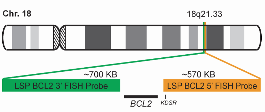
PROBE DESCRIPTION:The 5’ telomeric fragment of the BCL2 Break Apart FISH probe labeled with the fluorochrome CytoOrange covers sequences adjacent to the 5’ portion (beginning) of the BCL2. The 3’ centromeric fragment of the BCL2 Break apart FISH probe labeled with CytoGreen covers some distal sequences (3’ end) of the gene. Both marked loci are flanking sequences of the BCL2 gene, in which numerous cut-off points have been observed.

-
پروبهای فیش
پروب فیش BCL6 Break Apart FISH Probe
Rated 0 out of 5INTRODUCTION: Rearrangements of the BCL6 gene (also known as BCL5, LAZ3, BCL6A, ZNF51 or ZBTB27) have been described in lymphomas and B-cell leukemias. BCL6 is also unregulated in cases of multiple myeloma and several types of solid tumors. More than 30 different genes have been described as translocation pairs. In practice, the presence of the BCL6 translocation may have diagnostic value in follicular lymphomas grade 3B with negative BCL2 translocation, while in diffuse large B-cell lymphomas, along with BCL2 and MYC translocations, it presents a prognostic and therapeutic value.
INTENDED USE: The BCL6 Break Apart FISH probe is designed to detect rearrangements in the BCL6 human gene located in the chromosome band 3q27.3. Aside from detecting breaks that can lead to translocation of parts of the gene, to its inversion or to its fusion with other genes, the whole probe can also be used to identify other aberrations of BCL6, such as deletions or amplifications.
PROBE DESCRIPTION: The 5’ telomeric fragment of the BCL6 Break Apart FISH probe labeled with the fluorochrome CytoOrange covers some sequences adjacent to the 5’ portion (beginning) of the BCL6. The 3’ centromeric fragment of the BCL6 Break apart FISH probe labeled with CytoGreen covers some distal sequences (3’ end) of the gene. Both loci are flanking sequences of the BCL6 gene, in which numerous cut-off points have been observed.

-
پروبهای فیش
پروب فیش IGH Break Apart FISH Probe
Rated 0 out of 5INTRODUCTION: Rearrangements involving the IGH locus (or immunoglobulin heavy locus) have been observed in many types of leukemias and lymphomas. It represents the prototype for translocations in B-cell non-Hodgkin lymphomas, in which it has been detected in up to 50% of the cases, whereas reciprocal and specific translocations with different chromosomal loci have been characterized and considered as pathognomonic for different histological subtypes.
INTENDED USE: The IGH Break Apart FISH probe is designed to detect rearrangements in the human IGH locus located in the chromosome band 14q32.33. Aside from detecting breaks that can lead to translocation of parts of the locus, to its inversion or to its fusion with other genes, the probe can also be used to identify other aberrations of IGH such as deletions or amplifications.
PROBE DESCRIPTION: The telomeric 5’ Fragment of the IGH Break Apart FISH probe labeled with the fluorochrome CytoOrange covers the 5’ portion and the center of the locus of the IGH gene, whereas the centromeric 3’ fragment of the IGH Break Apart FISH probe labeled with CytorGreen covers some distal sequences (3’ end) of the gene.Both loci are flanking sequences of the BCL2 gene, in which numerous cut-off points have been observed.

-
پروبهای فیش
پروب فیش MYC Break Apart FISH Probe
Rated 0 out of 5INTRODUCTION: Rearrangements and abnormal expression of the MYC gene (also known as MRTL, MYCC, c-Myc or bHLHe39) has been described in Burkitt lymphoma, in which its positivity has diagnostic utility in a clinical, morphological and immunohistochemical context. On the other hand, diffuse large B-cell or non-classifiable lymphoma, the MYC translocation shows a prognostic value and dictates a more aggressive treatment of the neoplasia. On the other hand, along with the determination of the BCL2 and BCL6 genes translocation, it has the purpose of establishing the double or triple-hit character of the neoplasia. Other hematological neoplasias, myelomas, as well as breast, cervical, colon or ovarian tumors, among others, can show MYC translocation.
INTENDED USE: The MYC Break Apart FISH probe is designed to detect rearrangements in the MYC human gene located in the chromosome band 8q24.21. Aside from detecting breaks that can lead to translocation of parts of the gene, to its inversion or to its fusion with other genes, the probe can also be used to identify other aberrations of MYC such as deletions or amplifications.
PROBE DESCRIPTION: The 5’ centromeric fragment of the MYC Break Apart FISH probe labeled with the fluorochrome CytoOrange covers genomic sequences adjacent to the 5’ portion (beginning) of the MYC gene. The 3’ telomeric fragment of the MYC Break apart FISH probe labeled with CytoGreen covers some distal sequences (3’ end) of the gene.Both loci are flanking sequences of the MYC gene, in which variable cut-off points have been observed.

-
پروبهای فیش
پروب فیش MET/CEN7 FISH Probe
Rated 0 out of 5INTRODUCTION: The rearrangements and the abnormal expression of the MET gene (also known as HGFR, AUTS9, RCCP2 or c-Met) have been described in hereditary and sporadic renal tumors and other types of tumors. The amplification of the MET gene has been detected in lung, breast, gastrointestinal and prostate carcinomas as well as in gliomas and melanomas, among others. In these tumors, the amplification of the gene is a negative prognostic factor and, in some cases, it is associated with resistance to the treatment.
INTENDED USE: The MET/CEN7 FISH probe is designed to detect amplifications of the MET human gene located in the band of the chromosome 7q31.2, in relation to the centromere of the Chromosome 7.
PROBE DESCRIPTION: The MET FISH probe labeled with the CytoOrange fluorochrome covers a chromosome region that includes the whole MET gene. The CEN7 FISH probe labeled with the CytoGreen fluorochrome is derived from the alpha satellite region of the centromere of the chromosome 7 and is designed as a control to determine the number of copies of the chromosome 7 per cell.

-
پروبهای فیش
پروب فیش MDM2/CEN12 FISH Probe
Rated 0 out of 5INTRODUCTION: An abnormal expression of the MDM2 gene (also known as MDM2, hdm2 or ACTFS) has been described, generally associated by amplification of the same with a 7% of the human neoplastic processes. The neoplasms that more frequently show MDM2 gene amplification are atypical lipomatous tumors/well-differentiated liposarcomas, undifferentiated liposarcomas, intimal sarcomas and low-grade osteosarcomas (parosteal/intramedullary).
INTENDED USE: The MDM2/CEN12 FISH probe is designed to analyze the MDM2 human gene located in the band of the chromosome 12q15, in relation to the centromere of the Chromosome 12.
PROBE DESCRIPTION: The MDM2 FISH Probe labeled with the CytoOrange fluorochrome covers a chromosome region that includes the whole MDM2 gene. The CCP12 FISH Probe labeled with the CytoGreen fluorochrome is derived from the alpha satellite region of the centromere of the chromosome 12 and is designed as a control to determine the number of copies per cell of the chromosome 12.

-
پروبهای فیش
پروب فیش MALT1 Break Apart FISH Probe
Rated 0 out of 5INTRODUCTION: Rearrangements and abnormal expression of the MALT1 gene (also known as MLT, MLT1 or IMD12), in B-cell lymphomas and other malignant diseases have been described. There are two types of translocations that involve the MALT1 gene described in lymphomas: t(11;18)(q21;q21.32) that involves BIRC3/MALT1 (the most frequent) and t(14;18)(q32;q21.32) of the IGH/MALT1 genes. Up to 33% of the mucosa-associated lymphoid tissue B-cell lymphoma (MALT-type B-cell lymphoma) present translocation, whereas the vast majority of primary MALT lymphomas in lymph node or spleen are negative.
INTENDED USE: The MALT1 Break Apart FISH probe is designed to detect rearrangements in the MALT1 human gene located in the band of the chromosome 18q21.32. Aside from detecting breaks that can lead to translocation of parts of the gene, to its inversion or to its fusion with other genes, the probe can also be used to identify other aberrations of MALT1 such as deletions or amplifications.
PROBE DESCRIPTION: The 5’ centromeric fragment of the MALT1 Break Apart FISH probe labeled with the fluorochrome CytoOrange covers the 5’ portion (beginning) of the MALT1 gene and some adjacent genomic sequences. The 3’ telomeric fragment of the MALT1 Break apart FISH probe labeled with CytoGreen covers the 3’ part (end) and the adjacent and inferior sequences of the gene. Both probes are flanking sequences of the MALT1 gene, in which variable cut-off points have been observed.

-
پروبهای فیش
پروب فیش FUS Break Apart FISH Probe
Rated 0 out of 5INTRODUCTION: Rearrangements and abnormal expression of the FUS gene (also known as TLS, ALS6, ETM4, FUS1, POMP75 or HNRNPP2) have been described in the alveolar rhabdomyosarcoma, prostate carcinoma and other types of tumors such as myxoid liposarcoma, low-grade fibromyxoid sarcoma or some cases of Ewing’s sarcoma.
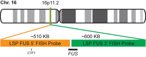
INTENDED USE: The FUS Break Apart FISH probe is designed to detect rearrangements in the FUS human gene located in the band of the chromosome 16p11.2. Aside from detecting breaks that can lead to the translocation of parts of the gene, to its inversion or to its fusion with other genes, the probe can also be used to identify other aberrations of FUS such as deletions or amplifications.
PROBE DESCRIPTION: The 5’ telomeric fragment of the FUS Break Apart FISH probe labeled with the fluorochrome CytoOrange covers the 5’ portion (beginning) of the FUS gene and some adjacent genomic sequences. The 3’ centromeric fragment of the FUS Break apart FISH probe labeled with CytoGreen covers the center and the 3’ part (end) as well as the distal sequences of the gene. Both probes are flanking sequences in the FUS gene, in which variable cut-off points have been observed.

-
پروبهای فیش
پروب فیش EWSR1 Break Apart FISH Probe
Rated 0 out of 5INTRODUCTION: The rearrangements and abnormal expression of the EWSR1 gene (also known as EWS or bK984G1.4) have been described in numerous soft-tissue lesions. Depending on the other gene involved in the reciprocal translocation, the break of the EWSR1 gene can be involved in the pathogenesis of the desmoplastic small-round-cell tumor, Ewing’s sarcoma/PNET, myxoid chondrosarcoma, soft-tissue myoepithelial tumor, myxoid liposarcoma, clear-cell sarcoma (soft-tissue melanoma), pulmonary myxoid sarcoma or hyalinizing clear cell carcinoma of salivary gland, among others. Thus, the presence of the EWSR1 gene rearrangement should be indicative, and the final diagnosis should be made by corroborating morphological, immunohistochemical and sometimes radiological facts.
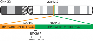
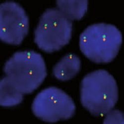
INTENDED USE: The EWSR1 Break Apart FISH probe is designed to detect rearrangements in the EWSR1 human gene located in the band of the chromosome 22q12.2. Aside from detecting breaks that can lead to translocation of parts of the gene, to its inversion or to its fusion with other genes, the probe can also be used to identify other aberrations of EWSR1 such as deletions or amplifications.
PROBE DESCRIPTION: The 5’ centromeric fragment of the EWSR1 Break Apart FISH probe labeled with the fluorochrome CytoOrange covers the 5’ portion (beginning) of the EWSR1 gene and some adjacent genomic sequences. The 3’ telomeric fragment of the EWSR1 Break apart FISH probe labeled with CytoGreen covers the 3’ part (end) as well as the adjacent and inferior sequences of the gene. Both probes are flanking sequences in the EWSR1 gene, in which variable cut-off points have been observed.
-
پروبهای فیش
پروب فیش SS18 Break Apart FISH Probe
Rated 0 out of 5INTRODUCTION: Abnormal expression of the SS18 gene (also known as SYT or SSXT) has been described in up to 80% of synovial sarcomas (regardless of their morphology) in which SYT-SSX1, SYT-SSX2, SYT-SSX4 translocations can appear. All of them are involved in their pathogenesis, but also in the prognosis of the neoplasm. The SYT-SSX1 one shows, in general, a less favorable prognosis. Rarely, translocation t(X-18) has been detected in pleomorphic sarcomas (previously named as malignant fibrous histiocytoma) or in fibrosarcomas.
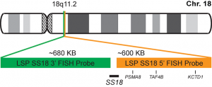
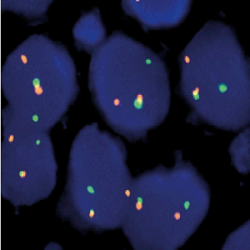
INTENDED USE: The SS18 Break Apart FISH probe is designed to detect rearrangements in the SS18 human gene located in the band of the chromosome 18q11.2. Aside from detecting breaks that can lead to translocation of parts of the gene, to its inversion or to its fusion with other genes, the probe can also be used to identify other aberrations of SS18 such as deletions or amplifications.
PROBE DESCRIPTION: The 5’ telomeric fragment of the SS18 Break Apart FISH probe labeled with the fluorochrome CytoOrange covers the 5’ portion (beginning) of the SS18 gene and some adjacent genomic sequences.The 3’ centromeric fragment of the SS18 Break apart FISH probe labeled with CytoGreen covers the 3’ part (end) and the adjacent and inferior sequences of the gene. Both probes are flanking sequences in the SS18 gene, in which variable cut-off points have been observed.

-
پروبهای فیش
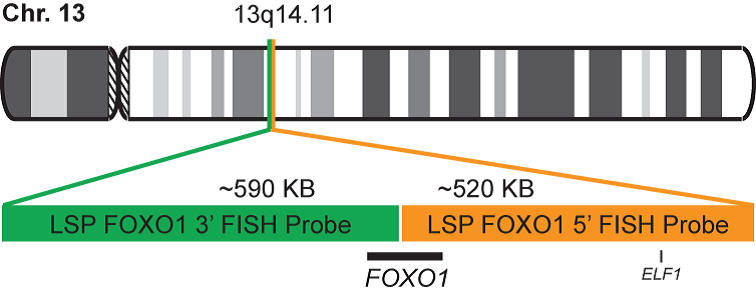
پروب فیش FOXO1 Break Apart FISH Probe
Rated 0 out of 5INTRODUCTION: Rearrangements and abnormal expression of the FOXO1 gene (also known as FKH1, FKHR or FOXO1A) have been observed in the alveolar rhabdomyosarcoma, prostate carcinoma and other types of tumors. In the case of alveolar rhabdomyosarcoma, fragments of the 3’ region of the FOXO1 gene translocate with fragments of the 5’ portions of the PAX3 and PAX7 genes, located in the chromosome 2 and 1 respectively. Those cases showing the PAX3-FOXO1 fusion in metastases present a worse prognosis, while in primary tumors, the development of the disease is independent of the type of translocation. In the prostate cancer, deletions of the FOXO1 gene have been detected in up to 30% of the cases.
INTENDED USE: The FOXO1 Break Apart FISH probe is designed to detect rearrangements in the FOXO1 human gene located in the band of the chromosome 13q14.11. Aside from detecting breaks that can lead to translocation of parts of the gene, to its inversion or to its fusion with other genes, the probe can also be used to identify other aberrations of the FOXO1 gene such as deletions or amplifications.
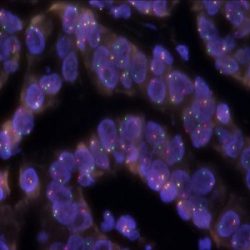
PROBE DESCRIPTION: The 5’ telomeric fragment of the FOXO1 Break Apart FISH probe labeled with the fluorochrome CytoOrange covers the center and the 5’ portion (beginning) of the FOXO1 gene and some adjacent genomic sequences. The 3’ centromeric fragment of the FOXO1 Break apart FISH probe labeled with CytoGreen covers the 3’ part (end) of the FOXO1 gene as well as the adjacent and inferior sequences of the gene. Both probes are flanking sequences of the FOXO1 gene, in which variable cut-off points have been observed.
-
پروبهای فیش
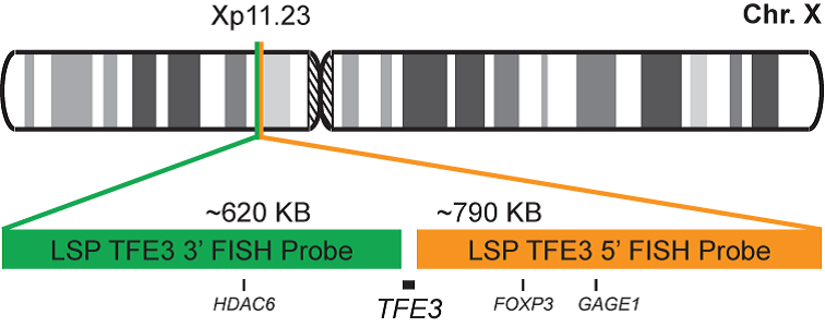
پروب فیش TFE3 Break Apart FISH Probe
Rated 0 out of 5INTRODUCTION: Rearrangements and abnormal expression of the TFE3 gene (also known as TFEA, RCCP2, RCCX1 or bHLHe33) have been described in a special subtype of renal cell carcinoma associated with Xp11.2 translocation, in alveolar sarcoma and other tumor types.
INTENDED USE: The TFE3 Break Apart FISH probe is designed to detect rearrangements in the TFE3 human gene located in the band of the chromosome Xp11.23. Aside from detecting breaks that can lead to translocation of parts of the gene, to its inversion or to its fusion with other genes, the probe can also be used to identify other aberrations of TFE3 such as deletions or amplifications.
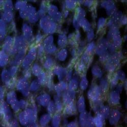
PROBE DESCRIPTION: The 5’ centromeric fragment of the TFE3 Break Apart FISH probe labeled with the fluorochrome CytoOrange covers the 5’ portion (beginning) of the TFE3 gene and some adjacent genomic sequences. The 3’ telomeric fragment of the TFE3 Break apart FISH probe labeled with CytoGreen covers the 3’ part (end) and the adjacent and inferior sequences of the gene. Both probes are flanking sequences in the TFE3 gene, in which variable cut-off points have been observed.

-
پروبهای فیش
پروب فیش DDIT3 (CHOP) Break Apart FISH Probe
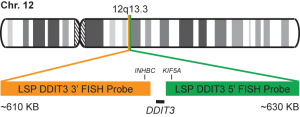
Rated 0 out of 5INTRODUCTION: Mixoid / round cell liposarcoma (MRCLS) is the most common liposarcoma. In comparison with other liposarcomas, MRCLS has a greater tendency to metastasize. Rearrangements and abnormal expression of the DDIT3 gene also known as CHOP, CEBPZ, CHOP10, CHOP-10, GADD153 in myxoid liposarcoma and other malignancies have been observed. One most common abnormality is the reciprocal translocation t (12; 16) (q13; p11) that is present in up to 95% of cases. The resulting FUS-DDIT3 fusion protein has oncogenic potential and interferes with adipogenic differentiation. EWSR1 is an alternative translocation partner of DDIT3 and the resulting fusion protein EWSR1-DDIT3, which originates at t (12; 22) (q13; q12), accounts for <5% of MRCLS cases. The DDIT3 rearrangements are highly specific for MRCLS due to the presence of the FUS-DDIT3 or EWSR1-DDIT3 fusion genes.
INTENDED USE: The DDIT3 Break Apart FISH probe is designed to detect rearrangements in the human DDIT3 gene located in the band of chromosome 12q13.3. In addition to detecting breaks that can lead to the translocation of parts of the gene, its inversion or its fusion to other genes, the probe can also be used to identify other aberrations of the DDIT3 gene such as deletions or amplifications.
PROBE DESCRIPTION: The telomeric 5 ‘fragment of the DDIT3 Break Apart FISH probe labeled with the CytoGreen fluorochrome covers some genomic sequences adjacent to the 5′ end of the DDIT3 geneThe 3’-centromeric fragment of the DDIT3 Break Apart FISH probe labeled with CytoOrange covers some genomic sequences of the 3 ‘end of the gene.The two probes are sequences that flank the DDIT3 gene region where several cut points have been observed.

-
پروبهای فیش
پروب فیش 1p36/1q25 FISH Probe
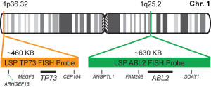
Rated 0 out of 5INTRODUCTION: Deletions and other abnormalities of the regions 1p and 19q have been observed in gliomas, astrocytomas, and other malignant diseases. The detection of the 1p/19q co-deletions represents a diagnostic criterion for oligodendrogliomas.
INTENDED USE: The FISH 1p36/1q25 probe is designed to simultaneously detect changes in the human TP73 and ABL2 genes, located in the 1p36.32 and 1q25.2 chromosome bands, respectively.
PROBE DESCRIPTION: The 1p36 FISH probe labeled with the CytoOrange fluorochrome covers a chromosome region that includes the whole TP73 gene located in the telomere region of the short arm of chromosome 1 and represents the target signal. The 1q25 FISH probe labeled with the CytoGreen fluorochrome covers a chromosome region that includes the whole ABL2 gene.

-
پروبهای فیش
پروب فیش 19q13/19p13 FISH Probe
Rated 0 out of 5INTRODUCTION: Deletions and other abnormalities of the regions 1p and 19q have been described in gliomas, astrocytomas, and other malignant diseases. The 1p/19q co-deletions represent a diagnostic criterion in the case of oligodendrogliomas.
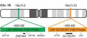
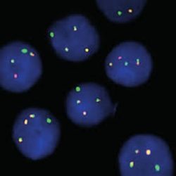
INTENDED USE: The FISH 19q13/19p13 is designed to simultaneously detect changes in the human GLTSCR1 and ZNF443 genes, located in the 19q13.33 and 19p13.2 chromosome bands, respectively.
PROBE DESCRIPTION: The 19q13 FISH probe labeled with the CytoOrange fluorochrome covers a chromosome region that includes the whole GLTSCR1 gene in the telomere region of the long arm of chromosome 19 and represents the target signal.The 19p13 FISH probe labeled with the CytoGreen fluorochrome covers a chromosome region that includes the whole ZNF443 gene.

-
پروبهای فیش
پروب فیش ATM/GLI1 FISH Probe
Rated 0 out of 5INTRODUCTION: Abnormalities in ATM – also known as AT1, ATA, ATC, ATD, ATE, ATDC, TEL1 or TELO1 – occur in ataxia telangiectasia, and have also been observed in chronic lymphocytic leukemia (CLL) and other malignancies. The GLI1 gene – also called GLI – is an oncogene that is expressed and sometimes amplified in several human cancers.
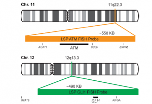
INTENDED USE: The ATM/GLI1 FISH Probe Kit is designed to detect rearrangements involving the human ATM and GLI1 genes located on chromosome bands 11q22.3 and 12q13.3, respectively.
PROBE DESCRIPTION:The ATM Probe of the ATM/GLI FISH Probe kit labeled with the fluorochrome CytoOrange covers a chromosomal region which includes the entire ATM gene. The GLI Probe of the ATM/GLI FISH Probe Kit labeled with CytoGreen covers a chromosomal region which includes the entire GLI1 gene. Both probes are designed to measure the copy number of the human ATM and GLI1 gene located on chromosome band 11q22.3 and 12q13.3, respectively.


