برای تماس کلیک کنید

به وب سایت سما تشخیص آریا خوش آمدید.

جستجوی پیشرفته
جستجوی عینی کلمه
جستجو در محتوا
جستجو در عنوان
دنبال محصول یا مطلبی میگردید؟ در این کادر جستجو کنید

Name: Antibody PU-1

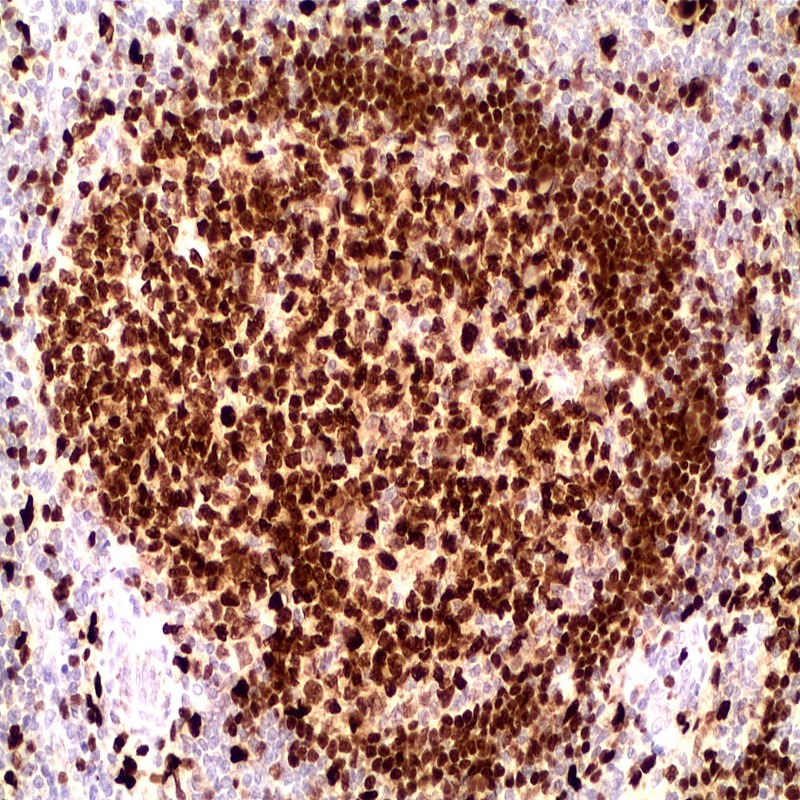
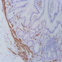
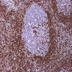
Description and applications: This antibody recognises a protein with a molecular mass of 42 kDa corresponding to PU.1 transcription factor, a member of the Ets family of transcription factors. It is required for the normal development and maturation of multiple hematopoietic lineages. PU.1 is a transcription factor restricted to the hematopoietic system, expressed in the myeloid series and in B cells but absent in mature T cells, the erythroid series, and in non-hematopoietic cells. Among the cells of the myeloid series, PU.1 is overexpressed in monocytes, histiocytes, and dendritic cells. Nodular lymphocyte-predominant Hodgkin lymphoma is consistently positive for PU.1, as opposed to classical Hodgkin lymphoma; therefore, PU.1, along with Pax-5, Oct-1, Oct-2, and BOB.1, is useful in establishing the differential diagnosis between both entities. PU.1 transcription factor and AML1, a DNAbinding subunit of the CBF transcription factor family, act as tumour suppressors in leukaemias. The function of PU.1 is down-regulated by AML1-ETO in myeloid leukaemias, while overexpression of PU.1 restores normal differentiation. High expression of PU.1 in follicular lymphomas gives them greater survival. PU.1 is a suitable marker to identify cutaneous neoplasms originating from histiocytes or from dendritic cells (reticulohistiocytomas, Langerhans cell histiocytosis, or juvenile xanthogranulomas).Dermatofibrosarcoma protuberans, dermatofibromas, fibrous papules, Spitz nevi, and melanomas are not stained with anti-PU.1.

Composition:Anti-human PU-1 rabbit monoclonal antibody purified from serum and prepared in 10mM PBS, pH 7.4, with 0.2% BSA and 0.09% sodium azide

Intended use : Immunohistochemistry (IHC) on paraffin embedded tissues. Not tested on frozen tissues or Western-Blotting

حجم موجود: 3 / 7 / 12 میلی‌لیتر

مورد استفاده: ایمونوهیستوشیمی روی بافت پارافین. بر روی بافتهای فروزن یا وسترن بلات تست نشده است.

کلون: EP18

ایزوتایپ: IgG

کنترل مثبت IHC: بافت لوزه ای

قابلیت دیده شدن: هسته ای

روش پیشنهادی IHC:

  • برش تهیه شده با ضخامت μm4 باید روی لام شارژ شده در طول شب و در دمای 60 درجه سانتی گراد خشک شود.
  • دپارافینه کردن، رهیدراته کردن و استفاده از حرارت جهت بازیابی اپی توپ : بافت را با استفاده از بافر EDTA pH 8 برای بیست دقیقه در دمای 95 درجه سانتی گراد بجوشانید. سپس 5-3 بار با استفاده از آب مقطر یا دیونیزه شده شسته و سپس بیست دقیقه در دمای اتاق خنک کنید.
  • بلاک پروکسیدازهای درونی : بلاک با استفاده از محلول پروکسیداز به مدت ده دقیقه
  • آنتی بادی اولیه: برای 10 دقیقه در انکوبه کنید پروتکل ممکن است بسته به تهیه نمونه و کاربرد خاص متفاوت باشد. شرایط استاندارد باتوجه به آزمایشگاه و کاربر متفاوت می باشد.
  • برای رنگ آمیزی ، می توانید از کیت Master Polymer Plus Detection یا کیتهای دیتکشن سایر کمپانی ها استفاده کنید.
  • رنگ میزی نهایی با هماتوکسیلین و فیکس نهایی اسلاید.

شرایط نگهداری و پایداری : حداکثر 18 ماه؛ در دمای 2-8 درجه سانتیگراد نگهداری شود. فریز نشود.


این آنتی‌بادی متعلق به پنل‌‌‌‌‌‌‌‌‌ تشخیصی LYMPHOID است.

دانلود کاتالوگ

Icon

PU-1 (EP18) 95.64 KB 4 downloads

...
.well.c2a3 .btn { padding: 11px 30px; font-size: 11pt; } .well.c2a3 .media-body { font-size: 11pt; } .well.c2a3 .wpdm_icon { height: 42px; width: auto; }

نقد و بررسی‌ها

هنوز بررسی‌ای ثبت نشده است.

اولین کسی باشید که دیدگاهی می نویسد “آنتی‌ بادی PU-1 (EP18)”

نشانی ایمیل شما منتشر نخواهد شد. بخش‌های موردنیاز علامت‌گذاری شده‌اند *

اسکرول به بالا