Category: IHC-Antibodies
Showing 421–440 of 923 results
فیلتر ها-
PSV
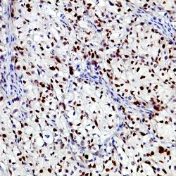
آنتی بادی Cyclin E1 کلون EP126 برند PathoSage
Rated 0 out of 5Intended use:
This antibody is intended for in vitro diagnostic (IVD) use. Primary Antibody is intended for professional laboratory use in formalin-fixed, paraffin-embedded (FFPE) tissue stained in manual qualitative immunohistochemistry (IHC) testing. A qualified pathologist must interpret the results using this product to aid diagnosis in conjunction with the patient’s relevant clinical history, other diagnostic tests, and proper controls.Presentation:
Anti-human Cyclin E1 rabbit monoclonal antibody purified from serum and prepared in 10mM PBS, pH 7.4, with 0.2% BSA and 0.09% sodium azide -
PSV
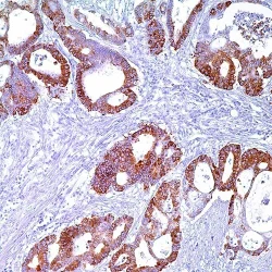
آنتی بادی COX2 کلون SP21 برند PathoSage
Rated 0 out of 5Intended use:
This antibody is intended for in vitro diagnostic (IVD) use. Primary Antibody is intended for professional laboratory use in formalin-fixed, paraffin-embedded (FFPE) tissue stained in manual qualitative immunohistochemistry (IHC) testing. A qualified pathologist must interpret the results using this product to aid diagnosis in conjunction with the patient’s relevant clinical history, other diagnostic tests, and proper controls.Presentation:
Anti-human COX2 rabbit monoclonal antibody purified from serum and prepared in 10mM PBS, pH 7.4, with 0.2% BSA and 0.09% sodium azide -
PSV
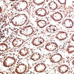
آنتی بادی Collagen 2 کلون 2B1.5 برند PathoSage
Rated 0 out of 5Intended use:
This antibody is intended for in vitro diagnostic (IVD) use. Primary Antibody is intended for professional laboratory use in formalin-fixed, paraffin-embedded (FFPE) tissue stained in manual qualitative immunohistochemistry (IHC) testing. A qualified pathologist must interpret the results using this product to aid diagnosis in conjunction with the patient’s relevant clinical history, other diagnostic tests, and proper controls.Presentation:
Anti-human Collagen II mouse monoclonal antibody purified from serum and prepared in 10mM PBS, pH 7.4, with 0.2% BSA and 0.09% sodium azide -
PSV
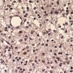
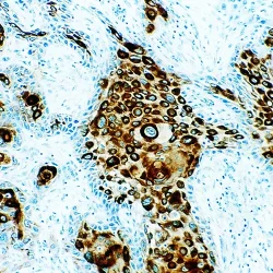
آنتی بادی C-Myc کلون Y69 برند PathoSage
Rated 0 out of 5Intended use:
This antibody is intended for in vitro diagnostic (IVD) use. Primary Antibody is intended for professional laboratory use in formalin-fixed, paraffin-embedded (FFPE) tissue stained in manual qualitative immunohistochemistry (IHC) testing. A qualified pathologist must interpret the results using this product to aid diagnosis in conjunction with the patient’s relevant clinical history, other diagnostic tests, and proper controls.Presentation:
Anti-human c-Myc rabbit monoclonal antibody purified from serum and prepared in 10mM PBS, pH 7.4, with 0.2% BSA and 0.09% sodium azide -
PSV
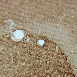
آنتی بادی CNPasa کلون 11-5B برند PathoSage
Rated 0 out of 5Intended use:
This antibody is intended for in vitro diagnostic (IVD) use. Primary Antibody is intended for professional laboratory use in formalin-fixed, paraffin-embedded (FFPE) tissue stained in manual qualitative immunohistochemistry (IHC) testing. A qualified pathologist must interpret the results using this product to aid diagnosis in conjunction with the patient’s relevant clinical history, other diagnostic tests, and proper controls.Presentation:
Anti-human CNPase mouse monoclonal antibody purified from serum and prepared in 10mM PBS, pH 7.4, with 0.2% BSA and 0.09% sodium azide -
PSV
آنتی بادی Cytokeratin 5/6/8/18 کلون EP24/EP67/B22.1&B23.1 برند PathoSage
Rated 0 out of 5Intended use:
This antibody is intended for in vitro diagnostic (IVD) use. Primary Antibody is intended for professional laboratory use in formalin-fixed, paraffin-embedded (FFPE) tissue stained in manual qualitative immunohistochemistry (IHC) testing. A qualified pathologist must interpret the results using this product to aid diagnosis in conjunction with the patient’s relevant clinical history, other diagnostic tests, and proper controls.Presentation:
Anti-human Cytokeratin 5/6/8/18 rabbit/mouse monoclonal antibody purified from serum and prepared in 10mM PBS, pH 7.4, with 0.2% BSA and 0.09% sodium azide -
PSV
آنتی بادی Cytokeratin کلون AE3 برند PathoSage
Rated 0 out of 5Intended use:
This antibody is intended for in vitro diagnostic (IVD) use. Primary Antibody is intended for professional laboratory use in formalin-fixed, paraffin-embedded (FFPE) tissue stained in manual qualitative immunohistochemistry (IHC) testing. A qualified pathologist must interpret the results using this product to aid diagnosis in conjunction with the patient’s relevant clinical history, other diagnostic tests, and proper controls.Presentation:
Anti-human Cytokeratin mouse monoclonal antibody purified from serum and prepared in 10mM PBS, pH 7.4, with 0.2% BSA and 0.09% sodium azide -
PSV
آنتی بادی Cytokeratin 5 کلون SP27 برند PathoSage
Rated 0 out of 5Intended use:
This antibody is intended for in vitro diagnostic (IVD) use. Primary Antibody is intended for professional laboratory use in formalin-fixed, paraffin-embedded (FFPE) tissue stained in manual qualitative immunohistochemistry (IHC) testing. A qualified pathologist must interpret the results using this product to aid diagnosis in conjunction with the patient’s relevant clinical history, other diagnostic tests, and proper controls.Presentation:
Anti-human Cytokeratin 5 rabbit monoclonal antibody purified from serum and prepared in 10mM PBS, pH 7.4, with 0.2% BSA and 0.09% sodium azide -
PSV
آنتی بادی Cytokeratin 17 کلون EP98 برند PathoSage
Rated 0 out of 5Intended use:
This antibody is intended for in vitro diagnostic (IVD) use. Primary Antibody is intended for professional laboratory use in formalin-fixed, paraffin-embedded (FFPE) tissue stained in manual qualitative immunohistochemistry (IHC) testing. A qualified pathologist must interpret the results using this product to aid diagnosis in conjunction with the patient’s relevant clinical history, other diagnostic tests, and proper controls.Presentation:
Anti-human Cytokeratin 17 rabbit monoclonal antibody purified from serum and prepared in 10mM PBS, pH 7.4, with 0.2% BSA and 0.09% sodium azide -
PSV
آنتی بادی Cytokeratin کلون CAM 5.2 برند PathoSage
Rated 0 out of 5Intended use:
This antibody is intended for in vitro diagnostic (IVD) use. Primary Antibody is intended for professional laboratory use in formalin-fixed, paraffin-embedded (FFPE) tissue stained in manual qualitative immunohistochemistry (IHC) testing. A qualified pathologist must interpret the results using this product to aid diagnosis in conjunction with the patient’s relevant clinical history, other diagnostic tests, and proper controls.Presentation:
Anti-human Keratin CAM5.2 mouse antibody purified from serum and prepared in 10mM PBS, pH 7.4, with 0.2% BSA and 0.09% sodium azide -
PSV
آنتی بادی Cytokeratin 13 کلون EP69 برند PathoSage
Rated 0 out of 5Intended use:
This antibody is intended for in vitro diagnostic (IVD) use. Primary Antibody is intended for professional laboratory use in formalin-fixed, paraffin-embedded (FFPE) tissue stained in manual qualitative immunohistochemistry (IHC) testing. A qualified pathologist must interpret the results using this product to aid diagnosis in conjunction with the patient’s relevant clinical history, other diagnostic tests, and proper controls.Presentation:
Anti-human Keratin 13 rabbit monoclonal antibody purified from serum and prepared in 10mM PBS, pH 7.4, with 0.2% BSA and 0.09% sodium azide -
PSV
آنتی بادی Cytokeratin 5/6 کلون EP24/EP67 برند PathoSage
Rated 0 out of 5Intended use:
This antibody is intended for in vitro diagnostic (IVD) use. Primary Antibody is intended for professional laboratory use in formalin-fixed, paraffin-embedded (FFPE) tissue stained in manual qualitative immunohistochemistry (IHC) testing. A qualified pathologist must interpret the results using this product to aid diagnosis in conjunction with the patient’s relevant clinical history, other diagnostic tests, and proper controls.Presentation:
Anti-human Cytokeratin 5/6 rabbit monoclonal antibody purified from serum and prepared in 10mM PBS, pH 7.4, with 0.2% BSA and 0.09% sodium azide -
PSV
آنتی بادی Cytokeratin 10 کلون SP99 برند PathoSage
Rated 0 out of 5Intended use:
This antibody is intended for in vitro diagnostic (IVD) use. Primary Antibody is intended for professional laboratory use in formalin-fixed, paraffin-embedded (FFPE) tissue stained in manual qualitative immunohistochemistry (IHC) testing. A qualified pathologist must interpret the results using this product to aid diagnosis in conjunction with the patient’s relevant clinical history, other diagnostic tests, and proper controls.Presentation:
Anti-human Cytokeratin 5 rabbit monoclonal antibody purified from serum and prepared in 10mM PBS, pH 7.4, with 0.2% BSA and 0.09% sodium azide -
PSV
آنتی بادی Cytokeratin 8/18/19 کلون B22.1&B23.1/BA17 برند PathoSage
Rated 0 out of 5Intended use:
This antibody is intended for in vitro diagnostic (IVD) use. Primary Antibody is intended for professional laboratory use in formalin-fixed, paraffin-embedded (FFPE) tissue stained in manual qualitative immunohistochemistry (IHC) testing. A qualified pathologist must interpret the results using this product to aid diagnosis in conjunction with the patient’s relevant clinical history, other diagnostic tests, and proper controls.Presentation:
Anti-human Cytokeratin 8/18/19 Mouse monoclonal antibody prepared in 10mM PBS, pH 7.4, with 0.2% BSA and 0.09% sodium azide -
PSV
آنتی بادی DOG1(Anoctamin 1) کلون SP31 برند PathoSage
Rated 0 out of 5Intended use:
This antibody is intended for in vitro diagnostic (IVD) use. Primary Antibody is intended for professional laboratory use in formalin-fixed, paraffin-embedded (FFPE) tissue stained in manual qualitative immunohistochemistry (IHC) testing. A qualified pathologist must interpret the results using this product to aid diagnosis in conjunction with the patient’s relevant clinical history, other diagnostic tests, and proper controls.Presentation:
Anti-human DOG-1 rabbit monoclonal antibody purified from serum and prepared in 10mM PBS, pH 7.4, with 0.2% BSA and 0.09% sodium azide -
PSV
آنتی بادی Digoxigenin کلون HY-A.1 برند PathoSage
Rated 0 out of 5Intended use:
This antibody is intended for in vitro diagnostic (IVD) use. Primary Antibody is intended for professional laboratory use in formalin-fixed, paraffin-embedded (FFPE) tissue stained in manual qualitative immunohistochemistry (IHC) testing. A qualified pathologist must interpret the results using this product to aid diagnosis in conjunction with the patient’s relevant clinical history, other diagnostic tests, and proper controls.Presentation:
Anti-human Digoxigenin mouse monoclonal antibody purified from serum and prepared in 10mM PBS, pH 7.4, with 0.2% BSA and 0.09% sodium azide -
PSV
آنتی بادی Cytokeratin 8 کلون EP17 برند PathoSage
Rated 0 out of 5Intended use:
This antibody is intended for in vitro diagnostic (IVD) use. Primary Antibody is intended for professional laboratory use in formalin-fixed, paraffin-embedded (FFPE) tissue stained in manual qualitative immunohistochemistry (IHC) testing. A qualified pathologist must interpret the results using this product to aid diagnosis in conjunction with the patient’s relevant clinical history, other diagnostic tests, and proper controls.Presentation:
Anti-human Cytokeratin 8 rabbit monoclonal antibody purified from serum and prepared in 10mM PBS, pH 7.4, with 0.2% BSA and 0.09% sodium azide -
PSV
آنتی بادی ER(Estrogen Receptor) کلون SP1 برند PathoSage
Rated 0 out of 5Intended use:
This antibody is intended for in vitro diagnostic (IVD) use. Primary Antibody is intended for professional laboratory use in formalin-fixed, paraffin-embedded (FFPE) tissue stained in manual qualitative immunohistochemistry (IHC) testing. A qualified pathologist must interpret the results using this product to aid diagnosis in conjunction with the patient’s relevant clinical history, other diagnostic tests, and proper controls.Presentation:
Anti-human ER rabbit monoclonal antibody purified from serum and prepared in 10mM PBS, pH 7.4, with 0.2% BSA and 0.09% sodium azide -
PSV
آنتی بادی EGFR(Epidermal Growth Factor Receptor) کلون EP22 برند PathoSage
Rated 0 out of 5Intended use:
This antibody is intended for in vitro diagnostic (IVD) use. Primary Antibody is intended for professional laboratory use in formalin-fixed, paraffin-embedded (FFPE) tissue stained in manual qualitative immunohistochemistry (IHC) testing. A qualified pathologist must interpret the results using this product to aid diagnosis in conjunction with the patient’s relevant clinical history, other diagnostic tests, and proper controls.Presentation:
Anti-human EGFR rabbit monoclonal antibody purified from serum and prepared in 10mM PBS, pH 7.4, with 0.2% BSA and 0.09% sodium azide -
PSV
آنتی بادی ERCC1 کلون SP68 برند PathoSage
Rated 0 out of 5Intended use:
This antibody is intended for in vitro diagnostic (IVD) use. Primary Antibody is intended for professional laboratory use in formalin-fixed, paraffin-embedded (FFPE) tissue stained in manual qualitative immunohistochemistry (IHC) testing. A qualified pathologist must interpret the results using this product to aid diagnosis in conjunction with the patient’s relevant clinical history, other diagnostic tests, and proper controls.Presentation:
Anti-human ERCC1 rabbit monoclonal antibody purified from serum and prepared in 10mM PBS, pH 7.4, with 0.2% BSA and 0.09% sodium azide

