-
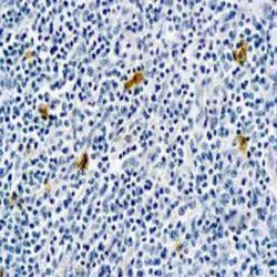
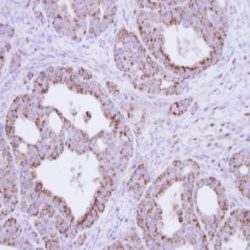 آنتی بادیهای ایمونوهیستوشیمی
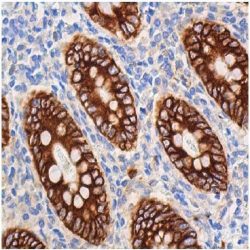
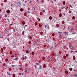
آنتی بادی Fumarate Hydratase (J-13)
Name: Mouse anti-human Fumarate Hydratase Antibody (Clon J-13)
Description and aplications: The fumarate hydratase protein (FH) is an enzyme of the Krebs cycle that catalyzes the reversible and sterospecific transformation of the fumarate to Fmalate.There exist two isoforms, the FH1 located a cytosolic level, where is responsible for the hydratio from the fumarate to L-malate, and the mitochondrial FH2, responsible for the dehydration from the Lmalate to fumarate. The secretion of the enzyme is coded by a gene located in the chromosome region 1q43. The lack of this gene determines severe metabolic disorders characterized by early hypotonia, psychomotor retardation and brain anomalies such as agenesis of the corpus callosus, anomalies in the gyri and ventriculomegaly.
Composition: Composition: anti-human Fumarate Hydratase mouse monoclonal antibody purified from serum and prepared in 10mM PBS, pH 7.4, with 0.2% BSA and 0.09% sodium azide
-
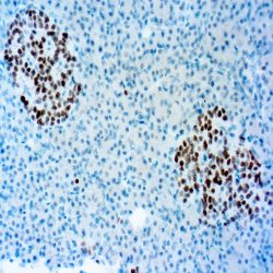
 آنتی بادیهای ایمونوهیستوشیمی
آنتی بادی HBc Antigen (CORE) (Polyclonal)
Name: Rabbit anti-human Hepatitis B Virus Core Antigen (HBVcAg) Polyclonal Antibody
Description and aplications: Hepatitis B virus is spherical in shape with a diameter of 42 nm. It contains a 27 nm partially double stranded DNA core enclosed within a lipoprotein coat. The antigenic activity of the nucleocapsid core is designated as hepatitis B core antigen. The antigens in the outer surface are called as hepatitis B virus surface antigens.
Core antigens are localized within the nuclei whereas the surface antigens are present in the cytoplasm of the infected cells. Antibodies to surface antigens appear in circulation at an early stage of infection whereas the antibodies to the core antigens are detected after several weeks. This antibody recognizes a protein in the core of hepatitis B virus.Composition: Anti-human HBVcAg rabbit polyclonal antibody purified from serum and prepared in 10mM PBS, pH
7.4, with 0.2% BSA and 0.09% sodium azide
Immunogen: Hepatitis B virus.
-
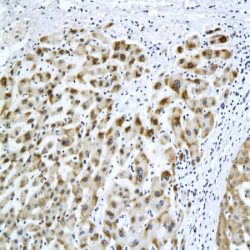
 آنتی بادیهای ایمونوهیستوشیمی
آنتی بادی HNF-1 beta (Hepatocyte Nuclear Factor 1 betaHomeobox A) (Polyclonal)
Name:HNF-1 beta (Hepatocyte Nuclear Factor 1 beta Homeobox A) (Polyclonal)
Description and aplications: Hepatocyte nuclear factors, a group of phylogenetically unrelated transcription factors
belonging to the group of hemoproteins, is made up of the molecules HNF-1 with its isoforms and , HNF-3 (, and ), HNF-4 (, and ) and HNF-6 which regulate the transcription of a wide group of genes and proteins which include among others clotting factors, and enzymes and transporters related to metabolism and glucose, cholesterol and fatty acids transport.Composition: Anti-human HNF-1 beta rabbit polyclonal antibody purified from serum and prepared in 10mM PBS, pH
7.4, with 0.2% BSA and 0.09% sodium azide
Immunogen: synthetic protein corresponding to the specific sequence of amino acids -
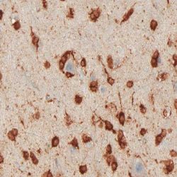
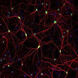 آنتی بادیهای ایمونوهیستوشیمی
آنتی بادی HuC/HuD Neuronal Protein (16A11)
Name: Mouse anti-human HuC/HuD Neuronal Protein Monoclonal Antibody (Clone 16A11)
Description and aplications: The Hu antigen is a protein of the Elav (embryonic lethal abnormal visual) family. This antibody recognises only some protein members of the Elav family (HuC, HuD, and Hel-N1), all of them neuronal proteins. It does not recognise HuR, another Elav family member that is present in all proliferating cells. This antibody can be considered as a neuron marker. This antibody identifies neuronal cells and neoplasms derived from them.
Composition:Anti-human HuC/HuD Neuronal Protein mouse monoclonal antibody purified from serum and prepared in 10mM PBS, pH 7.4, with 0.2% BSA and 0.09% sodium azide
-
 آنتی بادیهای ایمونوهیستوشیمی
آنتی بادی Human IgA (Polyclonal)
Name: Rabbit anti-human IgA Antibody (Polyclonal)
Description and aplications:Anti-IgA antibody reacts with surface immunoglobulin IgA alpha chains. It is useful when identifying leukemias, plasmacytomas, and B-cell lineage derived Hodgkin’s lymphomas. Due to the restricted expression of heavy and light chains in these diseases, demonstration of Bcell lymphoma/plasmacytoma is aided with this antibody.
Composition: Anti-human IgA rabbit polyclonal antibody purified from serum and prepared in 10mM PBS, pH 7.4, with
0.2% BSA and 0.09% sodium azide
-
 آنتی بادیهای ایمونوهیستوشیمی
آنتی بادی Human IgE (Polyclonal)
Name: Rabbit anti-human IgE Antibody (Polyclonal)
Description and aplications: This antibody recognises the human immunoglobulin epsilon heavy chain. The common feature of both plasmacytomas and certain types of non-Hodgkin lymphoma is the expression of a single class of heavy chain. The demonstration of clonality in lymphoproliferative processes indicates that the process is clonal and therefore malignant. This antibody is useful for the identification of leukaemias, plasmacytomas and some types of non-Hodgkin lymphoma. It does not cross-react with the heavy chains of immunoglobulins IgA, IgG, IgM and IgD, nor with T cells, monocytes, granulocytes or erythrocytes.
Composition:anti-human IgE rabbit polyclonal antibody purified from serum and prepared in 10mM PBS, pH 7.4, with 0.2% BSA and 0.09% sodium azide
Immunogen: IgE isolated from normal human serum.
-
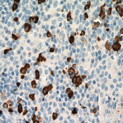
 آنتی بادیهای ایمونوهیستوشیمی
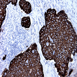
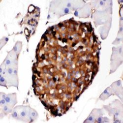
آنتی بادی INSM1 (A-8)
Name: Mouse anti-human INSM1 (Insulinoma-associated protein 1) Monoclonal Antibody (Clone A-8)
Description and aplications: INSM1 protein, also known as IA1, is a nuclear transcription factor encoded by INSM1 gene, located in the chromosome region 20p11.23 and composed of 510 amino acids with a molecular mass of approximately 52.8kDa. The protein, which shows a wide homology in different species, contains a Cterminal portion with 5 finger-binding domains and an N-terminal domain with four pro-hormone conversion sites as well as an amidation signal sequence. INSM1 factor plays an important role in neurogenesis and differentiation during the embryonic and fetal development of the pancreatic neuroendocrine, anterior pituitary, intestine cells and synapto-adrenal cells of the peripheral nervous system. INSM1 gene, which does not express in normal endocrine and non-endocrine tissues in adults, acts as a suppressor gene of the transmission pathways NEUROD1 and INS after interaction with Cyclin D1, inhibits the specific genes of the skeletal muscle in neuroendocrine cells of the pituitary and suppress neural factors such as HDAC1, HDAC2, HDAC3 and KDM1A (involved in the incorporation of chromatin-modifying factors). In addition to this, it suppresses the activity of the histone deacetylase RCOR1.
Composition: Anti-human INSM1 mouse monoclonal antibody purified from serum and prepared in 10mM PBS, pH
7.4, with 0.2% BSA and 0.09% sodium azide -
 آنتی بادیهای ایمونوهیستوشیمی
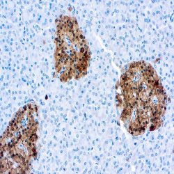
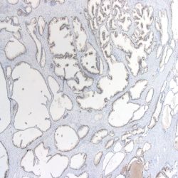
آنتی بادی Isocitrate Dehydrogenase 1 R132H point mutated form (IDH1 R132H) (H09)
Name: Mouse Anti-Human Isocitrate Dehydrogenase 1 R132H point mutated form (IDH1 R132H) (H09)
Description and aplications: Antibody clone H09 reacts specifically with the isocitrate dehydrogenase 1 (IDH1) R132H point mutated form in tissue sections from formalin-fixed brain tumor specimens. Heterozygous point mutations of IDH1 codon 132 are frequent in World Health Organization (WHO) grade II and III gliomas. IDH1 R132H mutations occur in approximately 70% of astrocytomas and oligodendroglial tumors. The high frequency and distribution of the IDH1 R132H mutation among specific brain tumor entities allow the highly sensitive and specific discrimination of various tumors by
immunohistochemistry, such as anaplastic astrocytoma from primary glioblastoma or diffuse astrocytoma WHO grade II from pilocytic astrocytoma or ependymoma. Noteworthy is the discrimination of the infiltrating edge of tumors with IDH1 mutation from reactive gliosis. This antibody is highly useful for tumor classification and in detecting single infiltrating tumor cells.
Composition:anti-Isocitrate Dehydrogenase 1 R132H (IDH1 R132H) mouse monoclonal antibody obtained from supernatant culture and prediluted in a tris buffered solution pH 7.4 containing 0.375mM sodium azide solution as bacteriostatic and bactericidal. The quantity of the active antibody was not determined.
Immunogen: Synthetic peptide, amino acid sequence CKPIIIGHHAYGD
-
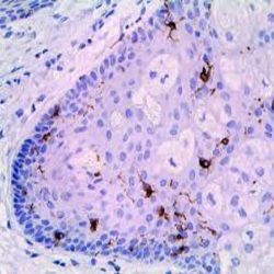 آنتی بادیهای ایمونوهیستوشیمی
آنتی بادی Langerin/CD207 (EPR15863)
Name: Rabbit anti-human Langerin/CD207 Monoclonal Antibody (Clone EPR15863)
Description and aplications: Calciumdependent lectin displaying mannose-binding specificity. Induces the formation of Birbeck granules (BGs); is a potent regulator of membrane superimposition and zippering. Binds to sulfated as well as mannosylated glycans, keratan sulfate (KS) and beta-glucans. Facilitates uptake of antigens and is involved in the routing and/or processing of antigen for presentation to T cells. Major receptor on primary Langerhans cells for Candida species, Saccharomyces species, and Malassezia furfur. Protects against human immunodeficiency virus-1 (HIV-1) infection. Binds to high-mannose structures present on the envelope glycoprotein which is followed by subsequent targeting of the virus to the Birbeck granules leading to its rapid degradation.
Composition: Anti-human Langerin/CD207 rabbit monoclonal antibody purified from serum and prepared in 10mM
PBS, pH 7.4, with 0.2% BSA and 0.09% sodium azide
Immunogen: Recombinant fragment within Human Langerin aa 200 to the C-terminus.
-
 آنتی بادیهای ایمونوهیستوشیمی
آنتی بادی Mucin 4 (8G7)
Name: Mouse anti-human Mucin 4 (MUC4) Monoclonal Antibody (Clone 8G7)
Description and aplications: Mucins are a group of high molecular weight glycoproteins consisting of a mucin core protein and O-linked carbohydrates. Mucin 4, a membrane-bound mucin, is the human homolog of the rat sialomucin complex (SMC). Mucin 4 protein consists of Mucin 4α, a large amino mucin type subunit and Mucin 4β, a transmembrane subunit containing three EGF-like domains. The Mucin 4 gene is the predominant mucin gene expressed in the normal urothelium and is also expressed in several normal tissues such as trachea, lung and testis.
Composition: Anti-human MUC4 mouse monoclonal antibody purified from serum and prepared in 10mM PBS, pH
7.4, with 0.2% BSA and 0.09% sodium azide
Immunogen: A synthetic peptide directed against the Mucin 4 tandem repeats of human origin.
-
 آنتی بادیهای ایمونوهیستوشیمی
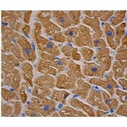
آنتی بادی Myosin – Skeletal Muscle (MY-32)
Name: Mouse anti-human Myosin – Skeletal Muscle Monoclonal Antibody (Clone MY-32)
Description and aplications: This antibody reacts against the skeletal muscle myosin heavy chain. Myosin is a 480-kDa protein present in muscle and non-muscle cells. This antibody may be used as a tool for the diagnosis of tumours of skeletal muscle origin. It may also be useful in studies of the architecture of thick filament cytoskeleton. It does not react with human or animal cardiac myosin or smooth muscle myosin. It reacts with myosin of human, rabbit, rat, murine, bovine, and guinea pig origin.
Composition: Anti-human Myosin – Skeletal Muscle mouse monoclonal antibody purified from serum and prepared in 10mM PBS, pH 7.4, with 0.2% BSA and 0.09% sodium azide
Immunogen: Rabbit myosin.
-
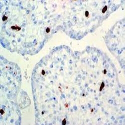
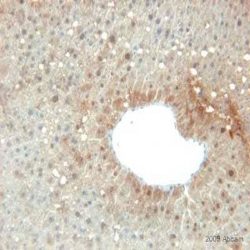
 آنتی بادیهای ایمونوهیستوشیمی
آنتی بادی Neuron Specific Enolase (NSE) (Polyclonal)
Name: Rabbit anti-human Neuron Specific Enolase (NSE) Polyclonal Antibody
Description and aplications: Enolase is a glycolytic enzyme catalyzing the reaction pathway between 2-phospho glycerate and phosphoenol pyruvate. In mammals, enolase molecules are dimmers composed of three distinct subunits (α,β and γ). The alpha-subunit is expressed in most tissues and the beta-subunit only in muscle. The gamma-subunit is expressed primarily in neurons, in normal and in neoplastic neuroendocrine cells. Co-expression of NSE and chromogranin A is common in neuroendocrine neoplasms.
Composition: Anti-human NSE rabbit polyclonal antibody purified from serum and prepared in 10mM PBS, pH 7.4, with
0.2% BSA and 0.09% sodium azide
Immunogen: Synthetic peptide derived from Cterminus of human NSE.
-
 آنتی بادیهای ایمونوهیستوشیمی
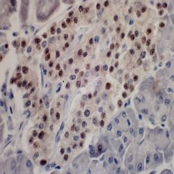
آنتی بادی NKX2.2 (EP336)
Name: Rabbit anti-NKX2.2 Monoclonal Antibody (clone EP336)
Description and aplications: The NKX2.2 antibody, also known as NK2 or NKX2B, is a nuclear transcription factor that belongs to the NK2 family of homeobox genes and is encoded in the chromosome region 20p11.22. This gene is involved in the development of beta cells of the pancreas that produce insulin, as well as in the neuroendocrine, glial, neuronal and oligodendroglial differentiation. Likewise, it is also involved in several process of diencephalic development and organization as well as in the control of the genes involved in the axonal orientation.
Composition: Rabbit anti-NKX2.2 monoclonal antibody obtained from purified ascitic fluid and prepared in 10mM PBS,
pH 7.4, with 0.2% BSA and 0.09% sodium azide.
Immunogen: synthetic peptide corresponding to the human NKX2.2 protein
-
 آنتی بادیهای ایمونوهیستوشیمی
آنتی بادی NKX3.1 (EP356)
Name: Rabbit Anti-Human NKX3.1 Monoclonal Antibody (EP356)
Description and aplications: NKX3.1 is a prostatespecific tumor suppressor protein that is encoded bythe homeobox gene NKX3.1 located in chromosomeregion 8p21 and whose expression is regulated byandrogens. The protein acts as an importanttranscription factor in the normNKX3.1 is a prostatespecifical development of theprostate since it regulates the proliferation of theglandular epithelium and the formation of excretoryducts. For this reason, the protein, which is located in the luminal cells (considered the stem of the prostate epithelium), intervenes in prostate regeneration and issusceptible to oncogenic transformation.
Composition: anti-NKX3.1 rabbit monoclonal antibody obtained from supernatant culture and prediluted in a tris buffered solution pH 7.4 containing 0.375mM sodium azide solution as bacteriostatic and bactericidal.
Immunogen: Synthetic peptide corresponding to residues in the human protein of the NKX3.1.
-
 آنتی بادیهای ایمونوهیستوشیمی
آنتی بادی Pan-Cytokeratin (BS5)
Name: Mouse anti-human Pan Cytokeratin Monoclonal Antibody (Clone BS5)
Description and aplications: Cytokeratins are structural intermediate filaments encoded in more than 49 different genes in the chromosomes 17 (I) and 12 (II). The nomenclature adopted in 1982 by Moll and Franke assigns the ranks 1 to 8 for the type II cytokeratins (neutral or basic) and between 9 and 21 for the type I (acid). Nowadays, an analogue nomenclature has been defined to name the hair keratin with the addition of the Ha and Hb letters to separate the ones of the group I from group II. Structurally speaking, cytokeratins share with the rest of intermediate filaments a central axis of 310 amino acids made up of four α-helices domains (1A, 1B, 2A and 2B) highly preserved that define the type of
intermediate filament that they will make up after its packing, separated by three non-helix binding sites (L1, L12 and L2) and two extreme domains highly different in size and sequence (head-1- and tail-2-), each of them with constant (E1/E2), variable (V1/V2) and homology regions (H1/H2), the last ones are characteristic of the type II keratins and absent of the type I. Usually, keratins assemble in heterodimers I/II and they are co-expressed in pairs specifically in each tissue.Composition:Anti-human Pan Cytokeratin mouse monoclonal antibody purified from serum and prepared in 10mM PBS, pH 7.4, with 0.2% BSA and 0.09% sodium azide
-
 آنتی بادیهای ایمونوهیستوشیمی
آنتی بادی Parvovirus B19 (R92F6)
Name: Mouse anti-Parvovirus B19 Monoclonal Antibody (Clone R92F6)
Description and aplications: Parvovirus B19 is one of the smallest viruses (20 kDa), whose nucleic acid is linear single-stranded DNA of negative polarity and without lipoprotein envelope. The viral genome encodes three proteins: the nonstructural protein NS1 and two viral capsid proteins, VP1 and VP2. The minor capsid protein VP1 has the same sequence of amino acids as VP2 plus 227 amino acids in the amino-terminus region, the VP1 unique region (VP1u). Recently, the phospholipase A2 (sPLA2) motif has been identified in VP1u in members of the Parvoviridae family, including B19, classified as an erythrovirus because of its ability to invade erythrocyte precursors in the bone marrow.
Composition: Anti-Parvovirus B19 mouse monoclonal antibody purified from serum and prepared in 10mM PBS, pH 7.4, with 0.2% BSA and 0.09% sodium azide
Immunogen: Amino acids 328-344 of the VP2 protein of the viral capsid
-
 آنتی بادیهای ایمونوهیستوشیمی
آنتی بادی PGP 9.5 (Polyclonal)
Name: Rabbit anti-human PGP 9.5 Antibody (Polyclonal)
Description and aplications: This antibody recognises a molecule with a molecular mass of 27 kDa known as “Protein Gene Product 9.5” or PGP 9.5 and encoded by the UCHL1 (ubiquitin carboxy-terminal hydrolase L1 isoenzyme) gene, located in the chromosomal region 4p13. This protein catalyses the hydrolysis of ubiquitin’s C-terminal esters and amides, playing an important role in protein degradation. Overexpression of the UCHL1 gene occurs in several neoplasms, including leukaemias, carcinomas, and several tumours of mesenchymal origin. In normal tissues, PGP 9.5, which shows mainly a cytoplasmic staining pattern, is present in neurons and nerve fibres of the central and peripheral nervous systems, as well as in numerous neuroendocrine cells, except those of the digestive tract, of which only the pancreatic islets of Langerhans show consistent positive staining. Other normal tissues that are usually stained are segments of renal tubules, spermatogonias and Leydig cells, smooth muscle, and germinal centres.
Composition: anti-human PGP 9.5 rabbit polyclonal antibody purified from serum and prepared in 10mM PBS, pH 7.4, with 0.2% BSA and 0.09% sodium azide
Immunogen: Immunogen: Recombinant protein corresponding to the total length of human protein PGP 9.5.
-
 آنتی بادیهای ایمونوهیستوشیمی
آنتی بادی Poly (ADP-Ribose) Polymerase 1 (Polyclonal)
Name: Rabbit anti-human Poly (ADP-Ribose) Polymerase 1 Antibody (Polyclonal)
Description and aplications: PARP (EC 2.4.2.30) is a nuclear protein with a molecular mass of 116 kDa and two zinc finger motifs which binds to DNA and specifically detects DNA nicks and breaks produced by different genotoxic agents. PARP catalises the ADP-ribosylation of proteins using NAD+ as a substrate. PARP activation is a consequence of ischemic injury and leads to intracellular depletion of NAD+, which can only be replaced by ATP consumption. Ischemia/Reperfusion (I/R) injury results in substantial DNA damage and cells need to consume large amounts of ATP to
sustain reparative poly-ADP-ribosylation. Proteolysis of PARP to its stable 85-kDa fragment is an early marker of programmed cell death (apoptosis), mediated by caspase 3 or CPP32. Cleavage occurs between Adp216 and Gly217, a site in PARP conserved across species. This antibody reacts with human, mouse and rat PARP.Composition:anti-human Poly (ADP-Ribose) Polymerase 1 rabbit polyclonal antibody purified from serum and prepared in 10mM PBS, pH 7.4, with 0.2% BSA and 0.09% sodium azide
Immunogen: Synthetic peptide derived from the Nterminal region of human protein PARP.
-
 آنتی بادیهای ایمونوهیستوشیمی
آنتی بادی Prealbumin (EPR3219)
Name: Rabbit anti-human Prealbumin Monoclonal Antibody (Clone EPR3219)
Description and aplications: This antibody recognizes a protein of 16 kDa, a tetramer of 127 amino acids, identified as prealbumin whose secretion is controlled by a gen located in the chromosome region 18q12.1. The prealbumin, also known as transthyretin (TTR), TBPA or PALB is a transport protein of hormones such as the thyroxine (T4) and retinol (vitamin A). Different forms of amyloidosis have been described: systemic amyloidosis which encompass secondary
(AA), primary (AL), senile and those associated with hemodialysis or hereditary cases. Localized amyloidosis is limited to an organ or tissue and includes cerebral amyloidosis, dystrophic (age-related or senile); endocrine, located or tumoral
Composition:Anti-human Prealbumin rabbit monoclonal antibody purified from serum and prepared in 10mM PBS, pH
7.4, with 0.2% BSA and 0.09% sodium azide
Immunogen: Synthetic peptide of the human prealbumin.
-
 آنتی بادیهای ایمونوهیستوشیمی
آنتی بادی Prohibitina (Mitochondrial Marker) (MTC02)
Name: Mouse anti- human Prohibitina (Mitochondrial Marker) Monoclonal Antibody (Clone MTC02)
Description and aplications: Prohibitina (Mitochondrial Marker) recognizes a 60kDa nonglycosylated protein component of mitochondria found in human cells. It can be used to stain the mitochondria in cell or tissue preparations
and can be used as a marker of the mitochondria in subcellular fractions. This monoclonal antibody is part of a new panel of reagents which recognizes subcellular organelles or compartments of human cells. These markers may be useful in identification of these organelles in cells, tissues, and biochemical preparations. Prohibitina (Mitochondrial Marker)
produces”spaghetti”-like staining pattern in the cytoplasm of normal and malignant human cells.Composition: anti-human Prohibitina (Mitochondrial Marker) Monoclonal Antibody purified from the ascites fluid by Protein G chromatography. Prepared in 10mM PBS, pH 7.4, with 0.2% BSA and 0.09% sodium azide
Immunogen: Semi purified mitocondrial preparation.
برای تماس کلیک کنید
سما تشخیص آریا ، عرضه کننده متنوع ترین و کامل ترین محصولات کیت های آزمایشگاهی