-
 آنتی بادیهای ایمونوهیستوشیمی
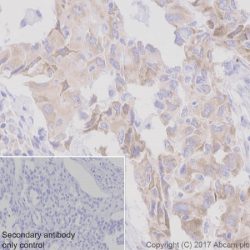
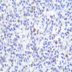
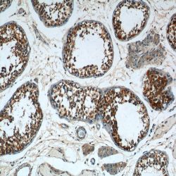
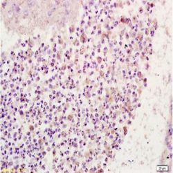
آنتی بادی ROS1 (Proto-Oncogene Tyrosine-Protein Kinase ROS)(D4D6)
Name: Proto-Oncogene Tyrosine-Protein Kinase Antibody clone D4D6
Description and applications: The transmembrane proto-oncogene tyrosine- kinase protein ROS, better known as ROS1, is a protein belonging to the insulin receptors tyrosine-kinase subfamily whose production is encoded by the ROS (MCF3) gene, located in the 6q22.1 chromosome region. After its activation, this gene intervenes in numerous molecular pathways related to the cell differentiation, proliferation, growth and survival including the activation of the PI3K-mTOR pathway. Chromosome aberrations affecting the ROS gene have been described in the glioblastoma multiforme through fusions of the C-terminal portion of ROS1 with the N-terminal domain of the FIG (Fused in Glioblastoma) encoded in the GOPC gene. The chimeric protein GOPC-ROS1 is located at the level of the Golgi apparatus and presents activity as tyrosine kinase receptor. Other fusions of ROS1 with different genes such as SLC34A2, CD74, EZR, LRIG3, SDC4, TPM3, CCDC6 or KDELR2 among others, have as a result the appearance and expression of several chimeric proteins in 1-3 % of cases of lung adenocarcinoma and represents the therapeutic target for Crizotinib and analog molecules. Non-specific staining on macrophages and reactive type II pneumocytes has also been described in isolated cases, whereas mucinous adenocarcinomas in general show low diffuse cytoplasmic staining in the absence of the translocation of the ROS1 gene. In order to identify the cases of lung carcinomas with translocations of ROS1, complex techniques of RT-PCR and FISH can be used, although the rabbit monoclonal antibody D4D6 has been recently validated as an useful screening tool for positive cases of immunostaining against ROS1, which in comparative studies through in situ hybridization techniques with break-apart probes has proven a sensitivity and specificity of over 95%. In order to consider a case as positive for ROS1, the immunostaining has to be strong or moderately intense on the membrane and/or the cytoplasm in more than 75% of tumor cells. Depending on the type of fusion of the ROS1 gene, other immunostaining patterns can be expressed as cytoplasmic punctiform in the CD74-ROS1 cases or cytoplasmic with linear accentuation in the lateral or apical membrane in the EZR-ROS1 cases. Although the morphology of the tumor cannot be considered as a criterion to select the positive ROS1 cases, the solid, micro-papillary, cribriform and signet ring cell growth patterns have been observed more frequently among the positive ROS1 cases. Focal and low-intensity staining can also be observed in up to 30% of the non-translocated cases. Therefore, the result must be confirmed with other analytical methods. 50% of the inflammatory myofibroblastic tumors also present translocation of the ALK gene, whereas the remaining ones show a little known genetic profile. It has been recently proven in a relatively low number of cases that up to 10% of these tumors show diffuse cytoplasmic or punctiform staining against the D4D6 clone (with translocation confirmed through FISH and RT-PCR techniques). In this same study, the antibody proved weak, focak nuclear staining in cases of gastrointestinal stromal tumors, myofibroblastic sarcomas, leiomyosarcomas and follicular dendritic cell sarcomas.Less than 1% of colorectal adenocarcinomas and up to 5% of stomach adenocarcinomas can show translocations of the ROS1 gene and consequential cytoplasmic staining. The low percentage of colorectal adenocarcinomas makes the gene as a potential therapeutic target, but in the case of the stomach adenocarcinomas, it can represent a therapeutic alternative, since, in general, the positive ROS1 cases show a non-amplified phenotype of HER2 and MET.
Composition: Anti-human ROS1 rabbit monoclonal antibody purified from serum and prepared in 10mM PBS, pH 7.4, with 0.2% BSA and 0.09% sodium azide
Immunogen: Human colon carcinoma extract.
Species reactivity: In vitro diagnostics in humans. Not tested in other species
-
 آنتی بادیهای ایمونوهیستوشیمی
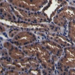
آنتی بادی SDHB (EP288)
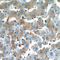
Name: SDHB Monoclonal Antibody clone EP288
Description and applications: Succinate dehydrogenase (SDH) is Complex II in the mitochondria, vital for mitochondrial electron transport, as well as Krebs cycle function. SDH catalyzes the oxidation of succinate to fumarate and transfers electrons to ubiquinone through the coordination of its four subunits (SDHA, SDHB, SDHC, and SDHD). The SDH complex functions as a tumor suppressor. Loss of any subunit proteins lead to destabilization of the complex and tumor formation. SDH subunit B (SDHB) is ubiquitously expressed in normal tissues. Germline mutations in SDHB, SDHC, or SDHD genes predisposes development of phaeochromocytoma, paraganglioma and gastrointestinal stromal tumor (GIST). SDHB immunohistochemistry is helpful in identification of phaeochromocytomas, paragangliomas or GIST with SDHB mutation.
Composition: Anti-human SDHB rabbit monoclonal antibody purified from serum and prepared in 10mM PBS, pH 7.4, with 0.2% BSA and 0.09% sodium azide
Intended use: Immunohistochemistry (IHC) on paraffin embedded tissues. Not tested on frozen tissues or Western-Blotting
-
 آنتی بادیهای ایمونوهیستوشیمی
آنتی بادی LEF1 (Lymphoid Enhancer-Binding Factor 1) (EP310)
Name: Lymphoid Enhancer-Binding Factor 1 Antibody clone EP310
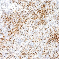
Description and applications: LEF1 is a nuclear protein of 42 kDa of molecular weight that is codified by the gen LEF1 located on the chromosomal region 4q25. It belongs to a family of regulatory proteins that share structural homology with high mobility group proteins (HMG1). LEF1 is expressed in pre-B and pre-T lymphocytes, where, through the Wnt signaling pathway, acts as an essential transcription factor for the proliferation and survival of these cells. The activations of the Wnt pathway leads to the accumulation of β-catenin and, eventually, to the gene transcription involved in the survival and cell cycle. The sustained activation with overexpression of LEF1, as it occurs in the chronic ymphoid leukemia, has been proven to play an important role in the molecular carcinogenesis, where its inhibition in experimental animal models leads to apoptosis of the neoplastic cells. In normal lymphoid tissues, the nuclear staining of LEF1 is observed to be predominant in T cells of the paracortical regions without being detected in the B cells, where their differentiation towards mature elements and plasmocytes is accompanied by the loss of the expression of LEF1.
In lymphoid neoplasias, the presence of over 10% of stained tumor cells is considered as positive staining and LEF1 is a marker with high specificity for the diagnosis of chronic lymphoid leukemia (CLL)/small lymphocytic lymphoma, including both the CD5 positive and CD5 negative cases if the cell morphology is concordant. The staining of LEF1 is in direct correlation with the expression of ZAP70 and implies a more favorable prognosis for the neoplasia without observing a correlation with the expression of CD38, the deletion of p53 or the trisomy 12. Nonetheless, and in probable relation with the sensitivity and specificity of the methods and antibodies used for the detection of LEF1, in other lymphomas, variable positive results have been obtained, so that the antibody has focal staining in up to 50% of the high grade follicular lymphomas and 40% of the diffuse large B-cell lymphomas, of which 18% are associated with the transformation of CLL in the Richter’s syndrome. In this later case, the staining is usually more intense in the areas with more atypia. Although most of publications confirm the negativity of the marginal and mantel cell lymphomas, more recent studies have proven focal staining in isolated cases. Additionally, and as LEF1 offers simultaneous staining in the reactive T lymphocytes often trapped within the clonal proliferative process, the correlation of the staining of LEF1 with other T marker as CD3, as well as other specific markers, is very advisable. LEF1 expression in other neoplasias, such as colon or pancreatic adenocarcinomas, has been mentioned in
isolated studies.Composition: Anti-human LEF1 rabbit monoclonal antibody purified from serum and prepared in 10mM PBS, pH 7.4, with 0.2% BSA and 0.09% sodium azide
Intended use: Immunohistochemistry (IHC) on paraffin embedded tissues. Not tested on frozen tissues or Western-Blotting
-
 آنتی بادیهای ایمونوهیستوشیمی
آنتی بادی Ep-CAM (EP155)
Name: Ep-CAM/Epithelial Specific Antigen Monoclonal Antibody clone EP-155
Description and Applications: Anti-MOC-31 reacts with a transmembrane glycoprotein present on most glandular epithelium and tumors originating from such epithelium. This antibody has been used to distinguish adenocarcinoma from mesothelioma and hepatocellular carcinoma. This antibody is also useful in distinguishing serous carcinomas of the ovary from mesothelioma.
Composition: Anti-human Ep-CAM mouse monoclonal antibody purified from serum and prepared in 10mM PBS, pH 7.4, with 0.2% BSA and 0.09% sodium azide
Intended use: Immunohistochemistry (IHC) on paraffin embedded tissues. Not tested on frozen tissues or Western-Blotting
IHC positive control:Breast, colon or lung adenocarcinoma
Visualization: Cell cytoplasm and membrane
-
 آنتی بادیهای ایمونوهیستوشیمی
آنتی بادی CD25 (4C9)
Name: CD25 antibody clone 4C9
Description and aplications: The CD25 molecule, also known as IK-2 receptor alpha, IL2RA, p55, T-cell growth factor receptor (TCGFR) or TAC antigen is an activation antigen present, along with CD4, in regulatory T cells. The gene that controls its expression, with 8 exons and over 25 kb, codes the alpha subunit of the cell surface receptor IL-2 and is located in the chromosome region 10p15.1. The regulatory T cells, since they suppress the activation of autoreactive T cells controlling the immune tolerance, prevent autoimmune diseases and, as negative collateral effects, avoid the destruction of tumor cells by cytotoxic T lymphocytes and act as suppressors of the NK cells. Partial deletions of the CD25 gene are responsible for immunodeficiency 41 (characterized by the association of various lymphoproliferative syndromes with autoimmune diseases) and insulin-dependent diabetes mellitus type 10, a variant of diabetes mellitus type I associated with autoimmune diseases and with typical familial aggregation. In normal tissues, CD25 can be expressed by activated B and T lymphocytes, macrophages and osteoblasts. Some thymocytes, myeloid precursors and oligodendrocytes can also show immunostaining. This molecule is not expressed in normal mastocytes. According to the classification system of the World Health Organization, the main diagnostic criterion for the involvement of bone marrow by systemic mastocytosis (SM) is the presence of dense aggregates (more than 15 cells) of mastocytes. For this reason, the aberrant expression of CD25 as a low affinity receptor for interleukin-2 (IL-2) by neoplastic mast cells is a good diagnostic tool to distinguish them from reactive proliferations of mast cells, and for this reason it has recently become a minor criterion for the diagnosis of SM, where aberrant staining of mastocytes aggregates by anti-CD25 antibody is diagnostic of SM. The anti-CD25 antibody has also been useful for the identification of mastocytes on skin biopsies in the context of urticaria pigmentosa as a predictor of systemic mastocytosis. Additionally, the quantification of regulatory T-cells (Treg) expressing CD25 in the context of hepatocellular carcinoma has been used as an independent predictor of tumor recurrence following liver resection of a previous hepatocellular carcinoma. In addition, the percentage of regulatory T cells FOXP3+ CD25+ infiltrating between melanoma tumor cells and in their periphery is significantly higher in melanomas with recurrent capacity than in their non-recurrent forms.
Finally, CD25 together with CD103 and CD123 is useful for completing the panel of markers of hairy cell leukemia, although the latter two antibodies have greater specificity and sensitivity for this diagnosis than CD25 itself.
Similarly, due to its general low specificity, the CD25 antibody’s positive staining should be evaluated within an antibody panel, not in isolation, and in correlation with the remaining morphological aspects of the lesions analyzed since many B lymphomas, T lymphomas, or even anaplastic large cell lymphoma may present staining against this marker.Composition: anti-CD25 mouse monoclonal antibody obtained from supernatant culture and prediluted in a tris buffered solution pH 7.4 containing 0.375mM sodium azide solution as bacteriostatic and bactericidal.
Intended use: Immunohistochemistry (IHC) on paraffin embedded tissues. Not tested on frozen tissues or Western-Blotting
Immunogen: Recombinant protein corresponding to the external domain of the human IL-2R
-
 آنتی بادیهای ایمونوهیستوشیمی
آنتی بادی PSMA (EP192)
Name: Prostate Specific Membrane Antigen Antibody clone EP192
Description and aplications: Glutamate Carboxypeptidase 2 (GCP2) or folate hydrolase 1 (FOLH1), also known as prostate specific membrane antigen (PSM or PSMA), is a type II homodimeric transmembrane protein of 750 amino acids of approximately 100 kDA of molecular mass that is encoded by the FOLH1 gene, which is located in the chromosome region 11p11.12. The FOLH1 protein is functionally characterized by an intense folase-hydrolase and dipeptidase activity on N-acetylated wastes of tri-alpha-glutamate peptides, regulating the absorption of folate in the intestine and the excitatory neurotransmission associated with the hydrolysis of the neuropeptide N-acetylaspartylglutamate in the CNS (central nervous system). In pathologic circumstances in the CNS, an excess of function of FOLH1 causes an excitotoxic effect due to the generation of high levels of glutamate that, as a consequence, leads to the death of motor cells in amyotrophic lateral sclerosis in both its sporadic and familial variants. In fact, FOLH1 inhibitors produce protective effects in this type of conditions when the glutamate levels decrease. In normal tissues, the protein is present in large amounts in the prostate epithelium, where it has an unknown function. However, it is known that in tumors that derive from this gland, FOLH1 is involved in the tumor progression. The protein can also be found in the small intestine (brush border), urinary epithelium, kidney, testicle, ovary, uterine tubes, breast, suprarenal gland, liver, esophagus, stomach, colon and brain (mainly brainstem and corpus striatum). In neoplastic tissues, FOLH1 can be present in tumors of the small intestine, brain, kidney, liver, spleen, trachea, spinal cord, and capillary endothelium. In the prostate, the PSMA molecule (FOLH1) can be found in both benign and malignant lesions, although in neoplasms, an increase in the intensity and number of stained cells in direct relation to the aggressiveness of the carcinoma can be observed. Therefore, it is also indirectly correlated with the expression of androgen receptors. In comparison with the prostate specific antigen (PSA), the determination of PSMA in tumors is a more sensitive procedure but less specific. Therefore, PSMA antibody is used in the diagnosis and prognosis of prostate tumors and as a possible marker for various neurodegenerative brain diseases such as schizophrenia and Alzheimer’s and Huntington’s diseases. Likewise, the Indium-labeled 7E11 anti-FOLH1 monoclonal antibody (ProstaScint®) is used for the diagnostic imaging of primary and metastatic prostate tumors.
Composition: Anti-human PSMA rabbit monoclonal antibody purified from culture supernatant, filtered, sterilized and prepared in 10mM PBS, pH 7.4, with 0.2% BSA and 0.09% sodium azide
Intended use: Immunohistochemistry (IHC) on paraffin embedded tissues. Not tested on frozen tissues or Western-Blotting
Immunogen: Synthetic peptide corresponding to the human PSMA.
-
 آنتی بادیهای ایمونوهیستوشیمی
آنتی بادی Oct2 (PT2)
Name: Mouse anti-human OCT-2 Monoclonal Antibody clone PT2
Description and aplications: Transcription factors Oct1 and OCT2 are located on chromosomal regions 1q22-q23 and 19 respectively, and belong to the POU family homeobox genes that bind DNA in monomeric and dimeric several configurations. Depending on the DNA sequence with which the dimers are joined, they are aligned in an accessible (MORE sequence) or inaccessible (PORE sequence) configuration for specific performance of the cofactor OBF1 of the B lymphocytes (Ocab, BOB1). OCT2 is an important element for the regulation of cellular and tissue specific transcription as well as transcription of numerous genes controllers. While Oct1 expression is an ubiquitous, OCT2 nuclear transcription factor expression is restricted to normal neurons of the central nervous system and B lymphocytes. This antibody recognizes the transcription factor Oct-2 of human, murine and rat origin and does not crossreact with other transcription factors. Oct-2 is expressed in normal B lymphocytes with greater intensity in the germinal center of lymphoid follicles. Small lymphocytes of the mantle areas have lower intensity and a small number of lymphocytes of the marginal and interfollicular areas also show intense nuclear staining. Since the expression of Oct-2 is related to the state of maturation of B cells, the antibody is primarily useful in the identification of B cell lymphomas originated in the germinal centre. In contrast to the L & H cells of lymphocyte-predominant Hodgkin’s disease (HD), the Reed-Sternberg (RS) cells of HD classical type are unable to transcribe immunoglobulins due to the absence of Oct-2. Therefore Oct-2 has been considered a new marker to identify and differentiate the L & H from HRS cells in Hodgkin’s disease. Immunostaining for Oct-2 is present intensively in nodular lymphocyte-predominant HD and with less intensity in the variants of the classic HD (HD rich in lymphocytes, HD mixed cellularity, HD nodular sclerosis) numerous cases being Oct-2 negative. In the nervous system it is considered that the expression of Oct-2 plays an important role in female sexual maturation in mammals regulated by the hypothalamus and the regulation of the latency period after infection with herpesvirus.
Composition: anti-human OCT2 mouse monoclonal antibody purified from serum and prepared in 10mM PBS, pH 7.4, with 0.2% BSA and 0.09% sodium azide
Immunogen: full length Oct-2 of human origin
Species reactivity: In vitro diagnostics in humans. Not tested in other species
-
 آنتی بادیهای ایمونوهیستوشیمی
آنتی بادی BRST-1/BCA-225 (CU-18)
(NAME: Mouse anti-human BRST-1/BCA-225 Monoclonal Antibody (Clone CU-18
DESCRIPTION AND APPLICATIONS:This antibody recognises BCA-225, a glycoprotein with a molecular mass of 225 kD secreted by the T47D breast carcinoma cell line that has subsequently been identified in various normal and neoplastic tissues
COMPOSITION:Anti-human BRST-1/BCA-225 mouse monoclonal antibody purified from serum and prepared in 10mM PBS, pH 7.4, with 0.2% BSA and 0.09% sodium azide
INTENDED USE: Immunohistochemistry (IHC) on paraffin embedded tissues. Not tested on frozen tissues or Western-Blotting
.IMMUNOGEN: BCA 225 protein secreted by the T47D (clone 11) human breast carcinoma cell line
-
 آنتی بادیهای ایمونوهیستوشیمی
آنتی بادی ALDH1A1 (Polyclonal)
Name: ALDH1A1 (Polyclonal)
Description and applications: ALDH1 isoform A1, also known as retinal (acetaldehyde) dehydrogenase 1 or aldehyde dehydrogenase 1 (ALDH1) has the mission to convert / oxidize the retinaldehyde in retinoic acid and is codified by a gene on the chromosome region 9q21.13.1.This enzyme belongs to a family of evolutionarily highly conserved ALDH enzymes comprising 19 isoforms localized in the cytoplasm, mitochondria or the nucleus and are involved in the metabolism of aldehydes units, retinol, certain xenobiotic compounds and likewise are involved in ethanol oxidation and Ras GTPase activity. Low levels of cytosolic together with normal levels of mitochondrial ALDH it’s seen in patients with alcoholic fatty liver.
Composition: anti-human ALDH1A1 goat polyclonal antibody purified by protein A/G in PBS/1% BSA buffer pH 7.6 with less than 0.1% sodium azide.
Immunogen: peptide mapping at the N-terminus of ALDH1A1 of human origin.
-
 آنتی بادیهای ایمونوهیستوشیمی
آنتی بادی Alpha-1 Antitrypsin (Polyclonal)
Name: Alpha-1 Antitrypsin (Polyclonal)
Description and applications: This antibody reacts with human alpha-1 antitrypsin, a glycoprotein responsible for the antiproteolytic activity of normal serum. Alpha-1-antitrypsin is mainly synthesized by hepatocytes, although human histiocytes and peripheral blood monocytes also express this protein. This antibody may be useful in the study of hereditary alpha-1 antitrypsin deficiency, benign and malignant liver tumours, embryonal carcinomas, and a variety of benign and malignant lesions of histiocytic origin. These antibody cross-reacts with horse, pig, dog, and rat alpha-1 antitrypsin.
Composition: anti-human Alpha-1 Antitrypsin rabbit polyclonal antibody purified from serum and prepared in 10mM PBS, pH 7.4, with 0.2% BSA and 0.09% sodium azide.
Immunogen: Human alpha-1 antitrypsin.
-
 آنتی بادیهای ایمونوهیستوشیمی
آنتی بادی Anexin A1 (29)
Name: Anexin A1 (29)
DESCRIPTION AND APPLICATIONS: Annexin I (a.k.a. lipocortin-1 or calpactin II) is a 38kDa protein that is part of a calcium-dependent phospholipid-binding protein family. Members of these family, share a common core domain, but each has a unique Nterminal tail that imparts its functional specificity. It is believed that phosphorylation of this region in
annexin I regulates its functions.COMPOSITION: Anti-human Annexin 1 mouse monoclonal antibody purified from serum and prepared in 10mM PBS, pH 7.4, with 0.2% BSA and 0.09% sodium azide INTENDED USE.
IMMUNOGEN: Aminoacids 1-346 of bovine annexin.
-
 آنتی بادیهای ایمونوهیستوشیمی
آنتی بادی ATRX (POLYCLONAL)
Name: ATRX (POLYCLONAL)
Description and aplications: ATRX, also known as ATP-dependent helicase ATRX, Xlinked helicase II,X-linked nuclear protein or Znf-HX, is encoded by a gene located in the chromosomal region Xq21.1, which undergoes an inactivation and encodes a nuclear and homologous NTP protein with several types of helicases II present at the membrane level. It belongs to the superfamily of proteins similar to the SNF2 subgroup and features a C-terminal region rich in glutamine similar to other nuclear transcription factors. Mutations of the gene cause a severe mental retardation syndrome linked to alphathalassemia (ATR-X syndrome) characterized by severe psychomotor retardation, characteristic facial features, urogenital abnormalities and alpha thalassemia with H hemoglobin inclusions at the erythrocyte level. Other syndromes related to mutations of the ATRX gene have been described such as X-linked mental retardation-hypotonic faciessyndrome or alpha thalassemia myelodysplastic syndrome. After interacting with the DAXX domain and potential binding with the EZH2, the ATRX protein plays several roles in mitosis, rearrangement of the chromatin and transcription as well as in the biology of the telomeric regions.
Composition: Rabbit anti-ATRX plyclonal antibody obtained from purified ascitic fluid and prepared in 10mM PBS, pH
7.4, with 0.2% BSA and 0.09% sodium azide.Immunogen: Recombinant protein corresponding to the alpha thalassemia X-linked intellectual disability syndrome (ATR-X) subunit.
-
 آنتی بادیهای ایمونوهیستوشیمی
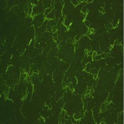
آنتی بادی Borrelia Burgdorferi (Polyclonal)
Name: Borrelia Burgdorferi
Description and aplications: This antibody recognises B. burgdorferi and shows cross-reactivity with Treponema pallidum, B. hermsii and B. parkeri. Lyme disease is a zoonosis related to infection by
Borrelia burgdorferi. It may induce different skin manifestations (erythema migrans, acrodermatitis chronica atrophicans or Herxheimer disease, or lymphocytoma cutis). Some primary cutaneous marginal zone B-cell lymphomas have been associated with Borrelia burgdorferi infection in European countries.Composition: anti-human Borrelia Burgdorferi rabbit polyclonal antibody purified from serum and prepared in 10mM PBS, pH 7.4, with 0.2% BSA and 0.09% sodium azide.
Immunogen: Prepared from strain B31 ATC#35210
-
 آنتی بادیهای ایمونوهیستوشیمی
آنتی بادی BRG1 (EPNCIR111A)
Name: BRG1 (EPNCIR111A)
Description and aplications: BRG1, also known as SMARCA4 or BAF190A, SNF2B and SNF2L4, is a member of the enzymatic chromatin remodeling complex SWI/SNF, where it encodes the catalytic subunit that intervenes in the configuration of nucleosomes, making the transcriptional activation of the complex more accessible. The gene encoding the protein is located in the chromosome region 19p13.2, whose alterations have primarily been associated with the rhabdoid tumor predisposition syndrome-2 and Coffin-Siris syndrome 4. The former is characterized by a predisposition to develop renal or extrarenal rhabdoid tumors as well as a variety of brain tumors among which are choroid plexus carcinoma, meduloblastoma and PNET. CoffinSiris syndrome 4 presents both autosomal recessive and dominant transmission and is characterized by intellectual disability, psychomotor retardation, coarse facial features and multiple congenital malformations such as aplasia or hypoplasia of thedistal phalanx or fifth finger nail. Additionally, cardiac, gastrointestinal, genitourinary or central nervous system malformations may be associated.
Composition: Anti-human BRG1 rabbit monoclonal antibody purified from serum and prepared in 10mM PBS, pH 7.4, with 0.2% BSA and 0.09% sodium azide
Immunogen: Synthetic peptide corresponding to the fraction of amino acids 200-300 of the mouse BRG1.
-
 آنتی بادیهای ایمونوهیستوشیمی
آنتی بادی Cadherin E (HECD-1)
Name: Cadherin E (HECD-1)
Description and aplications: E-cadherin is a transmembrane glycoprotein that mediates epithelial cell-to-cell adhesion. The loss of Ecadherin can result in the disruption of cell clusters. It is therefore, postulated that E-cadherin may function as a tumor suppressor protein. The loss of E-cadherin has been associtated with metastasis and poor prognosis in invasive breast cancer and can help differentiate between ductal and lobular neoplasms of the breast. It has also been shown to be an independent predictor in the disease progression in bladder cancer.
Composition: Anti-human Cadherin E mouse monoclonal antibody purified from serum and prepared in 10mM PBS, pH
7.4, with 0.2% BSA and 0.09% sodium azide -
 آنتی بادیهای ایمونوهیستوشیمی
آنتی بادی CD35 (EP197)
Name: Rabbit anti-human CD35 Monoclonal Antibody (Clone EP197)
Description and aplications: Anti-CD35, also named as erythrocyte complement receptor 1 (CR1), is a member of the complement activation (RCA) family and is located in the ‘cluster RCA’ region of chromosome 1. CD35 is considered a mature B-cell marker which labels follicular dendritic reticulum cells and tumors derived from, such as follicular dendritic cell tumor/sarcoma. CD35 antigen is found on erythrocytes, B cells, a subset of T cells, monocytes, as well as eosinophils, and neutrophils. This antibody labels also the dendritic cells in tonsil and spleen and glomerular podocytes in kidney.
Composition: Anti-human CD35 rabbit monoclonal antibody purified from serum and prepared in 10mM PBS, pH 7.4, with 0.2% BSA and 0.09% sodium azide
Immunogen: A synthetic peptide corresponding to residues of human CD35 protein.
-
 آنتی بادیهای ایمونوهیستوشیمی
آنتی بادی CD64 (EPR4624)
Name: Rabbit anti-human FCGR1A (CD64) Monoclonal Antibody (Clone EPR4624)
Description and aplications:This antibody, also known as HAM56, recognizes the immunoglobulin gamma receptor 1 (FcγRI/CD64) expressed in healthy people in approximately 10% of the circulating monocytes and encoded by a gene
located in the chromosome region 1q21.2. There are three types of FcγRs expressed in circulating monocytes, the high-affinity FcγRs/CD64 receptor, being expressed constitutively; the low-affinity FcγRII/CD32 with two isoforms functionally different and the moderate-affinity receptor FcγRIII/CD16, expressed in 10%-15% of circulating monocytes. This antibody can be used for the detection of some leukemias with monocytic differentiation such as the acute myeloid leukemia (AML) without maturation
Composition:Anti-human FCGR1A (CD64) rabbit monoclonal antibody purified from serum and prepared in 10mM PBS, pH 7.4, with 0.2% BSA and 0.09% sodium azide.
Immunogen: Synthetic peptide corresponding to the C-terminus of the human FCGRA1
-
 آنتی بادیهای ایمونوهیستوشیمی
آنتی بادی CD74 (LN-2)
Name: Mouse anti-human CD74 Monoclonal Antibody (Clone LN2)
Description and aplications: This antibody reacts with a 35-kD antigen related to the HLA complex invariant chain. The antigen is expressed in mantle and germinal centre B cells. It is also expressed in monocytes, macrophages,
interdigitating reticular cells, endothelium, and some other epithelia. This antibody may be used for the identification of B lymphomas (except for plasmacytomas). It also reacts with some T lymphomas containing activated neoplastic cells and Reed-Sternberg cells.
Composition: Anti-human CD74 mouse monoclonal antibody purified from serum and prepared in 10mM PBS, pH 7.4, with 0.2% BSA and 0.09% sodium azide.
Immunogen: Lymphoma cell line SU-DHL-4.
-
 آنتی بادیهای ایمونوهیستوشیمی
آنتی بادی Digoxigenin (HY-A.1)
Name: Mouse anti-human Digoxigenin Monoclonal Antibody (Clone HY-A.1)
Description and aplications: This product is recommended for the qualitative detection of DNA probes marked with digoxigenin, on formalin-fixed paraffin-embedded tissue sections.
Composition: Anti-human Digoxigenin mouse monoclonal antibody purified from serum and prepared in 10mM PBS, pH 7.4, with 0.2% BSA and 0.09% sodium azide
-
 آنتی بادیهای ایمونوهیستوشیمی
آنتی بادی Epithelial Specific Antigen (MOC-31)
Name: Mouse anti-human Ep-CAM/Epithelial Specific Antigen Monoclonal Antibody (Clone MOC-31)
Description and aplications: This antibody reacts with the 40 kDa transmembrane protein, which is present in most of normal and tumor epithelia. This antibody shows a wide pattern of reactivity with human epithelial tissues: simple epithelia, pseudostratified epithelia and transitional epithelia, but not with mature squamous epithelium. In paraffin-embedded formalin-fixed tissue sections, staining for MOC-31 is observed in the kidney epithelium, endometrial cells, mammary acini, hepatic biliary ducts, prostatic glandular epithelium, acini, ducts and pancreatic islets of Langerhans.
Composition: Anti-human Ep-CAM mouse monoclonal antibody purified from serum and prepared in 10mM PBS, pH 7.4, with 0.2% BSA and 0.09% sodium azide
Immunogen: Neuraminidase treated cells from a variant small cell lung carcinoma cell line (GLS-1).
برای تماس کلیک کنید
سما تشخیص آریا ، عرضه کننده متنوع ترین و کامل ترین محصولات کیت های آزمایشگاهی