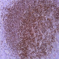
Name: Lymphoid Enhancer-Binding Factor 1 Antibody clone EP310
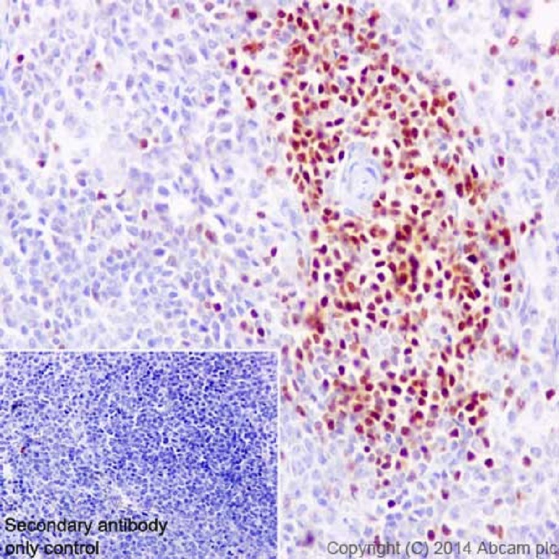
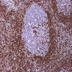
Description and applications: LEF1 is a nuclear protein of 42 kDa of molecular weight that is codified by the gen LEF1 located on the chromosomal region 4q25. It belongs to a family of regulatory proteins that share structural homology with high mobility group proteins (HMG1). LEF1 is expressed in pre-B and pre-T lymphocytes, where, through the Wnt signaling pathway, acts as an essential transcription factor for the proliferation and survival of these cells. The activations of the Wnt pathway leads to the accumulation of β-catenin and, eventually, to the gene transcription involved in the survival and cell cycle. The sustained activation with overexpression of LEF1, as it occurs in the chronic ymphoid leukemia, has been proven to play an important role in the molecular carcinogenesis, where its inhibition in experimental animal models leads to apoptosis of the neoplastic cells. In normal lymphoid tissues, the nuclear staining of LEF1 is observed to be predominant in T cells of the paracortical regions without being detected in the B cells, where their differentiation towards mature elements and plasmocytes is accompanied by the loss of the expression of LEF1.
In lymphoid neoplasias, the presence of over 10% of stained tumor cells is considered as positive staining and LEF1 is a marker with high specificity for the diagnosis of chronic lymphoid leukemia (CLL)/small lymphocytic lymphoma, including both the CD5 positive and CD5 negative cases if the cell morphology is concordant. The staining of LEF1 is in direct correlation with the expression of ZAP70 and implies a more favorable prognosis for the neoplasia without observing a correlation with the expression of CD38, the deletion of p53 or the trisomy 12. Nonetheless, and in probable relation with the sensitivity and specificity of the methods and antibodies used for the detection of LEF1, in other lymphomas, variable positive results have been obtained, so that the antibody has focal staining in up to 50% of the high grade follicular lymphomas and 40% of the diffuse large B-cell lymphomas, of which 18% are associated with the transformation of CLL in the Richter’s syndrome. In this later case, the staining is usually more intense in the areas with more atypia. Although most of publications confirm the negativity of the marginal and mantel cell lymphomas, more recent studies have proven focal staining in isolated cases. Additionally, and as LEF1 offers simultaneous staining in the reactive T lymphocytes often trapped within the clonal proliferative process, the correlation of the staining of LEF1 with other T marker as CD3, as well as other specific markers, is very advisable. LEF1 expression in other neoplasias, such as colon or pancreatic adenocarcinomas, has been mentioned in
isolated studies.
Composition: Anti-human LEF1 rabbit monoclonal antibody purified from serum and prepared in 10mM PBS, pH 7.4, with 0.2% BSA and 0.09% sodium azide
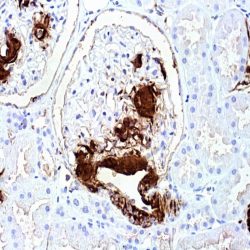
Intended use: Immunohistochemistry (IHC) on paraffin embedded tissues. Not tested on frozen tissues or Western-Blotting
Reviews
There are no reviews yet.