Category: IHC-Antibodies
Showing 801–820 of 923 results
فیلتر ها-
PSW
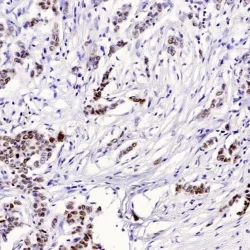
آنتی بادی TRPS1 کلون 8131R برند PathoSage
Rated 0 out of 5Intended use:
This antibody is intended for research use only (RUO). It is designed for professional laboratory use in formalin-fixed, paraffin-embedded (FFPE) tissues stained in manual qualitative immunohistochemistry (IHC) testing. In addition to FFPE tissues, this antibody may be tested on frozen tissues or applied in Western blot analyses.Presentation:
Bioreactor Concentrate with 0.05% Azide -
PSW
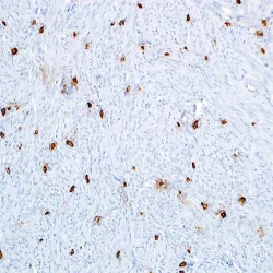
آنتی بادی Tryptase کلون G3 برند PathoSage
Rated 0 out of 5Intended use:
This antibody is intended for research use only (RUO). It is designed for professional laboratory use in formalin-fixed, paraffin-embedded (FFPE) tissues stained in manual qualitative immunohistochemistry (IHC) testing. In addition to FFPE tissues, this antibody may be tested on frozen tissues or applied in Western blot analyses.Presentation:
– -
PSW
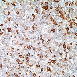
آنتی بادی TSH(Thyroid Stimulating Hormone) کلون Polyclonal برند PathoSage
Rated 0 out of 5Intended use:
This antibody is intended for research use only (RUO). It is designed for professional laboratory use in formalin-fixed, paraffin-embedded (FFPE) tissues stained in manual qualitative immunohistochemistry (IHC) testing. In addition to FFPE tissues, this antibody may be tested on frozen tissues or applied in Western blot analyses.Presentation:
Anti-TSH is a rabbit polyclonal antibody purified from rabbit anti-sera diluted in tris buffered saline, pH 7.3- -
PSW
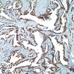
آنتی بادی TTF1 کلون 8G7G3/1 برند PathoSage
Rated 0 out of 5Intended use:
This antibody is intended for research use only (RUO). It is designed for professional laboratory use in formalin-fixed, paraffin-embedded (FFPE) tissues stained in manual qualitative immunohistochemistry (IHC) testing. In addition to FFPE tissues, this antibody may be tested on frozen tissues or applied in Western blot analyses.Presentation:
Bioreactor Concentrate with 0.05% Azide, the ready-to-use antibody is diluted in Tris Buffer, pH 7.3-7.7, with -
PSW
آنتی بادی TTF1 کلون SPT24 برند PathoSage
Rated 0 out of 5Intended use:
This antibody is intended for research use only (RUO). It is designed for professional laboratory use in formalin-fixed, paraffin-embedded (FFPE) tissues stained in manual qualitative immunohistochemistry (IHC) testing. In addition to FFPE tissues, this antibody may be tested on frozen tissues or applied in Western blot analyses.Presentation:
It is a liquid tissue culture supernatant containing 15mM sodium azide as a preservative -
PSW
آنتی بادی Tyrosinase کلون T311 برند PathoSage
Rated 0 out of 5Intended use:
This antibody is intended for research use only (RUO). It is designed for professional laboratory use in formalin-fixed, paraffin-embedded (FFPE) tissues stained in manual qualitative immunohistochemistry (IHC) testing. In addition to FFPE tissues, this antibody may be tested on frozen tissues or applied in Western blot analyses.Presentation:
Bioreactor Concentrate with 0.05% Azide -
PSW
آنتی بادی Vimentin کلون V9 برند PathoSage
Rated 0 out of 5Intended use:
This antibody is intended for research use only (RUO). It is designed for professional laboratory use in formalin-fixed, paraffin-embedded (FFPE) tissues stained in manual qualitative immunohistochemistry (IHC) testing. In addition to FFPE tissues, this antibody may be tested on frozen tissues or applied in Western blot analyses.Presentation:
Monoclonal antibody in TBS, pH 7.6, containing 1% BSA and 0.09% sodium azide. -
PSW
آنتی بادی YAP کلون 63.7 برند PathoSage
Rated 0 out of 5Intended use:
This antibody is intended for research use only (RUO). It is designed for professional laboratory use in formalin-fixed, paraffin-embedded (FFPE) tissues stained in manual qualitative immunohistochemistry (IHC) testing. In addition to FFPE tissues, this antibody may be tested on frozen tissues or applied in Western blot analyses.Presentation:
YAP1 in PBS with less than 0.1% sodium azide and 0.1% gelatin -
PSW
آنتی بادی ZAP70 کلون 2F3.2 برند PathoSage
Rated 0 out of 5Intended use:
This antibody is intended for research use only (RUO). It is designed for professional laboratory use in formalin-fixed, paraffin-embedded (FFPE) tissues stained in manual qualitative immunohistochemistry (IHC) testing. In addition to FFPE tissues, this antibody may be tested on frozen tissues or applied in Western blot analyses.Presentation:
Bioreactor Concentrate with 0.05% Azide -
Quartett
آنتی بادی ALK/p80(CD246) کلون QR017 برند Quartett
Rated 0 out of 5Anaplastic lymphoma kinase (ALK) belongs to the insulin receptor superfamily acting as a transmembrane receptorprotein-tyrosine kinase. In normal tissues, ALK protein is expressed only few cells within the developing and mature nervous system. ALK can be active in cancer through multiple mechanisms. The most common mechanism is through the formation of a fusion protein from chromosomal translocations. The protein expression by tumor cells is an independent prognostic factor that predicts a favorable outcome. ALK staining is positive in 50-60% of anaplastic large cell lymphoma (ALCL). NSCLC is found to express ALK 3-7%.
-
Quartett
آنتی بادی P504S(AMACR) کلون QR108 برند Quartett
Rated 0 out of 5AMACR, also known as P504S or alpha-methylacyl-CoAracemase, is an essential enzyme in the b-oxidation ofbranched-chain fatty acids and their derivatives. The enzyme is expressed in mitochondria and peroxisomes of numerous tissues such as liver, biliary tract, kidney and lung. AMACR is overexpressed in patients with prostate cancer compared to benign tissue. Highest expression is found in localized prostate cancer and decreases as soon as the carcinoma forms metastases. It therefore makes sense to use AMACR as a biomarker for the aggressiveness of a prostate carcinoma and its course. The antibody may be particularly helpful in the panel with HMW cytokeratin and/or p63.
-
Quartett
آنتی بادی BCL2 کلون QR062 برند Quartett
Rated 0 out of 5Bcl2 (B-cell lymphoma 2) plays an important role in regulation of apoptosis. It is expressed in different cells and tissues. Overexpression due to chromosomal translocation, gene amplification, increased gene transcription and/or altered post-translational processing is found in many cancer types. These include B-cell malignancies, such as B-cell lymphoma, follicular lymphoma (FL) and chronic lymphocytic leukemia (CLL), as well as some T-cell lymphomas, breast cancer, lung cancer, ovarian cancer, and prostat a cancer.
-
Quartett
آنتی بادی BOB1 کلون QR072 برند Quartett
Rated 0 out of 5BOB.1 is a lymphoid-specific transcriptional coactivator, which interacts with the transcription factors Oct-1 andOct-2. BOB.1 is expressed in germinal center B-cells, mantle B-cells and plasma cells. It detects a variety of B-cell lymphomas and Hodgkin’s lymphoma.
-
Quartett
آنتی بادی ARG1(Arginase1) کلون QR083 برند Quartett
Rated 0 out of 5Arginase (ARG) is a manganese metalloenzyme that is part of the urea cycle. It catalyses the conversion of L-arginine to L-ornithine and urea. ARG-1 and ARG-2 are the two isoforms of ARG. ARG-1 is a cytosolic enzyme expressed in liver. The ARG-1 antibody is a sensitive marker of hepatic differentiation and is useful in the detection of hepatocytes in normal tissue as well as granulocytes in peripheral blood. For the identification of hepatocellular carcinoma ARG-1 is a sensitive and specific marker.
-
Quartett
آنتی بادی BCL6 کلون QR047 برند Quartett
Rated 0 out of 5Bcl6 is a regulatory gene encoding a zinc finger protein. It plays a key role in the formation of the germinal center (GC) and acts as a regulator of B lymphocyte growth and development by protecting GC-B cells from DNA damage-induced apoptosis. Bcl-6 is mainly expressed in GC-B cells. Surrounding mantle-zone and marginal-zone B cells, plasma cells, and progenitor B cells are negative for Bcl-6. The antibody Bcl-6 detects GC cells in lymphoid follicles and a variety of lymphomas including follicular lymphomas, diffuse large B-cell lymphomas (DLBCL) and Burkitt’s lymphomas. In contrast, Bcl-6 is not expressed in hairy cell leukemias, mantle cell or peripheral lymphomas. Bcl-6 is not restricted to the B cell line, but could also be detected in anaplastic large cell lymphoma. Furthermore, Bcl-6 is involved in mammary epithelial differentiation, which could play a potential role in carcinogenesis.
-
Quartett
آنتی بادی C4d کلون QR053 برند Quartett
Rated 0 out of 5The complement system is part of the human innate immune system and mediates responses to inflammatory triggers. Complement 4d (C4d) is an inactive split product of Complement 4, activated by the classical or lectin complement pathway. After the cleavage a highly reactive thioester bond remains and C4d can bind covalently to the surrounding tissue to rest at sites of complement activation. This “footprint” remains much longer than the connection between tissue and antibody. Therefore, C4d is a reliable marker for an antibody-mediated rejection (AMR) in kidney, but also in heart and pancreas allografts. Complement 4d can easily be determined by immunohistochemical techniques in frozen or paraffin-embedded tissues and it is applied for the detection of humoral mediated alloresponses in histological sections.
-
Quartett
آنتی بادی Beta Catenin 1 کلون QR095 برند Quartett
Rated 0 out of 5Anti-human antibody for immunohistochemical use. The primary antibody is intended for qualitative detection of antigens in formalin-fixed, paraffin-embedded (FFPE) tissue sections. The antibody may be used manually or with any automated staining platform. Authorized and skilled personnel may only use the product. The clinical interpretation of any test results should be evaluated within the context of the patient’s medical history and other diagnostic laboratory test results. A qualified pathologist must perform evaluation.
-
Quartett
آنتی بادی Cytokeratin 20 کلون Ks20.8 برند Quartett
Rated 0 out of 5Keratins are cytoplasmic intermediate filament proteins expressed by epithelial cells and their neoplasms. Cytokeratin 20 is a type I keratin mainly found in the gastric and intestinal mucosa. Antibody Cytokeratin 20 is useful in the detection of a variety of adenocarcinomas arising from epithelia, such as colorectal carcinomas, transitional-cell and Merkel cell carcinomas, gastric and pancreatic tumors. CK20 is often used in a panel with antibody Cytokeratin 7.
-
Quartett
آنتی بادی CD3e کلون QR004 برند Quartett
Rated 0 out of 5CD3 is a protein complex that is composed of five chains (gamma, delta, epsilon, zeta, eta) which associate with theT-cell receptor (TCR). CD3 is first expressed in the cytoplasm of developing T-cells, then as they mature it moves to the membrane. CD3e is a membrane protein that plays a role in TCR-CD3 complex assembly and signal transduction regulating T-cell proliferation, apoptosis and thymocyte development. CD3e immunostaining is only seen in T-cells. These predominantly occur in lymphatic organs such as thymus, lymph nodes and tonsils but are regularly also seen – at lower density – in many other tissue types. In malignant lymphomas, CD3 is a pan-T-cell lineage-restricted antigen, detected in 72-95% of the T-cell lymphomas. As B-cell lymphomas are mostly negative forCD3e, the antibody can be used to distinguish between T-cell and B-cell lymphomas.
-
Quartett
آنتی بادی CD117(c-kit) کلون QR012 برند Quartett
Rated 0 out of 5CD117, also known as c-Kit, encodes a receptor tyrosine kinase that is activated by its ligand stem cell factor (also known as mast cell growth factor). CD117 plays an important role in vital functions, such as homeostasis and melanogenesis. In normal tissues, expression is found in interstitial cells of Cajal, mast cells, melanocytes, various epithelia, testicular and ovarian interstitial cells, neurons of the central nervous system, immature myeloid cells, trophoblastic cells, fetal (but not mature) endothelial cells and fetal (but not mature) basal cells of the skin. CD117 does not occur in smooth muscle cells.
CD117 is a proto-oncogene. Mutations and overexpression are associated with several cancer types, including gastrointestinal stromal tumors (GIST) with 80 95% expression and seminoma with > 90% expression.
CD117 is expressed in around 15-30% of melanomas, although this can vary depending on the type of melanoma. About 30% of lung adenocarcinoma show expression of CD117.

