-
 آنتی بادیهای ایمونوهیستوشیمی
آنتی بادی Borrelia Burgdorferi (Polyclonal)
Name: Borrelia Burgdorferi
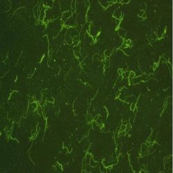
Description and aplications: This antibody recognises B. burgdorferi and shows cross-reactivity with Treponema pallidum, B. hermsii and B. parkeri. Lyme disease is a zoonosis related to infection by
Borrelia burgdorferi. It may induce different skin manifestations (erythema migrans, acrodermatitis chronica atrophicans or Herxheimer disease, or lymphocytoma cutis). Some primary cutaneous marginal zone B-cell lymphomas have been associated with Borrelia burgdorferi infection in European countries.Composition: anti-human Borrelia Burgdorferi rabbit polyclonal antibody purified from serum and prepared in 10mM PBS, pH 7.4, with 0.2% BSA and 0.09% sodium azide.
Immunogen: Prepared from strain B31 ATC#35210
-
 آنتی بادیهای ایمونوهیستوشیمی
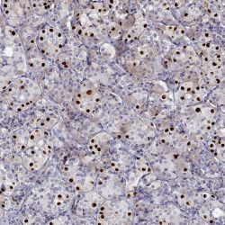
آنتی بادی BRG1 (EPNCIR111A)
Name: BRG1 (EPNCIR111A)
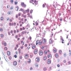
Description and aplications: BRG1, also known as SMARCA4 or BAF190A, SNF2B and SNF2L4, is a member of the enzymatic chromatin remodeling complex SWI/SNF, where it encodes the catalytic subunit that intervenes in the configuration of nucleosomes, making the transcriptional activation of the complex more accessible. The gene encoding the protein is located in the chromosome region 19p13.2, whose alterations have primarily been associated with the rhabdoid tumor predisposition syndrome-2 and Coffin-Siris syndrome 4. The former is characterized by a predisposition to develop renal or extrarenal rhabdoid tumors as well as a variety of brain tumors among which are choroid plexus carcinoma, meduloblastoma and PNET. CoffinSiris syndrome 4 presents both autosomal recessive and dominant transmission and is characterized by intellectual disability, psychomotor retardation, coarse facial features and multiple congenital malformations such as aplasia or hypoplasia of thedistal phalanx or fifth finger nail. Additionally, cardiac, gastrointestinal, genitourinary or central nervous system malformations may be associated.
Composition: Anti-human BRG1 rabbit monoclonal antibody purified from serum and prepared in 10mM PBS, pH 7.4, with 0.2% BSA and 0.09% sodium azide
Immunogen: Synthetic peptide corresponding to the fraction of amino acids 200-300 of the mouse BRG1.
-
 آنتی بادیهای ایمونوهیستوشیمی
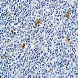
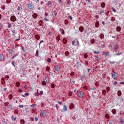
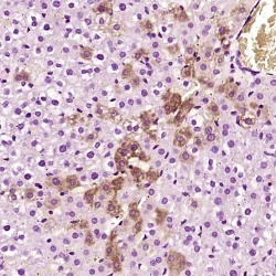
آنتی بادی HBc Antigen (CORE) (Polyclonal)
Name: Rabbit anti-human Hepatitis B Virus Core Antigen (HBVcAg) Polyclonal Antibody
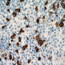
Description and aplications: Hepatitis B virus is spherical in shape with a diameter of 42 nm. It contains a 27 nm partially double stranded DNA core enclosed within a lipoprotein coat. The antigenic activity of the nucleocapsid core is designated as hepatitis B core antigen. The antigens in the outer surface are called as hepatitis B virus surface antigens.
Core antigens are localized within the nuclei whereas the surface antigens are present in the cytoplasm of the infected cells. Antibodies to surface antigens appear in circulation at an early stage of infection whereas the antibodies to the core antigens are detected after several weeks. This antibody recognizes a protein in the core of hepatitis B virus.Composition: Anti-human HBVcAg rabbit polyclonal antibody purified from serum and prepared in 10mM PBS, pH
7.4, with 0.2% BSA and 0.09% sodium azide
Immunogen: Hepatitis B virus.
-
 آنتی بادیهای ایمونوهیستوشیمی
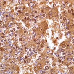
آنتی بادی Human IgA (Polyclonal)
Name: Rabbit anti-human IgA Antibody (Polyclonal)
Description and aplications:Anti-IgA antibody reacts with surface immunoglobulin IgA alpha chains. It is useful when identifying leukemias, plasmacytomas, and B-cell lineage derived Hodgkin’s lymphomas. Due to the restricted expression of heavy and light chains in these diseases, demonstration of Bcell lymphoma/plasmacytoma is aided with this antibody.
Composition: Anti-human IgA rabbit polyclonal antibody purified from serum and prepared in 10mM PBS, pH 7.4, with
0.2% BSA and 0.09% sodium azide
-
 آنتی بادیهای ایمونوهیستوشیمی
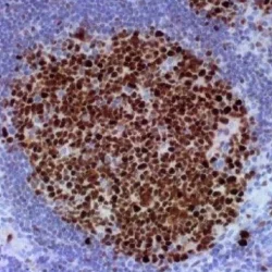
آنتی بادی Human IgE (Polyclonal)
Name: Rabbit anti-human IgE Antibody (Polyclonal)
Description and aplications: This antibody recognises the human immunoglobulin epsilon heavy chain. The common feature of both plasmacytomas and certain types of non-Hodgkin lymphoma is the expression of a single class of heavy chain. The demonstration of clonality in lymphoproliferative processes indicates that the process is clonal and therefore malignant. This antibody is useful for the identification of leukaemias, plasmacytomas and some types of non-Hodgkin lymphoma. It does not cross-react with the heavy chains of immunoglobulins IgA, IgG, IgM and IgD, nor with T cells, monocytes, granulocytes or erythrocytes.
Composition:anti-human IgE rabbit polyclonal antibody purified from serum and prepared in 10mM PBS, pH 7.4, with 0.2% BSA and 0.09% sodium azide
Immunogen: IgE isolated from normal human serum.
-
 آنتی بادیهای ایمونوهیستوشیمی
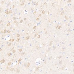
آنتی بادی Isocitrate Dehydrogenase 1 R132H point mutated form (IDH1 R132H) (H09)
Name: Mouse Anti-Human Isocitrate Dehydrogenase 1 R132H point mutated form (IDH1 R132H) (H09)
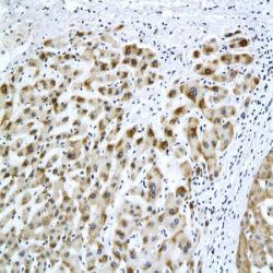
Description and aplications: Antibody clone H09 reacts specifically with the isocitrate dehydrogenase 1 (IDH1) R132H point mutated form in tissue sections from formalin-fixed brain tumor specimens. Heterozygous point mutations of IDH1 codon 132 are frequent in World Health Organization (WHO) grade II and III gliomas. IDH1 R132H mutations occur in approximately 70% of astrocytomas and oligodendroglial tumors. The high frequency and distribution of the IDH1 R132H mutation among specific brain tumor entities allow the highly sensitive and specific discrimination of various tumors by
immunohistochemistry, such as anaplastic astrocytoma from primary glioblastoma or diffuse astrocytoma WHO grade II from pilocytic astrocytoma or ependymoma. Noteworthy is the discrimination of the infiltrating edge of tumors with IDH1 mutation from reactive gliosis. This antibody is highly useful for tumor classification and in detecting single infiltrating tumor cells.
Composition:anti-Isocitrate Dehydrogenase 1 R132H (IDH1 R132H) mouse monoclonal antibody obtained from supernatant culture and prediluted in a tris buffered solution pH 7.4 containing 0.375mM sodium azide solution as bacteriostatic and bactericidal. The quantity of the active antibody was not determined.
Immunogen: Synthetic peptide, amino acid sequence CKPIIIGHHAYGD
-
 آنتی بادیهای ایمونوهیستوشیمی
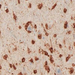
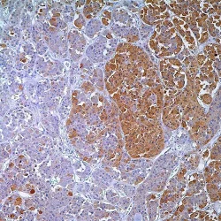
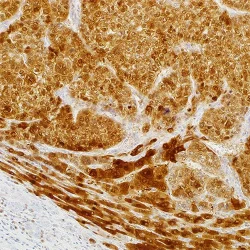
آنتی بادی Neuron Specific Enolase (NSE) (Polyclonal)
Name: Rabbit anti-human Neuron Specific Enolase (NSE) Polyclonal Antibody
Description and aplications: Enolase is a glycolytic enzyme catalyzing the reaction pathway between 2-phospho glycerate and phosphoenol pyruvate. In mammals, enolase molecules are dimmers composed of three distinct subunits (α,β and γ). The alpha-subunit is expressed in most tissues and the beta-subunit only in muscle. The gamma-subunit is expressed primarily in neurons, in normal and in neoplastic neuroendocrine cells. Co-expression of NSE and chromogranin A is common in neuroendocrine neoplasms.
Composition: Anti-human NSE rabbit polyclonal antibody purified from serum and prepared in 10mM PBS, pH 7.4, with
0.2% BSA and 0.09% sodium azide
Immunogen: Synthetic peptide derived from Cterminus of human NSE.
-
 آنتی بادیهای ایمونوهیستوشیمی
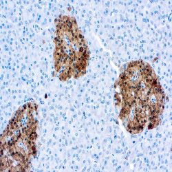
آنتی بادی PGP 9.5 (Polyclonal)
Name: Rabbit anti-human PGP 9.5 Antibody (Polyclonal)
Description and aplications: This antibody recognises a molecule with a molecular mass of 27 kDa known as “Protein Gene Product 9.5” or PGP 9.5 and encoded by the UCHL1 (ubiquitin carboxy-terminal hydrolase L1 isoenzyme) gene, located in the chromosomal region 4p13. This protein catalyses the hydrolysis of ubiquitin’s C-terminal esters and amides, playing an important role in protein degradation. Overexpression of the UCHL1 gene occurs in several neoplasms, including leukaemias, carcinomas, and several tumours of mesenchymal origin. In normal tissues, PGP 9.5, which shows mainly a cytoplasmic staining pattern, is present in neurons and nerve fibres of the central and peripheral nervous systems, as well as in numerous neuroendocrine cells, except those of the digestive tract, of which only the pancreatic islets of Langerhans show consistent positive staining. Other normal tissues that are usually stained are segments of renal tubules, spermatogonias and Leydig cells, smooth muscle, and germinal centres.
Composition: anti-human PGP 9.5 rabbit polyclonal antibody purified from serum and prepared in 10mM PBS, pH 7.4, with 0.2% BSA and 0.09% sodium azide
Immunogen: Immunogen: Recombinant protein corresponding to the total length of human protein PGP 9.5.
-
 آنتی بادیهای ایمونوهیستوشیمی
آنتی بادی Poly (ADP-Ribose) Polymerase 1 (Polyclonal)
Name: Rabbit anti-human Poly (ADP-Ribose) Polymerase 1 Antibody (Polyclonal)
Description and aplications: PARP (EC 2.4.2.30) is a nuclear protein with a molecular mass of 116 kDa and two zinc finger motifs which binds to DNA and specifically detects DNA nicks and breaks produced by different genotoxic agents. PARP catalises the ADP-ribosylation of proteins using NAD+ as a substrate. PARP activation is a consequence of ischemic injury and leads to intracellular depletion of NAD+, which can only be replaced by ATP consumption. Ischemia/Reperfusion (I/R) injury results in substantial DNA damage and cells need to consume large amounts of ATP to
sustain reparative poly-ADP-ribosylation. Proteolysis of PARP to its stable 85-kDa fragment is an early marker of programmed cell death (apoptosis), mediated by caspase 3 or CPP32. Cleavage occurs between Adp216 and Gly217, a site in PARP conserved across species. This antibody reacts with human, mouse and rat PARP.Composition:anti-human Poly (ADP-Ribose) Polymerase 1 rabbit polyclonal antibody purified from serum and prepared in 10mM PBS, pH 7.4, with 0.2% BSA and 0.09% sodium azide
Immunogen: Synthetic peptide derived from the Nterminal region of human protein PARP.
-
 آنتی بادیهای ایمونوهیستوشیمی
آنتی بادی T-PIT/TBX19 (polyclonal)
Name: Rabbit Anti-Human T-PIT/TBX19 Polyclonal Antibody
Description and aplications: The TPIT molecule, also known as T-Box 19 or TBX19, is a gene transcription factor of the T-box gene family that is mainly involved in the regulation of embryonic development. In the case of TPIT, whose coding gene consists of 8 exons and is located in the chromosomal region 1q24.2, its expression is restricted to the two pituitary cell lines (corticotroph and melanotroph), in which it activates the POMC gene responsible for the secretion of proopiomelanocortin. This molecule, and its derivative opiomelanocortin, are the precursors of ACTH synthesis as well as of some molecules related to appetite regulation, mainly lipotropin beta.
Composition: Anti-human T-PIT/TBX19 rabbit polyclonal antibody obtained from ascitic fluid and prepared in 10mM PBS, pH 7.4, with 0.2% BSA and 0.09% sodium azide.
Immunogen: Recombinant protein corresponding to human T-PIT/TBX19.
-
 آنتی بادیهای ایمونوهیستوشیمی
آنتی بادی PIT-1/POU1F1 (polyclonal)
Name: Rabbit Anti-Human PIT-1/POU1F1 Polyclonal Antibody
Description and aplications: The PIT-1 molecule (pituitary-specific transcription factor 1, also known as POU1F1 or GHF-1) belongs to the POU group of transcription factors involved in development together with OCT1 and Unc-06. It is
encoded by a gene located on chromosomal region 3p11.2. PIT-1 is involved in the differentiation and development of anterior pituitary cells, secreting hormones with lactotropic, somatotropic and
thyrotropic activity (beta subunit). It is also involved in the activation of genes controlling growth hormones and prolactin. Heterogeneous (homo- and heterozygous) mutations of PIT-1 lead to combined pituitary deficiency type 1 which is clinically characterized by irreversible mental and growth retardation and presents deficient plasma levels of growth hormone, combined mainly with low levels of prolactin or TSH. In contrast, ACTH, LH or FSH levels remain within normal values in this disease. Acquired combined pituitary deficiency has also been described, in which patients do not have mutations of the PIT-1 gene but nevertheless have low plasma levels of free TSH and free T4 along with undetectable levels of growth hormone and prolactin. In these cases, the disease is associated with the presence of autoantibodies to PIT-1, microsomes, thyroglobulin, thyroid peroxidase, GAD, and parietal cells of the stomach. PIT-1 staining of pituitary adenomas is similar to that of the normal gland. Although PIT-1 is not routinely used to characterize these neoplasms, in the case of non-functioning adenomas that have tested negative for the first panel of antibodies of
pituitary hormones, the combined use of PIT-1, T-PIT and SF-1 antibodies can differentiate PIT-1-positive adenomas from true null adenomas.
Composition: Anti-human PIT-1/POU1F1 rabbit polyclonal antibody obtained from ascitic fluid and prepared in 10mM PBS, pH 7.4, with 0.2% BSA and 0.09% sodium azide.
Immunogen: Recombinant protein corresponding to human PIT-1/POU1F1.
-
 PSV
PSVآنتی بادی ACTH(Adrenocorticotropic Hormone) کلون Polyclonal برند PathoSage
Intended use:
This antibody is intended for in vitro diagnostic (IVD) use. Primary Antibody is intended for professional laboratory use in formalin-fixed, paraffin-embedded (FFPE) tissue stained in manual qualitative immunohistochemistry (IHC) testing. A qualified pathologist must interpret the results using this product to aid diagnosis in conjunction with the patient’s relevant clinical history, other diagnostic tests, and proper controls.Presentation:
Anti-human ACTH rabbit polyclonal antibody purified from serum and prepared in 10mM PBS, pH 7.4, with 0.2% BSA and 0.09% sodium azide -
 PSV
PSVآنتی بادی Adipophilin کلون Polyclonal برند PathoSage
Intended use:
This antibody is intended for in vitro diagnostic (IVD) use. Primary Antibody is intended for professional laboratory use in formalin-fixed, paraffin-embedded (FFPE) tissue stained in manual qualitative immunohistochemistry (IHC) testing. A qualified pathologist must interpret the results using this product to aid diagnosis in conjunction with the patient’s relevant clinical history, other diagnostic tests, and proper controls.Presentation:
Anti-human Adipophilin rabbit polyclonal antibody purified from serum and prepared in 10mM PBS, pH 7.4, with 0.2% BSA and 0.09% sodium azide -
 PSV
PSVآنتی بادی ALK/p80 کلون 5A4 برند PathoSage
Intended use:
This antibody is intended for in vitro diagnostic (IVD) use. Primary Antibody is intended for professional laboratory use in formalin-fixed, paraffin-embedded (FFPE) tissue stained in manual qualitative immunohistochemistry (IHC) testing. A qualified pathologist must interpret the results using this product to aid diagnosis in conjunction with the patient’s relevant clinical history, other diagnostic tests, and proper controls.Presentation:
Anti-human ALK/p80 mouse monoclonal antibody purified from serum and prepared in 10mM PBS, pH 7.4, with 0.2% BSA and 0.09% sodium azide -
 PSV
PSVآنتی بادی Amyloid P کلون EP1018Y برند PathoSage
Intended use:
This antibody is intended for in vitro diagnostic (IVD) use. Primary Antibody is intended for professional laboratory use in formalin-fixed, paraffin-embedded (FFPE) tissue stained in manual qualitative immunohistochemistry (IHC) testing. A qualified pathologist must interpret the results using this product to aid diagnosis in conjunction with the patient’s relevant clinical history, other diagnostic tests, and proper controls.Presentation:
Anti-human Amyloid P rabbit monoclonal antibody purified from serum and prepared in 10mM PBS, pH 7.4, with 0.2% BSA and 0.09% sodium azide -
 PSV
PSVآنتی بادی Amyloid A کلون MC1 برند PathoSage
Intended use:
This antibody is intended for in vitro diagnostic (IVD) use. Primary Antibody is intended for professional laboratory use in formalin-fixed, paraffin-embedded (FFPE) tissue stained in manual qualitative immunohistochemistry (IHC) testing. A qualified pathologist must interpret the results using this product to aid diagnosis in conjunction with the patient’s relevant clinical history, other diagnostic tests, and proper controls.Presentation:
Anti-human Amyloid-A rabbit monoclonal antibody purified from Bioreactor Concentrate by Protein A/G and diluted in a pH7.6 buffer containing stabilizing protein and sodium azide as bacteriostatic and bactericidal agent -
 PSV
PSVآنتی بادی Alpha1 Antichymotrypsin کلون Polyclonal برند PathoSage
Intended use:
This antibody is intended for in vitro diagnostic (IVD) use. Primary Antibody is intended for professional laboratory use in formalin-fixed, paraffin-embedded (FFPE) tissue stained in manual qualitative immunohistochemistry (IHC) testing. A qualified pathologist must interpret the results using this product to aid diagnosis in conjunction with the patient’s relevant clinical history, other diagnostic tests, and proper controls.Presentation:
Anti-human Alpha-1-Antichymotrypsin rabbit polyclonal antibody purified from serum and prepared in 10mM PBS, pH 7.4, with 0.2% BSA and 0.09% sodium azide -
 PSV
PSVآنتی بادی Alpha Fetoprotein کلون EP209 برند PathoSage
Intended use:
This antibody is intended for in vitro diagnostic (IVD) use. Primary Antibody is intended for professional laboratory use in formalin-fixed, paraffin-embedded (FFPE) tissue stained in manual qualitative immunohistochemistry (IHC) testing. A qualified pathologist must interpret the results using this product to aid diagnosis in conjunction with the patient’s relevant clinical history, other diagnostic tests, and proper controls.Presentation:
Anti-human AFP rabbit monoclonal antibody purified from serum and prepared in 10mM PBS, pH 7.4, with 0.2% BSA and 0.09% sodium azide -
 PSV
PSVآنتی بادی BCL6 کلون LN22 برند PathoSage
Intended use:
This antibody is intended for in vitro diagnostic (IVD) use. Primary Antibody is intended for professional laboratory use in formalin-fixed, paraffin-embedded (FFPE) tissue stained in manual qualitative immunohistochemistry (IHC) testing. A qualified pathologist must interpret the results using this product to aid diagnosis in conjunction with the patient’s relevant clinical history, other diagnostic tests, and proper controls.Presentation:
Anti-human Bcl-6 mouse monoclonal antibody purified from serum and prepared in 10mM PBS, pH 7.4, with 0.2% BSA and 0.09% sodium azide -
 PSV
PSVآنتی بادی ARG1 کلون EP261 برند PathoSage
Intended use:
This antibody is intended for in vitro diagnostic (IVD) use. Primary Antibody is intended for professional laboratory use in formalin-fixed, paraffin-embedded (FFPE) tissue stained in manual qualitative immunohistochemistry (IHC) testing. A qualified pathologist must interpret the results using this product to aid diagnosis in conjunction with the patient’s relevant clinical history, other diagnostic tests, and proper controls.Presentation:
Anti-human ARG-1 rabbit monoclonal antibody purified from serum and prepared in 10mM PBS, pH 7.4, with 0.2% BSA and 0.09% sodium azide
برای تماس کلیک کنید
سما تشخیص آریا ، عرضه کننده متنوع ترین و کامل ترین محصولات کیت های آزمایشگاهی