برای تماس کلیک کنید

به وب سایت سما تشخیص آریا خوش آمدید.

جستجوی پیشرفته
جستجوی عینی کلمه
جستجو در محتوا
جستجو در عنوان
دنبال محصول یا مطلبی میگردید؟ در این کادر جستجو کنید

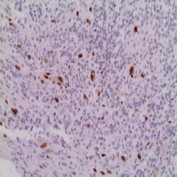
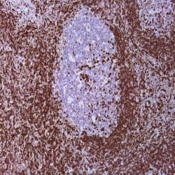
Name: CD7 Antibody-Clone-EP132

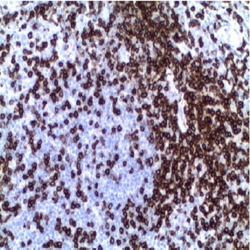
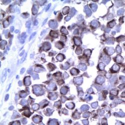
Description and aplications: This antibody recognizes a glycoprotein of 40kD molecular weight known as CD7 (also known as TP40 and Leu9). CD7 is positioned at 17q25.2-q25.3 and present on thymocytes, mature T cells, and NK cells. During T cell differentiation, CD7 is one of the earliest lineage markers to appear, therefore, the marker is considered the most clinically useful for acute lymphoid leukemia of T origin. In contrast, the CD7 molecule is absent in some cases as severe combined immunodeficiency. As the CD7 antigen is expressed in mature and immature T cells and NK cells, including those with mixed immuophenotype myeloid (precursor NK/myeloid cells leukemia), the anti-CD7 antibody is useful for the identification of lymphoid neoplasms derived therefrom. However it should be noted that in the peripheral T-cell lymphomas, frequent deletion or pre- and post-transcriptional regulation can lead to the loss of CD7 expression in the cell membrane, therefore their absence in this case does not rule out the origin of the T neoplasia. It is also not unusual in benign inflammatory dermatoses that reactive T cells do not express CD7, therefore, these histopathological features must be considered in the differential diagnosis.

Composition: anti-CD7 rabbit monoclonal antibody obtained from supernatant culture and prediluted in a tris buffered solution pH 7.4 containing 0.375mM sodium azide solution as bacteriostatic and bactericidal.

Immunogen: A synthetic peptide corresponding to residues of human CD7 protein.


حجم موجود3 / 7 / 12 میلی‌لیتر

مورد استفاده: ایمونوهیستوشیمی روی بافت پارافین. بر روی بافتهای فروزن یا وسترن بلات تست نشده است.

کلونEP132

ایزوتایپ: Rabbit IgG

کنترل مثبت IHC: تونسیل

قابلیت دیده شدن:غشای سلولی و سیتوپلاسمی

روش پیشنهادی IHC:

  • برش تهیه شده با ضخامت μm4 باید روی لام شارژ شده در طول شب و در دمای 60 درجه سانتی گراد خشک شود.
  • دپارافینه کردن، رهیدراته کردن و استفاده از حرارت جهت بازیابی اپی توپ : بافت را با استفاده از بافر EDTA pH 8 برای بیست دقیقه در دمای 95 درجه سانتی گراد بجوشانید. سپس 5-3 بار با استفاده از آب مقطر یا دیونیزه شده شسته و سپس بیست دقیقه در دمای اتاق خنک کنید.
  • بلاک پروکسیدازهای درونی : بلاک با استفاده از محلول پروکسیداز به مدت ده دقیقه
  • آنتی بادی اولیه: برای 10 دقیقه در انکوبه کنید پروتکل ممکن است بسته به تهیه نمونه و کاربرد خاص متفاوت باشد. شرایط استاندارد باتوجه به آزمایشگاه و کاربر متفاوت می باشد.
  • برای رنگ آمیزی ، می توانید از کیت Master Polymer Plus Detection یا کیتهای دیتکشن سایر کمپانی ها استفاده کنید.
  • رنگ میزی نهایی با هماتوکسیلین و فیکس نهایی اسلاید.

شرایط نگهداری و پایداری : حداکثر 18 ماه؛ در دمای 2-8 درجه سانتیگراد نگهداری شود. فریز نشود.


این آنتی‌بادی متعلق به پنل‌ تشخیصی LYMPHOID است.


دانلود کاتالوگ

Icon

CD7 (EP132) 464.89 KB 5 downloads

...
.well.c2a3 .btn { padding: 11px 30px; font-size: 11pt; } .well.c2a3 .media-body { font-size: 11pt; } .well.c2a3 .wpdm_icon { height: 42px; width: auto; }

Reviews

There are no reviews yet.

Be the first to review “آنتی­ بادی CD7 (EP132)”

نشانی ایمیل شما منتشر نخواهد شد. بخش‌های موردنیاز علامت‌گذاری شده‌اند *

پیمایش به بالا