برای تماس کلیک کنید

به وب سایت سما تشخیص آریا خوش آمدید.

جستجوی پیشرفته
جستجوی عینی کلمه
جستجو در محتوا
جستجو در عنوان
دنبال محصول یا مطلبی میگردید؟ در این کادر جستجو کنید

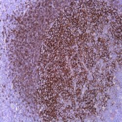
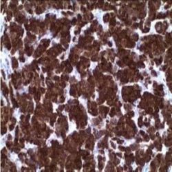
Name: P62 Antibody clone LCK LIGAND 3/P62

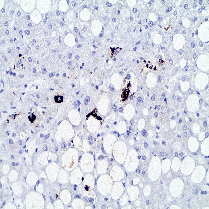
Description and applications: This antibody recognises phosphotyrosine p62, a member of the c-src family of cytoplasmic kinases, which binds as a ligand to the SH2 domains of p56 (Lcr) in the absence of phosphotyrosine. p62/SQSTM1 is an adapter protein targeting the protein aggregates ubiquitinated by lysosomal degradation. p62/SQSTM1 is selectively degraded via the autophagic pathway. Mallory’s hyaline and intracellular hyaline bodies are associated with chronic non-neoplastic liver diseases (steatohepatitis, related or not to alcohol abuse; toxic or metabolic chronic cholestasis) and with neoplastic liver diseases such as hepatocellular carcinoma. These inclusions are related to and contain keratin aggregates, especially keratin 8, ubiquitin, heat shock proteins, and the stress adaptor protein p62, stabilized by bonds catalysed by transglutaminases. P62 binds to ubiquitin and acts as an adaptor to bind ubiquitinated proteins. Autophagy is an evolutionary highly conserved degradative mechanism affecting the components of the cytoplasm that contributes to homoeostasis by making organelle replacement possible. Unlike the ubiquitin-proteosome system, the autophagy mechanism occurs in multiple steps that include the formation of the phagophore, which traps proteins and organelles for its degradation, and the autophagosome, which, through mechanisms involving cytoskeleton’s microtubules, fuses with lysosomes to form autolysosomes, where the material is eventually degraded. This antibody is useful for the identification of p62, found in Mallory’s hyaline and intracellular hyaline bodies present in chronic liver diseases and hepatocellular carcinomas. It is also useful to detect p62 in autophagic vacuoles present in various neurodegenerative, neoplastic infectious, and inflammatory neuromuscular diseases.

Composition: Anti-human P62 mouse monoclonal antibody purified from serum and prepared in 10mM PBS, pH 7.4, with 0.2% BSA and 0.09% sodium azide

حجم موجود: 3 / 7 / 12 میلی‌لیتر

مورد استفاده: ایمونوهیستوشیمی روی بافت پارافین. بر روی بافتهای فروزن یا وسترن بلات تست نشده است.

کلون: 3/P62 LCK LIGAND

ایزوتایپ: IgG1

کنترل مثبت IHC: برش کبد با hyaline.سرطان سلولهای کبدی

قابلیت دیده شدن: سیتوپلاسمی

روش پیشنهادی IHC:

  • برش تهیه شده با ضخامت μm4 باید روی لام شارژ شده در طول شب و در دمای 60 درجه سانتی گراد خشک شود.
  • دپارافینه کردن، رهیدراته کردن و استفاده از حرارت جهت بازیابی اپی توپ : بافت را با استفاده از بافر EDTA pH 8 برای بیست دقیقه در دمای 95 درجه سانتی گراد بجوشانید. سپس 5-3 بار با استفاده از آب مقطر یا دیونیزه شده شسته و سپس بیست دقیقه در دمای اتاق خنک کنید.
  • بلاک پروکسیدازهای درونی : بلاک با استفاده از محلول پروکسیداز به مدت ده دقیقه
  • آنتی بادی اولیه: برای 10 دقیقه در انکوبه کنید پروتکل ممکن است بسته به تهیه نمونه و کاربرد خاص متفاوت باشد. شرایط استاندارد باتوجه به آزمایشگاه و کاربر متفاوت می باشد.
  • برای رنگ آمیزی ، می توانید از کیت Master Polymer Plus Detection یا کیتهای دیتکشن سایر کمپانی ها استفاده کنید.
  • رنگ میزی نهایی با هماتوکسیلین و فیکس نهایی اسلاید.

شرایط نگهداری و پایداری : حداکثر 18 ماه؛ در دمای 2-8 درجه سانتیگراد نگهداری شود. فریز نشود.


این آنتی‌بادی متعلق به پنل‌‌‌ تشخیصی DIGESTIVE است.


دانلود کاتالوگ

Icon

P62 (3/P62 LCK LIGAND) 96.97 KB 3 downloads

...
.well.c2a3 .btn { padding: 11px 30px; font-size: 11pt; } .well.c2a3 .media-body { font-size: 11pt; } .well.c2a3 .wpdm_icon { height: 42px; width: auto; }

دیدگاهها

هیچ دیدگاهی برای این محصول نوشته نشده است.

اولین نفری باشید که دیدگاهی را ارسال می کنید برای “آنتی‌ بادی P62 (3/P62 LCK LIGAND)”

نشانی ایمیل شما منتشر نخواهد شد. بخش‌های موردنیاز علامت‌گذاری شده‌اند *

اسکرول به بالا