-
 آنتی بادیهای ایمونوهیستوشیمی
آنتی بادی LH (Luteinizing Hormone) (Polyclonal)
Name: LH (Luteinizing Hormone) Antibody (Polyclonal)
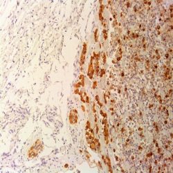
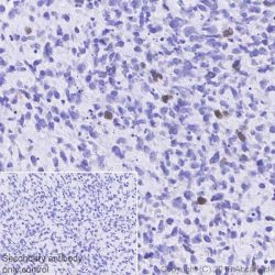
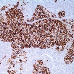
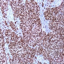
Description and applications: This antibody reacts with the human luteinizing hormone (LH). The adenohypophysis produces six main hormones: prolactin (PRL), growth hormone (GH), adrenocorticotropic hormone (ACTH), thyroid stimulating hormone (TSH), follicle-stimulating hormone (FSH) and luteinizing hormone (LH). The
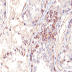
producing cells (approximately 10%) of the two latest hormones are located in the pars tuberalis of the hypophysis. The glycoprotein hormones LH, FSH and TSH are heterodimers with a common alpha subunit and a solely beta subunit that gives a biochemical, immunological and functional specificity as hormone. The LH induces the secretion of sex steroids in the gonads of both sexes; the Leydig cells of the testicle respond synthesizing testosterone and in the ovary, the theca cells respond synthesizing estrogens and inducing ovulation. The luteinizing hormone (LH) is a glycoprotein secreted by both normal and neoplasic hypophysary cells. Therefore, it can be detected in hypophysary adenomas derived from these cells that correspond to chromophobe cells. They represent approximately 6% of hypophysary adenomas.
The LH/FSH secreting adenomas have a slow growth and, at the moment of the diagnosis, are usually large, although they are rarely associated with high serum levels of gonadotropins. The adenomas that only secrete LH are rare. The null cell adenomas and oncocytomas, that do not prove clinically nor biochemically hormone production, show focal expression of LH, FSH, TSH and alpha subunit.Composition: anti-human LH (Luteinizing Hormone) rabbit polyclonal antibody purified from serum and prepared in 10mM PBS, pH 7.4, with 0.2% BSA and
0.09% sodium azide.Immunogen: Purified luteinizing hormone from the human hypophysis.
-
 آنتی بادیهای ایمونوهیستوشیمی
آنتی بادی Cytokeratin 18 (DC-10)
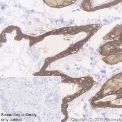
Name: Keratin 18 Antibody Clone DC-10
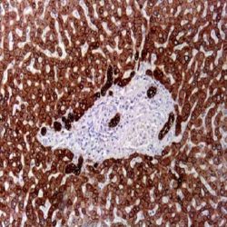
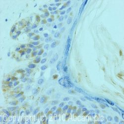
Description and applications: The nomenclature coined in 1982 by Moll and Franke assigned ranges from 1 to 8 for keratin type II (neutral or basic) and between 9 and 21 for type I (acidic). However and because of the high homology among the different molecules is common for a single monoclonal antibody to react with different types of keratins , for example keratin brand AE1 keratins 10, 14 , 15, 16 and 19. This antibody reacts with the acid intermediate filament protein of 45 kDa keratin, identified as Keratin 18. Keratin 18 is generally co-expressed with the keratin 8 in most simple gland epithelia. In general, the keratin 18 does not react with squamous multilayered epithelium. In neoplastic tissues, this antibody reacts with different benign and malignant epithelial lesions. Most adenocarcinomas and basal cell carcinomas express this keratin, whereas it is absent in squamous cell carcinomas.
Composition: anti-Keratin 18 mouse monoclonal antibody obtained from supernatant culture and prediluted in a tris buffered solution pH 7.4 containing 0.375mM sodium azide solution as bacteriostatic and bactericidal. The amount of the active antibody in the sample is 3,3 μg/mL..
Immunogen: Human breast cancer PMC 42 cells.
-
 آنتی بادیهای ایمونوهیستوشیمی
آنتی بادی LIN-28 (EP150)
Name: LIN-28 Antibody Clone EP150
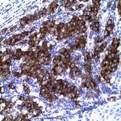
Description and applications: LIN28 is a highly conserved, RNA-binding protein (RBP). It plays an important role as a translational enhancer, leading specific mRNAs to polysomes and therefore increasing the competence of protein synthesis. LIN28 was identified as a negative regulator of miRNA biogenesis and suggested to play a central role in blocking miRNAmediated differentiation in stem cells and certain cancers. LIN28 is expressed by various undifferentiated embryonic cell types. Anti- LIN28 has been used as a sensitive marker for germ cell tumors. The positive staining of LIN28 in yolk sac tumors showed an advantage over OCT4, which is negative in these tumors.
Composition: Anti-human LIN-28 rabbit monoclonal antibody purified from serum and prepared in 10mM PBS, pH 7.4, with 0.2% BSA and 0.09% sodium azide.
-
 آنتی بادیهای ایمونوهیستوشیمی
آنتی بادی Lysozyme (Polyclonal)
Name: Lysozyme Antibody (Polyclonal)
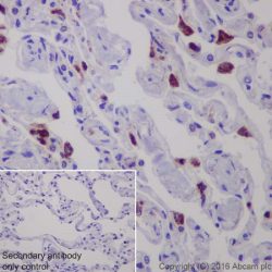
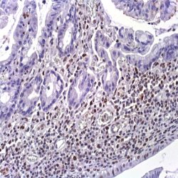
Description and applications: Lysozyme is one of the anti-microbial agents found in human milk, and is also present in spleen, lung, kidney, white blood cells,
plasma, saliva, and tears. Missense mutations in LYZ have been identified in heritable renal amyloidosis. Lysozyme is synthesized predominantly in reactive histiocytes rather than in resting, unstimulated phagocytes. Anti-lysozyme stains myeloid cells, histiocytes, granulocytes, macrophages, and monocytes. It is an important marker that may demonstrate the myeloid or monocytic nature of acute leukemia. Anti-lysozyme may aid in the identification of histiocytic neoplasias, large lymphocytes, and classifying lymphoproliferative disorders.Composition: anti-human Lysozyme rabbit polyclonal antibody purified from serum and prepared in 10mM PBS, pH 7.4, with 0.2% BSA and 0.09% sodium azide
-
 آنتی بادیهای ایمونوهیستوشیمی
آنتی بادی Cytokeratin 5 (SP27)
Name: Cytokeratin 5 Antibody Clone SP27
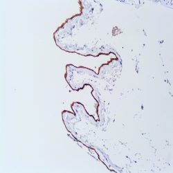
Description and applications:Twenty human keratins are divided into acidic (pI <5.7) and basic (pI >6.0) subfamilies. Members of the acidic and basic subfamilies are found together in pairs. The composition of keratin pairs varies with the epithelial cell type, stage of differentiation, cellular growth environment, and disease state. Many studies have shown the usefulness of keratins as markers in
cancer research and tumor identification. Point mutations in keratin 5 gene can cause various types of epidermolysis bullosa simplex. Keratin 5 is expressed in most epithelial cells, prostate basal cells and epithelial and biphasic mesotheliomas.Composition: Anti-human Cytokeratin 5 rabbit monoclonal antibody purified from serum and prepared in 10mM PBS, pH 7.4, with 0.2% BSA and 0.09% sodium azide.
Immunogen: Synthetic peptide from Cterminus of human cytokeratin 5.
-
 آنتی بادیهای ایمونوهیستوشیمی
آنتی بادی MAP-2 (AP20)
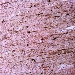
Name: Mouse anti-human MAP-2 Monoclonal Antibody clone AP20
Description and applications: MAP-2 (microtubule associated protein-2) is one of several high molecular weight proteins that play an important role in brain neuron microtubule assembly. In addition to its association with microtubules, MAP- 2 associates with neurofilaments and actin filaments suggesting that it may guide interaction among microtubules, other cytoskeletal elements, and cytoplasmic organelles. MAP-2 is a stringent marker for neurons. In addition, MAP-2 displays intracellular specificity. In the central nervous system, MAP-2 is confined to neuronal cell bodies and dendrites. There are exceptions, however, where some axons stain positive for small amounts of MAP-2. MAP-2 is uniformly distributed throughout the cell when first expressed in cultured neurons but becomes selectively localized as dendritic development proceeds.
Composition: Anti-human MAP-2 mouse monoclonal antibody purified from serum and prepared in 10mM PBS, pH 7.4, with 0.2% BSA and 0.09% sodium azide
Intended use: Immunohistochemistry (IHC) on paraffin embedded tissues. Not tested on frozen tissues or Western-Blotting
-
 آنتی بادیهای ایمونوهیستوشیمی
آنتی بادی MyoD1 (EP212)
Name: Rabbit anti-human Myo-D1 Monoclonal Antibody clone EP212
Description and applications: MyoD1 is a protein with a key role in regulating muscle differentiation. It regulates muscle cell differentiation by inducing cell cycle arrest, a prerequisite for myogenic initiation. The protein is also involved in muscle regeneration. MyoD1 is expressed in developing skeletal muscle tissue but faintly in adult skeletal muscle. In abnormal tissues, it labels tumour cell in rhabdomyosarcoma. MyoD1 is one of the earliest markers of myogenic commitment. Antibody to MyoD1 has been useful to differentiate rhabdomyosarcomas from other tumors. It is a sensitive and specific marker for myogenic differentiation
Composition: anti-human Myo-D1 rabbit monoclonal antibody purified from ascites. Prepared in 10mM PBS, pH 7.4, with 0.2% BSA and 0.09% sodium azide
Intended use: Immunohistochemistry (IHC) on paraffin embedded tissues. Not tested on frozen tissues or Western-Blotting
-
 آنتی بادیهای ایمونوهیستوشیمی
آنتی بادی Mast Cell Tryptase (EP259)
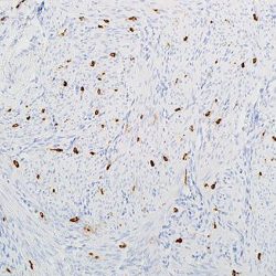
Name: Mast Cell Tryptase Antibody Clone EP259
Description and applications: Human mast cell tryptase, comprises a family of trypsin-like neutral serine proteinases that are predominantly expressed in mast cells. Tryptase has effects on peptides, proteins, cells, and tissues, and many of these actions can ultimately contribute to asthma symptoms. Mast-cell tryptase is found in mast-cell granules and has also been reported to be expressed by peripheral blood basophils at low level. Tryptase has been used as a marker of mast cell activation.
Composition: Anti-human Mast Cell Tryptase rabbit monoclonal antibody purified from serum and prepared in 10mM
PBS, pH 7.4, with 0.2% BSA and 0.09% sodium azide. -
 آنتی بادیهای ایمونوهیستوشیمی
آنتی بادی Myogenin (EP162)
Name: Myogenin-Clone EP162
Description and applications: acid-rich region and a helix-loop-helix (HLH) structure, which can promote muscle development and maintain muscle-specific gene expression by transactivation. Myogenin, one of the myogenic regulatory factors, plays a key role in determining the commitment and differentiation of primitive mesenchymal cells into skeletal muscle. The expression of Myogenin is restricted to cells of skeletal muscle origin, but it is not detected in adult skeletal muscles. It is therefore considered to be an extremely reliable and specific marker for diagnosing rhabdomyosarcomas. Myogenin is positive in virtually 100% of alveolar, sclerosing and botryoides rhabdomyosarcoma and in over 60% of pleomorphic rhabdomyosarcoma and is useful in recognizing adult spindle cell rhabdomyosarcomas. Less than 20% of Wilms tumors, congenital infantile fibrosarcoma, childhood myofibromatosis, synovial sarcoma and desmoid tumors have been reported positive. Ewing sarcoma, rhabdoid tumor, epithelioid sarcoma, leiomyosarcoma, miofibrosarcomas and nodular fasciitis show no reactivity to myogenin.
Composition:anti-human Myogenin rabbit monoclonal antibody purified by protein affinity and prepared in 10mM PBS, pH 7.4, with 0.2% BSA and 0.09% sodium azide.
Immunogen: A synthetic peptide corresponding to residues in human Myogenin was used as an immunogen.
-
 آنتی بادیهای ایمونوهیستوشیمی
آنتی بادی MDM2 (IF2)
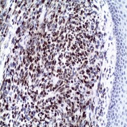
Name: Mouse anti-human MDM2 Monoclonal Antibody clone IF2
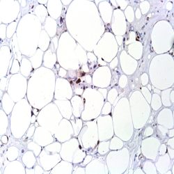
Description and aplications: MDM2 (murine double minute 2) or E3 ubiquitinprotein ligase, is a protein encoded by the chromosomal region 12q13-15, and mediates the ubiquitination of p53 / TP53 leading to its degradation by the proteasome. Due to MDM2 binding to the transcriptional activation domain of p53 / p73 TP53- and p73/ TP73- inhibits G1 arrest of the cell cycle and apoptosis. It also acts as an E3 ubiquitin ligase and ARRB1 itself. Allows nuclear export of p53 / TP53. It promotes ubiquitin-dependent proteasomeindependent degradation of the retinoblastoma protein (RB1). This monoclonal antibody reacts specifically with human MDM2 protein. Immunohistochemical detection of MDM2 and CDK4 overexpression may be helpful to distinguish between well-differentiated liposarcomas and benign tumors of adipocytes (sensitivity / specificity 100% / 58.8% and 68.4% / 88.2%, respectively) and between dedifferentiated liposarcoma from other malignant mesenchymal tumors. MDM2 and CDK4 are expressed in 100% and 93% of dedifferentiated liposarcoma and in only 18% of malignant tumors of peripheral nerve sheaths, mixofibrosarcomas, embryonal rhabdomyosarcoma and 1-6% of poorly differentiated sarcomas (malignant fibrous histiocytoma and leiomyosarcoma). Overexpression of MDM2 / CDK4 proteins is also a highly sensitive marker for the diagnosis of low grade osteosarcomas.
Composition: anti-human MDM2 mouse monoclonal antibody purified from ascites fluid by Protein A chromatography. Prepared in 10mM PBS, pH 7.4, with 0.2% BSA and 0.09% sodium azide
Intended use : Immunohistochemistry (IHC) on paraffin embedded tissues. Not tested on frozen tissues or Western-Blotting
Immunogen: Synthetic peptide derived from the Nterminal region of human MDM2
-
 آنتی بادیهای ایمونوهیستوشیمی
آنتی بادی MELAN A/MART-1 (EP43)
Name: Melan A/Mart-1 Antibody Clone EP43
Description and applications: MART-1, also known as Melan-A,is a melanocyte lineage-specific protein (MART-1; melanoma antigen recognized by T cells 1) recognized by the T lymphocytes of patients with established malignancy. Antibody to MART-1 labels both normal melanocyte and diseased cell with melanocyte differentiation. It is useful for diagnosis of tumors with melanocyte differentiation, especially metastatic melanoma. Identification of MART-1 also opens possibilities for the development of immunotherapies for patients with melanoma.
Composition: Anti-human Melan A/Mart-1 rabbit monoclonal antibody purified from serum and prepared in 10mM PBS, pH 7.4, with 0.2% BSA and 0.09% sodium azide INTENDED USE Immunohistochemistry (IHC) on paraffin embedded tissues. Not tested on frozen
tissues or Western-Blotting. -
 آنتی بادیهای ایمونوهیستوشیمی
آنتی بادی Melanoma-Associated Antigen-1 (MAGE-1) (6C1)
Name: Melanoma-Associated Antigen-1 (MAGE-1)Antibody Clone 6C1
Description and applications: Human malignant neoplasms carry rejection antigens that are recognized by the patients’ autologous,
tumor-directed and specific, cytolytic, CD8+ T lymphocyte clones (CTL). An important group of antigens is coded by the MAGE family of genes. It was identified that melanomas and primary glial brain tumors express common melanoma associated antigens (MAAs). Because MAGE-1 is expressed on a significant proportion of human neoplasms of various histological types (melanoma, brain tumors of glial origin, neuroblastoma, non-small cell lung cancer, breast, gastric, colorectal, ovarian, renal cell carcinomas) and not on normal tissues, the encoded antigen may serve as a marker of early detection and target for cancer immunotherapy.Composition: Anti-human MAGE-1 mouse monoclonal antibody purified from serum and prepared in 10mM PBS, pH 7.4, with 0.2% BSA and 0.09% sodium azide.
-
 آنتی بادیهای ایمونوهیستوشیمی
آنتی بادی MELANOMA (HMB45)
Name:Melanoma Antibody Clone HMB-45
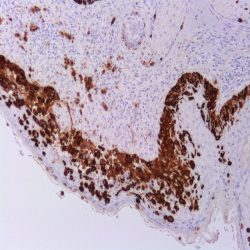
Description and applications:This antibody gives a qualitative assessment of malignant melanoma, which can be extremely difficult
to diagnose. Metastatic amelanotic melanoma can often be confused with a variety of poorly diferentiated carcinomas, large cell lymphomas, and sarcomas. It is also difficult to differentiate melanoma from spindle cell carcinomas and various types of mesenchymal neoplasms. Melanoma antibody stains fetal and neonatal melanocytes, junctional and blue nevus cells, and malignant melanocytes. Typically, a keratin negative, vimentin rich neoplasm, that immunoreacts with antibody to S-100 protein and this melanoma antibody is, with rare exception, a melanoma.As a marker of melanosomes, careful should be taken as malanofages can also be stained.Composition: Anti-human Melanoma mouse monoclonal antibody purified from serum and prepared in 10mM PBS, pH 7.4, with 0.2% BSA and 0.09% sodium azide.
-
 آنتی بادیهای ایمونوهیستوشیمی
آنتی بادی Melanoma Associated Antigen (KBA.62)
Name: anti-Melanoma Associated Antigen Antibody Clone KBA.62
Description and applications: Anti-KBA.62 (Melanoma Associated Antigen) is a novel antimelanoma antibody. Studies thus far have shown a similar sensitivity to melanocytic proliferations as that seen with S-100 protein staining, which is somewhat higher than that seen with HMB-45. This has been con#rmed by one study on a series of 215 sentinel lymph nodes. Moreover, anti-KBA.62 identi#ed 6 patients (3%) who had con#rmed sentinel lymph node metastasis but stained negative for HMB-45. In this setting, the resolution appears to be higher than S- 100 protein in that the staining pattern (membranous) is quite distinct. Interestingly, most cases of desmoplastic and spindle cell melanomas show strongly positive results with anti-KBA.62, unlike that seen with other melanocyte markers. It should be noted that anti-KBA.62 will label occasional endothelial cells which can serve as an internal positive control. A small percentage of well differentiated squamous cell carcinomas of the skin (and lung) have also been noted to stain with this antibody; however, the poorly-differentiated forms of carcinoma do not, thus resolving a greater practical problem in differential diagnosis. Therefore, anti-KBA.62 is a useful additional marker for melanoma, specifically in desmoplastic/spindle cell cases and in the context of micro-metastasis in sentinel lymph node.
Composition: anti-human KBA.62 mouse monoclonal antibody purified from serum and prepared in 10mM PBS, pH 7.4, with 0.2% BSA and 0.09% sodium azide.
-
 آنتی بادیهای ایمونوهیستوشیمی
آنتی بادی Mesothelin (EP140)
Name: Mesothelin Antibody Clone EP140
Description and aplications: Mesothelin gene encodes a 69 kDa precursor protein that progress to a protein of 40-kDa bound to a glycosylphosphatidylinositol (GPI), the mature mesothelin, present on the cell surface. Its biological function is not well understood, but recent studies have shown it forms a strong and specific complex with the MUC16, a union that has been suggested as the basis of ovarian cancer metastasis. Mesothelin is present on the surface of normal mesothelium that coats the pleura, peritoneum and pericardium. Mesothelin is overexpressed in patients with epithelial ovarian carcinoma and malignant mesotheliomas and may be useful to distinguish mesothelioma from other solid tumors. Its overexpression has been observed in other tumors too, including pancreatic carcinomas and cholangiocarcinoma.
Composition: anti-human Mesothelin rabbit monoclonal antibody obtained from supernatant culture and prediluted in a tris buffered solution pH 7.4 containing 0.375mM sodium azide solution as bacteriostatic and bactericidal.
Immunogen: A synthetic peptide corresponding to residues at the C-terminus of human Mesothelin protein
-
 آنتی بادیهای ایمونوهیستوشیمی
آنتی بادی Smooth muscle myosin, heavy chain (EP166)
Name: Myosin, heavy chain Antibody-Clone EP1663
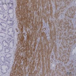
Description and applications: Myosin heavy chain 11 (MYH11) is a smooth muscle myosin belonging to the myosin heavy chain family. It is a subunit of a hexameric protein that consists of two heavy chain subunits and two pairs of non-identical light chain subunits. Myosin heavy chain functions as a major contractile protein, converting chemical energy into mechanical energy through the hydrolysis of ATP. An aberration in this protein is associated with acute myeloid leukemia of the M4Eo subtype. MYH labels smooth muscle cells and myoepithelial cells in various tissues. The immunoreactivity in glial cells of the cerebellum and spermatocytes in the testis is also observed. MYH has been a useful marker for myoepithelial cell as well as smooth muscle cell differentiation. MYH together with p63 are one of the most useful antibodies in establishing the invasive character of breast lesions.
Composition: Anti-human Smooth muscle myosin, heavy chain rabbit monoclonal antibody purified from serum and prepared in 10mM PBS, pH 7.4, with 0.2% BSA and 0.09% sodium azide.
-
 آنتی بادیهای ایمونوهیستوشیمی
آنتی بادی MITF (Microphthalmia-Associated Transcription Factor) (C5+D5)
Name: MITF (Microphthalmia-Associated Transcription Factor) Antibody Clone C5+D5
Description and applications: This antibody reacts with human microphthalmia transcription factor (MITF). The MITF gene (microphthalmia transcription factor, MITF) encodes a nuclear helix-loop-helix transcription factor involved in pigmentation and mast and bone cell development. The MITF gene is essential for the normal development of melanocytes and is responsible for the regulation of the expression of other genes related to the melanocytic lineage: tyrosinase genes, SILV/PMEL17/GP100 (encoding HMB45), MLANA/MART1 (coding for Melan A), TRP-1, and TRP-2. Mutation of the MITF gene in humans causes Waardenburg syndrome IIA and Tietz syndrome, characterized by pigmentation disorders and deafness, the latter due to melanocyte deficiency in the inner ear. There are two known isoforms of MITF that differ in 66 amino acid residues from the amino terminus. The shortest isoform of MITF is melanocyte-specific. MITF-M is expressed in both normal and malignant melanocytes. The other isoforms, MITF-A, MITF-C, and MITF-H, differ structurally from MITF-M at the amino terminus and are expressed in osteoclasts, mast and heart cells, and in B16 melanoma cell lines (although not in other melanoma cell lines). The anti-MITF antibody reacts against the melanocytic and non melanocytic isoforms of the MITF gene, and although it does not distinguish between benign and malignant melanocytic lesions, it shows high sensitivity and specificity as a histopathological marker of melanocytes and melanocyte-derived neoplasms (melanomas). Using MITF and Melan A in the same panel of melanoma markers, the sensitivity (95%) and specificity (100%) values obtained are higher than those obtained using the S100 protein and HMB45, even in cytological samples. MITF, Melan-A, and tyrosinase are sensitive markers for the diagnosis of epithelioid melanomas. Low levels of Melan-A and MITF have been shown to correlate with a poorer prognosis. MITF has demonstrated 100% sensitivity and specificity to detect invasive malignancies of melanocytic origin and to study sentinel lymph nodes.
Composition: Anti-human MITF mouse monoclonal antibody purified from serum and prepared in 10mM PBS, pH 7.4, with 0.2% BSA and 0.09% sodium azide.
-
 آنتی بادیهای ایمونوهیستوشیمی
آنتی بادی MLH1 کلون MLH1/6467 برند Vitro
Name: MLH1 Antibody Clone MLH1/6467
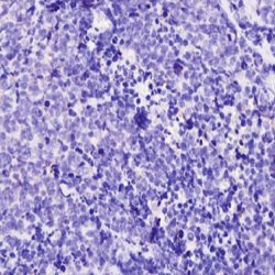
Description and aplications: MLH1 is a mismatch repair gene that is deficient in a high proportion of patients with microsatellite instability (MSI-H). This finding is associated with the autosomal dominant condition known as Hereditary Non-Polyposis Colon Cancer (HNPCC). The anti-MLH1 antibody is useful in screening patients and families for this condition. Colon cancers that are microsatellite unstable have a better prognosis than their microsatellite stable counterparts. Along with PMS2, MSH2 and MSH6, MLH1 antibody is helpful in diagnosis of MSI. An IHC study conducted by Mayo clinic on 535 cases with MSI-high, 90% of the tumors showed loss of MLH1, MSH2 and/or MSH6 expression, while 70% of the remaining cases showed isolated loss of PMS2 expression. Endometrial carcinomas are the most common non-colorectal cancers occur in HNPCC. The most common IHC abnormality in endometrial carcinomas with MSI was concurrent loss of MLH1/PMS2. Adding of PMS2 and MSH6 to MLH1 and MSH2 antibodies increased sensitivity for diagnosis of MSI. Tumors with low-level MSI show unfavourable pathological characteristics compared to tumours with no and tumours with high-level MSI.
Composition: anti-human MLH1 mouse monoclonal antibody obtained from supernatant culture and prediluted in a tris buffered solution pH 7.4 containing 0.375mM sodium azide solution as bacteriostatic and bactericidal.
-
 آنتی بادیهای ایمونوهیستوشیمی
آنتی بادی Napsin A (BS10)
Name: Napsin A Antibody-Clone BS10
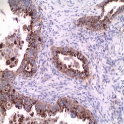
Description and aplications: Napsin is a pepsin-like aspartic proteinase, in the A1 group of the AA class of proteinases. There are two closely related napsins, napsin A and napsin B. Napsin A is expressed as a single chain protein with the molecular weight of approximately 38 kDa. Immunohistochemical studies revealed high expression levels of napsin A in human lung and kidney but low expression in spleen. Napsin A is expressed in type II pneumocytes and in adenocarcinomas of lung. The high specificity expression of napsin A in adenocarcinomas of lung is useful to distinguish primary lung adenocarcinomas from adenocarcinomas of other organs. Napsin A is also useful for the study of renal papillary adenocarcinomas and clear cell adenocarcinomas as it might be positive in high percentage of the cases. Conversely it is expressed by less than 5% of papillary carcinomas of the thyroid.
Composition: anti-Napsin A mouse monoclonal antibody obtained from supernatant culture and prediluted in a tris buffered solution pH 7.4 containing 0.375mM sodium azide solution as bacteriostatic and bactericidal.
-
 آنتی بادیهای ایمونوهیستوشیمی
آنتی بادی MPCyV Large T-Antigen (CM2B4)
Name:MPCyV Large T-Antigen Antibody Clone CM2B4
Description and applications: Merkel cells are round neuroendocrine cells found in skin where they form synaptic complexes with
somatosensory afferent fibres. These complexes are responsible for fine touch and produce various neuroactive substances that act as fast-acting skin neurotransmitters or neuromodulators. In pathological conditions, Merkel cells can proliferate and generate a rare but very aggressive form of skin cancer known as Merkel cell carcinoma (CCM). Around 80% of CMMs are caused by the recently described Merkel cell polyomavirus (MCPyV or MCV), which integrates into the genome of Merkel cells and proliferates clonally. The MCPyV virus is a nonencapsulated DNA virus consisting of two early units (two T antigens, one large and one small) and three late units (VP1, 2, and 3). The large T antigen is the MCPyV’s functional nuclear protein, which has a molecular mass of 125 kDa and is expressed in tumour cells, where it binds to cell cycle suppressor molecules, degrades and sequesters them, promoting their S phase. The large T antigen has natural truncated mutations that also produce functionally active proteins of smaller size. The CM2B4 antibody, which shows no cross-staining with other types of polyomavirus and recognises the MCPyV large T antigen, has been synthesized against a peptide derived from exon 2 of the mentioned antigen’s locus, for which it is highly specific, as well as for its 57kT isoforms; however, it may not detect some of its minor truncated forms. CM2B4, assessed along with a panel of markers including cytokeratins 20 and 7, p63, TTF1, neuronspecific enolase, S100 protein, HMB45, and CD45, facilitates the diagnosis of approximately 80% of classical Merkel cell carcinoma cases. However, most CCM cases with eccrine, squamous, rhabdomyoblastic, leiomyosarcomatous, or neuroblastic divergent differentiation are MCPyV negative and are associated with a worse prognosis of the disease. In comparison with MCPyV level detection using PCR, the antibody has a sensitivity of 95% and a specificity of 83% since, as mentioned above, MCPyV negative CCM cases have been observed, as well as occasional positivity of uncertain meaning in mature lymphocytes, sweat glands, and vascular endothelium.Composition: Anti-human MPCyV Large T-Antigen mouse monoclonal antibody purified from serum and prepared in 10mM PBS, pH 7.4, with 0.2% BSA and 0.09% sodium azide.
برای تماس کلیک کنید
سما تشخیص آریا ، عرضه کننده متنوع ترین و کامل ترین محصولات کیت های آزمایشگاهی