-
 آنتی بادیهای ایمونوهیستوشیمی
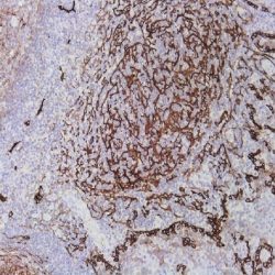
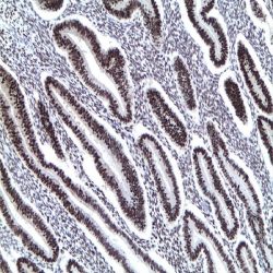
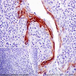
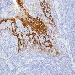
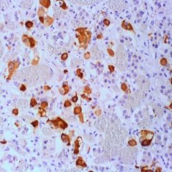
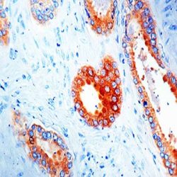
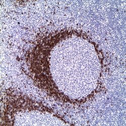
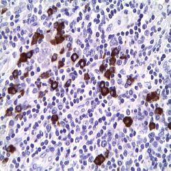
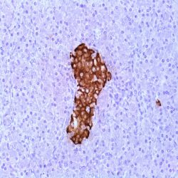
آنتی بادی Podoplanin (D2-40)
Name: Podoplanin Antibody Clone D2-40
Description and applications: D2-40 recognizes the M2A antigen encoded by the gene Prox1 localised on the chromosomal region 1q32.2–q32.3. It consists in a sialoglycoprotein of 40 kDa, containing mucin-type carbohydrate structure required to preserve the antigenicity of this molecule. M2A is expressed in lymphatic vessels and is absent in venous and arterial vascular endothelium. Other normal tissue that express Podoplanin are myoepithelial cell, mesothelial cells, dendritic cells, cells of the biliary ducts, osteocytes, chondrocytes, granulosa cells, squamous epithelium basal cells etc. It is expressed in various types of vascular tumors, seminomas and in some degree embryonal carcinomas, mesotheliomas, haemangioblastomas, schwanomas, dermatofibromas, thricoblastomas etc.
Composition: anti-Podoplanin mouse monoclonal antibody obtained from supernatant culture and prediluted in a tris buffered solution pH 7.4 containing 0.375mM sodium azide solution as bacteriostatic and bactericidal.
-
 آنتی بادیهای ایمونوهیستوشیمی
آنتی بادی Cyclin E1 (EP126)
Name: Rabbit anti-human Cyclin E1 Monoclonal Antibody clone EP126
Description and applications: This antibody reacts with human protein cyclin E. E-type cyclins (cyclin E1 and cyclin E2) are expressed during the late G1 phase of the cell cycle and until the end of the S phase. Cyclin E activity is limited by the passage of the cells through the “R” restriction point, which marks a “point of no return” for cells going into division from a quiescent state (G0) or through G1 and S phases. Cyclin E expression is mainly regulated by gene transcription by members of the E2F transcription factor family and by degradation through the proteasome catabolic pathway. Cyclin E binds and activates Cdk2 kinase and, through phosphorylation of this kinase’s substrate, the socalled pocket proteins, the cyclin/Cdk2 complex regulates the entry into the S phase by initiating a cascade of events that allows the specific expression of S-phase genes. In addition, cyclin E plays a direct role in the initiation of DNA replication and in the control of genomic and centrosome stability. Overexpression of cyclin E shortens the G1 phase of the cell cycle, accelerating progression to the S phase. High levels of cyclin E expression have been associated with the initiation and progression of different human neoplasms, particularly breast cancer, leukaemias, and lymphomas, among others.
Composition: Anti-human Cyclin E1 rabbit monoclonal antibody purified from serum and prepared in 10mM PBS, pH 7.4, with 0.2% BSA and 0.09% sodium azide
Intended use: Immunohistochemistry (IHC) on paraffin embedded tissues. Not tested on frozen tissues or Western-Blotting
Immunogen: Recombinant protein corresponding to residues from human protein cyclin E.
-
 آنتی بادیهای ایمونوهیستوشیمی
آنتیبادی PARP-1 – پلیکلونال
Name: Poly (ADP-Ribose) Polymerase 1 Antibody
Description and applications: PARP (EC 2.4.2.30) is a nuclear protein with a molecular mass of 116 kDa and two zinc finger motifs which binds to DNA and specifically detects DNA nicks and breaks produced by different genotoxic agents. PARP catalises the ADP-ribosylation of proteins using NAD+ as a substrate. PARP activation is a consequence of ischemic injury and leads to intracellular depletion of NAD+, which can only be replaced by ATP consumption. Ischemia/Reperfusion (I/R) injury results in substantial DNA damage and cells need to consume large amounts of ATP to sustain reparative poly-ADP-ribosylation. Proteolysis of PARP to its stable 85-kDa fragment is an early marker of programmed cell death (apoptosis), mediated by caspase 3 or CPP32. Cleavage occurs between Adp216 and Gly217, a site in PARP conserved across species. This antibody reacts with human, mouse and rat PARP. PARP-1 determination may be useful for detecting cells undergoing DNA replication or repair. In ischemia/reperfusion situations, it may serve as an early marker of cell death. Studies on chemical carcinogenesis have involved PARP on its pathogenesis. Recently, PARP has been considered a therapeutic target in some types of malignancies such as breast carcinomas with BRCA1/2 mutation.
Composition: anti-human Poly (ADP-Ribose) Polymerase 1 rabbit polyclonal antibody purified from serum and prepared in 10mM PBS, pH 7.4, with 0.2% BSA and 0.09% sodium azide
Intended use: Immunohistochemistry (IHC) on paraffin embedded tissues. Not tested on frozen tissues or Western-Blotting
Immunogen: Synthetic peptide derived from the Nterminal region of human protein PARP.
-
 آنتی بادیهای ایمونوهیستوشیمی
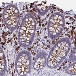
آنتی بادی Cytokeratin (CAM 5.2)
Name: Keratin CAM5.2 Antibody Clone CAM5.2
Description and applications: The nomenclature coined in 1982 by Moll and Franke assigned ranges from 1 to 8 for keratin type II (neutral or basic) and between 9 and 21 for type I (acidic). However and because of the high homology among the different molecules is common for a single monoclonal antibody to react with different types of keratins. Keratin CAM 5.2 antibody reacts with 38-50 kD molecular weight keratins present in most simple and ductal epithelia. This antibody is useful for identifying normal and neoplastic epithelial cells in histological sections. Positive stain is obtained in adenocarcinomas of the breast, bowel, liver, renal, neuroendocrine carcinomas (eg Merkel cell tumor), mesotheliomas , germ cell tumors (except seminoma), synovial and epithelioid sarcoma. Some tissues of non-epithelial origin can be immunostained as smooth muscle, some breast sarcomas, meningiomas and malignant neuroblastomas. CAM 5.2 doesn´t stain the stratified epitheliums and does not react with carcinomas arising in the esophagus, vagina and cervix. Negative results were also obtained in lymphomas, melanomas, meningiomas, sarcomas and seminomas.
Composition: IgG2a immunoglobulin, obtained from mouse ascites purified by chromatography and diluted in Tris Buffer, pH 7.3-7.7, with 1% BSA and <0.1% Sodium Azide.
Imunogen: human colorectal carcinoma cell line HT24.
-
 آنتی بادیهای ایمونوهیستوشیمی
آنتی بادی Progesterone Receptor (16)
Name: Mouse Anti-Progesterone Receptor (PR) Monoclonal Antibody clone 16
Description and applications: This mouse monoclonal antibody clone 16 it has been demonstrated to react with both A and B forms of human progesterone receptor by Western blotting procedure. In normal conditions, the nuclear expression of PR occurs in the acinary cells of the breast, endometrium (proliferative phase glands and stroma are strongly positive while absence or variable expression is noted in the secretory phase glands and stroma) myometrium and superficial cervical stroma. PR along with estrogen receptors, Cerb-B2, Ki-67 and p53 are considered to have prognostic value in breast cancer. As the neoplastic growth is hormone dependent, the increased expression of PR is associated with a better response to Tamoxifen-treatment in breast cancers. PR determination is also useful in gynecologic pathology where is positive in endometrial adenocarcinoma, stromal sarcomas or leiomyomas. Meningiomas also show positivity for the PR.
Composition: anti-PR mouse monoclonal antibody obtained from supernatant culture and prediluted in a tris buffered solution pH 7.4 containing 0.375mM sodium azide solution as bacteriostatic and bactericidal.
Immunogen: Recombinant protein encoding N-terminal region of human progesterone receptor.
-
 آنتی بادیهای ایمونوهیستوشیمی
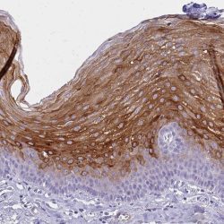
آنتی بادی Cytokeratin 10 (SP99)
Name: Cytokeratin 10 Antibody Clone SP99
Description and applications: Cytokeratin 10 is a type I cytokeratin, expressed by suprabasal cells of stratified and cornified epithelia. Its defects are implicated in bullous congenital ichthyosiform rythroderma, epidermolytic hyperkeratosis and diffuse non-epidermolytic palomoplantar keratoderma.
Composition: Anti-human Cytokeratin 10 rabbit monoclonal antibody purified from serum and prepared in 10mM PBS, pH 7.4, with 0.2% BSA and 0.09% sodium azide,
Immunogen: Synthetic peptide corresponding to the C-terminus of human keratin 10.
-
 آنتی بادیهای ایمونوهیستوشیمی
آنتی بادی Cytokeratin 13 (EP69)
Name: Keratin 13 Monoclonal Antibody Clone EP69
Description and aplications: The nomenclature coined in 1982 by Moll and Franke assigned ranges from 1 to 8 for cytokeratin type II (neutral or basic) and between 9 and 21 for type I (acidic). However and because of the high homology among the different molecules is common for a single monoclonal antibody to react with different types of keratins , for example keratin brand AE1 keratins 10, 14 , 15, 16 and 19. The 54 kD keratin 13 is the major acidic keratin that is assembled in vivo with type 4 and is expressed in the suprabasal cells of the multilayered keratinizing epithelium of mucous membranes, transitional epithelium, pseudostratified epithelium and myoepithelial cells. The monoclonal anti- keratin 13 antibody has been used as a marker for non-keratinized squamous epithelium and is expressed in various squamous metaplasias, but is down regulated in squamous dysplasia and squamous carcinoma. It is useful to identify epithelial derived tumors of the trachea, apocrine and eccrine sweat glands, salivary glands, stem cells of the endocervical glands, bladder, exocervix , tongue, esophagus, anus and basal layer of the epidermis.In addition, mutations in the gene sequence of keratin 13 have been implicated in genetic disease known as autosomal dominant white sponge nevus of the oral cavity.
Composition: anti-Keratin 13 rabbit monoclonal antibody obtained from supernatant culture and prediluted in a tris buffered solution pH 7.4 containing 0.375mM sodium azide solution as bacteriostatic and bactericidal. The amount of the active antibody was not determined.
Immunogen: Synthetic peptide corresponding to human keratin 13 residues.
-
 آنتی بادیهای ایمونوهیستوشیمی
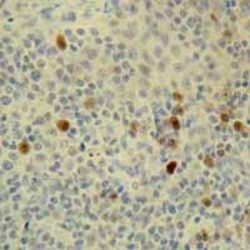
آنتی بادی Prolactin (EP193)
Name: Prolactin Antibody clone EP193
Description and applications: Prolactin is a peptide hormone secreted by the anterior pituitary that is necessary for the proliferation and differentiation of the mammary glands. Prolactin also acts in a cytokine-like manner and as an important regulator of the immune system. Prolactin has important cell cycle related functions as a growth, differentiating and antiapoptotic factor. Prolactin is secreted by lactotrophs in the anterior pituitary. Prolactin producing cells make up approximately 20 percent of the pituitary. Elevated counts of these cells have been observed in pregnant women, newborns and in multiparous women. An antibody to prolactin is useful for the identification of pituitary tumors.
Composition: Anti-human Prolactin rabbit monoclonal antibody purified from serum and prepared in 10mM PBS, pH 7.4, with 0.2% BSA and 0.09% sodium azide
Immunogen: A synthetic peptide corresponding to residues of human Prolactin protein
-
 آنتی بادیهای ایمونوهیستوشیمی
آنتی بادی Prostate-specific antigen (PSA) (ER-PR8)
Name: PSA antibody Clone ER-PR8
Description and applications: PSA is an antigen present in prostatic tissue and in the majority of prostatic carcinomas. This antibody recognizes primary and metastatic prostatic neoplasms and rarely in tumors of nonprostatic origin. These include breast and a minority of salivary gland tumors. The antigen is a 33-34 kD glycoprotein that is restricted to epithelial cells of the prostate. Because of the high specificity of PSA for prostate glandular epithelium, the antibody is useful to identify prostatic carcinomas of the prostate and their frequent infiltration of the surrounding organs (eg .: rectum and urinary bladder). Rectal adenocarcinomas, urothelial carcinoma or adenocarcinoma of the urinary bladder did not express PSA. The loss of immunoreactivity for PSA which can occur in poorly differentiated neoplasms cannot completely exclude the diagnosis of prostate cancer with a negative PSA. Another important use is for differential diagnosis and determine the origin of the primary tumor with metastases of unknown origin, especially lymph node and bone. PSA can be used together with the Prostein, which has at least the same sensitivity and is slightly more specific tha PSA.
Composition: anti-human PSA mouse monoclonal antibody purified from supernatant diluted in tris bffered saline, pH 7.3-7.7, with protein base, and preserved with sodium azide.
-
 آنتی بادیهای ایمونوهیستوشیمی
آنتی بادی Prostatic Acid Phosphatase (PASE/4LJ)
Name: Prostatic Acid Phosphatase Antibody Clone PASE/4LJ
Description and Applications: Anti-PSAP reacts with prostatic acid phosphatase in the glandular epithelium of normal and hyperplastic prostate, carcinoma of the prostate and metastatic cells of prostatic carcinoma. This marker may be helpful in pinpointing the site of origin in cases of metastatic carcinoma of the prostate, and is considered a more sensitive marker than PSA. However, it also offers less specificity. Nevertheless, PSAP complements PSA in the right clinical context.
Composition: Anti-human Prostatic Acid Phosphatase mouse monoclonal antibody purified from serum and prepared in 10mM PBS, pH 7.4, with 0.2% BSA and 0.09% sodium azide
-
 آنتی بادیهای ایمونوهیستوشیمی
آنتی بادی Human IgD (Polyclonal)
Name: IgD Antibody (Polyclonal)
Description and applications: Anti-IgD antibody reacts with surface immunoglobulin IgD delta chains. This antibody is useful when identifying leukemias, plasmacytomas, and B-cell lineage derived lymphomas (in particular Marginal Zone Lymphoma). Cytoplasmic staining is easily identified on paraffin tissue. Surface immunoglobulin is dificult to demonstrate on paraffin sections but can be demonstrated on frozen sections.
Composition: anti-human IgD rabbit polyclonal antibody purified from serum and prepared in 10mM PBS, pH 7.4, with 0.2% BSA and 0.09% sodium azide
-
 آنتی بادیهای ایمونوهیستوشیمی
آنتی بادی Human IgG (E20-V)
Name: IgG Antibody Clone E20-V
Description and applications: The antibody labels the IgGs contained in the plasma cells and their precursors. In general, membrane bound immunoglobulins, extracellular immunoglobulins bound to connective tissue or blood vessels, and immunocomplexes can only be demonstrated in frozen tissues. Plasma cells may not
be intensely stained in frozen tissue since the immunoglobulins are distributed diffusely through the cytoplasm of these cells. This antibody is useful for identifying leukemias, plasmacytomas and B cells derived from Hodgkin lymphomas. Due to the restrictions on the expression of light and heavy chains in these diseases, demonstration of B-cell lymphomas is possible with clonal rearangements studies. Another application isComposition: Anti-human IgG rabbit monoclonal antibody purified from serum and prepared in 10mM PBS, pH 7.4, with 0.2% BSA and 0.09% sodium azide.
-
 آنتی بادیهای ایمونوهیستوشیمی
آنتی بادی Human IgM (Polyclonal)
Name: IgM Antibody (Polyclonal)
Description and applications: Anti-IgM antibody reacts with surface immunoglobulin IgM mu chains. IgM is one of the predominant surface immunoglobulins on B-lymphocytes. This antibody is useful when identifying lymphomas, plasmacytomas, and B-cell lineage derived Hodgkin’s lymphomas. Due to the restricted expression of heavy and light chains in these diseases, demonstration of B-cell lymphomas is possible with clonal gene rearrangement studies.
Composition: Anti-human IgM rabbit polyclonal antibody purified from serum and prepared in 10mM PBS, pH 7.4, with 0.2% BSA and 0.09% sodium azide
-
 آنتی بادیهای ایمونوهیستوشیمی
آنتی بادی IgG4 (EP138)
Name: IgG4 Antibody Clone EP138
Description and applications: Human IgG4, one of four subclasses of IgG, contains a gamma 4 heavy chain and a hinge region that is shorter than that of IgG1. No allotypes have been detected on the heavy chains of IgG4. Its two primary effector functions are activating complements and binding to the FcgR of effector cells to initiate phagocytosis. Human IgG4 accounts for less than 6% of the total IgG serum level. Recent studies show that serum levels and immunohistochemistry staining with IgG4 antibody is a useful diagnosis marker for IgG4-related sclerosing diseases. A new concept of IgG4-related systemic disease (ISD) has been established recently. The ISD is characterized by elevated serum IgG4 levels and extensive IgG4+ plasma cell infiltrate in pancreas and/or in other organs, including peripancreatic tissue, bile duct, gallbladder, portal area of the liver, gastric mucosa, colonic mucosa, salivary glands, lymph nodes, and bone marrow. Immunohistochemistry analysis of IgG4 is useful for identifying ISD.
Composition: anti-human IgG4 rabbit monoclonal antibody purified from serum and prepared in 10mM PBS, pH 7.4, with 0.2% BSA and 0.09% sodium azide.
-
 آنتی بادیهای ایمونوهیستوشیمی
آنتی بادی Immunoglobulin J chain (SP105)
Name: J Chain Immunoglobulin Antibody Clone SP105
Description and applications: J chain is a small glycopeptide and is structurally unrelated to heavy or light chains, but is synthesized by all plasma cells that secrete polymeric immunoglobulins. J chain is linked to IgA and IgM by disulfide bonds, however, it has been detected in IgG- and IgD-containing cells. J chains are present in a large proportion of the immunoglobulin-positive cells in the germinal centers of the tonsil and lymph node. B cells secrete J chain at an early stage of differentiation with the expression persisting in those cells destined to produce IgA or IgM. It has been shown that a significant proportion of mesangial immunoglobulin deposits of IgA nephropathy are dimeric and therefore positive for this antibody.
Composition: anti-human J Chain immunoglobulin rabbit monoclonal antibody purified and prepared in 10mM PBS, pH 7.4, with 0.2% BSA and 0.09% sodium azide.
-
 آنتی بادیهای ایمونوهیستوشیمی
آنتی بادی IMP 3 (EP286)
Name: IMP 3 Antibody Clone EP286
Description and applications: IMP-3, known as Insulin-like growth factor 2 (IGF-II) mRNA-binding protein 3, is an oncofetal protein that stabilizes IGF-II mRNA for trafficking and plays an important role in cell growth and migration. This 65- 70 kDa protein is expressed normally in developing tissues during early embryogenesis in a variety of fetal tissues including the liver, lung kidney, thymus, and placenta, but at low or undetectable levels in normal adult tissues. Recent studies have demonstrated IMP-3 expression in various malignant tumors of the lung, gastrointestinal tract, liver, endometrium, and bladder, while undetectable in adjacent benign tissues. IMP-3 may have a critical role in tumor proliferation, invasion and metastasis, and has been suggested to be an independent marker for poor prognosis in patients with clear cell carcinomas.
Composition: Anti-human IMP 3 rabbit monoclonal antibody purified from serum and prepared in 10mM PBS, pH 7.4, with 0.2% BSA and 0.09% sodium azide.
-
 آنتی بادیهای ایمونوهیستوشیمی
آنتی بادی Inhibin Alpha (R1)
Name: Inhibin Alpha Antibody (Clone R1)
Description and applications: This antibody recognizes the 32kDa alpha subunit of human inhibin. Inhibin alpha is expressed in a range of tissues including prostate, brain, adrenal gland, testis and ovary. The antibody may be of value in the differentiation of adrenocortical tumors, placental and gestational trophoblastic lesions and sex cord stromal tumors.
Composition: anti-human Inhibin alpha mouse monoclonal antibody purified from ascites. Prepared in 10mM PBS, pH 7.4, with 0.2% BSA and 0.09% sodium azide.
-
 آنتی بادیهای ایمونوهیستوشیمی
آنتی بادی Insulin (EP125)
Name: Insulin Antibody Clone EP125
Description and applications: Insulin is a hormone that regulates glucose homeostasis. It increases cell permeability to monosaccharides, amino acids and fatty acids, and it accelerates glycolysis, the pentose phosphate cycle, and glycogen synthesis in liver. It is synthesized in the beta cell of the pancreas. The antibody labels both normal and neoplastic insulinproducing cells. It is useful in identifying insulinoma.
Composition: Anti-human Insulin rabbit monoclonal antibody purified from serum and prepared in 10mM PBS, pH 7.4, with 0.2% BSA and 0.09% sodium azide.
-
 آنتی بادیهای ایمونوهیستوشیمی
آنتی بادی Involucrin (SY5)
Name: Involucrin Antibody Clone SY5
Description and applications: Involucrin is expressed in a range of stratified squamous epithelia, including the cornea. In normal epidermis, it is first expressed in the upper spinous layers, and in keratinocyte cultures it is expressed by all cells that have left the basal layer. Involucrin expression is abnormal in squamous cell carcinomas and premalignant lesions, and is reduced in severe dysplasias of the larynx and cervix.
Composition: Anti-human Involucrin mouse monoclonal antibody purified from serum and prepared in 10mM PBS, pH 7.4, with 0.2% BSA and 0.09% sodium azide.
-
 آنتی بادیهای ایمونوهیستوشیمی
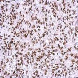
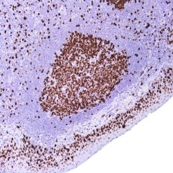
آنتی بادی KI 67 (SP6)
Name: Rabbit Anti-Human Ki-67 Monoclonal Antibody clone SP6
Description and aplications: This rabbit monoclonal antibody (clone SP6) reacts with a nuclear antigen which is expressed in every proliferating human cell. It is expressed mainly during the cell cycle phases, including late G1, S, G2 and M phase. The cells in the G0 phase (quiescent cells) are not immunostained. This antibody is useful to identify the degree of cell proliferation. Is a marker to define the growth fraction in benign and malignant tissues, such as prostate, breast or lymphoid tissues. Therefore, it can play an important role as a predictor of malignancy.
Composition: anti-Ki67 rabbit monoclonal antibody obtained from supernatant culture and prediluted in a tris buffered solution pH 7.4 containing 0.375mM sodium azide solution as bacteriostatic and bactericidal.
Intended use : Immunohistochemistry (IHC) on paraffin embedded tissues. Not tested on frozen tissues or Western-Blotting
Immunogen: Synthetic peptide derived from the Cterminus of human Ki67 protein
Species reactivity: In vitro diagnostics in humans. Not tested in other species
برای تماس کلیک کنید
سما تشخیص آریا ، عرضه کننده متنوع ترین و کامل ترین محصولات کیت های آزمایشگاهی