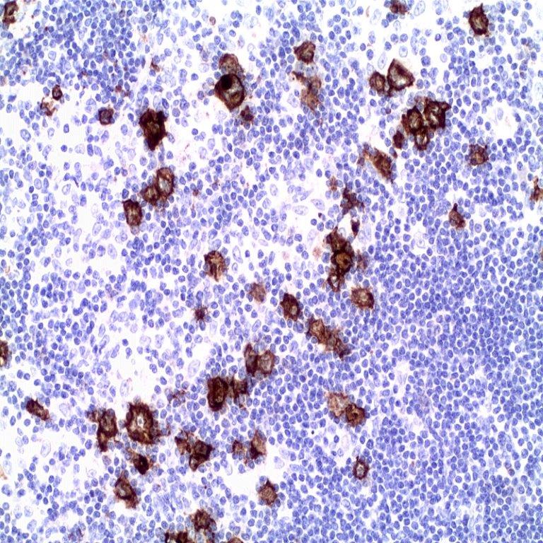
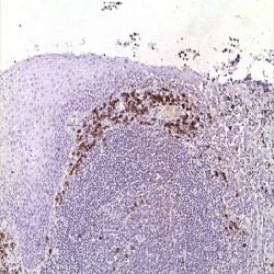
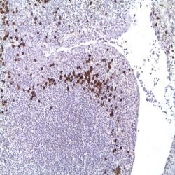
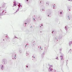
Specificity: The Epstein-Barr virus (EBV) also called human herpes virus type 4, which was discovered by Epstein, Achong and Barr in 1964 in cell cultures of Burkitt’s lymphoma, is a virus of the herpes family of 120-180 nm in diameter composed of a double-stranded DNA of 192,000 bp containing 85 genes. It is one of the virus with the major capacity of infection in humans in which, once incorporated persists throughout the individual’s life with a prevalence of 90% in adults. Structurally, the viral genome is contained in a nucleocapsid that in turn, is surrounded by a protein coat containing a lipid component and the glycoprotein gp350. Like other herpes virus, EBV infects cells that are not actively dividing, in this case resting mature B lymphocytes. For this, the gp350 envelope virus initiates adhesion to the surface of the host cell by binding to the CR2 molecule (CD21), which facilitates activation of the glycoprotein gp42 which, together with the HLA class II and gp25 and gp85 proteins of the virus form a complex which is used for penetration on B lymphocytes. Once the virus enters the cell, virus capsid dissolves and the viral genome is transported to the nucleus where it is inserted by the action of DNA polymerase of the own host cells. Once in the nucleus of the host cell (usually the memory B cells), the virus enters in a period of latency during which no new virions are produced and expresses only a limited number of their genes (about 11) encoding various molecules. Among them there are two types of messenger non-coding RNA, six nuclear proteins and two membrane proteins. This remarkable surface protein expression makes difficult the recognition by cytotoxic T cells of the infected cells. EBV nuclear antigen (EBNA) 1 is the protein that allows the virus to remain within the B cell as a circular DNA episome. By contrast, the antigen EBNA 2 regulates the expression of latent membrane protein (LMP) 1 and 2 of the EBV as well as the proteins contributing to the growth and transformation of B lymphocytes. LMP-1 protein acts as an oncogene that mimics the function of the CD40 molecule on the surface of the lymphocytes and is able to bind to various receptors of tumor necrosis factor, resulting in the activation of nuclear factor kB and c-jun and numerous other cell adhesion molecules; this way EBV enhance the cytokine production capacity and generates a powerful cellular proliferation signal. Additionally, LMP-2 EBV protein, which prevents EBV reactivation from latency stage by blocking the phosphorylation of tyrosine kinases, guarantees the persistency of a sufficient population of infected cells to ensure the survival of the virus. EBER 1 and 2 RNAs of EBV, which do not encode proteins and therefore cannot be transcribed, play an important role in oncogenesis and the resistance of infected cells to programmed cell death and apoptosis. EBER genes are transcribed by the viral RNA polymerase and are most abundant EBV molecules in Burkitt’s lymphoma and other latently infected cells. Functionally the type EBER RNAs act as transcription factors of interleukin-10 which is perhaps the most important existing autocrine growth factor in Burkitt lymphoma. At certain times, or only in certain patients from the beginning, the virus can enter a lytic phase or of productive infection that results in the generation of large numbers of infectious virions. This requires that the virus genome passes from circular to linear with the early activation of many genes, including the viral DNA polymerase itself. Primary EBV infections usually occur in the childhood and are asymptomatic or cause mild nonspecific symptoms. On the contrary the adolescent´s and adult´s infections quite often cause infectious mononucleosis, which can occasionally cause serious complications such as haemolytic anaemia, thrombocytopenia, myocarditis, hepatitis, splenic rupture, or neurological complications such as Guillain-Barré syndrome or encephalitis. In these cases the virus is transmitted through saliva and initially infects epithelial cells of the oropharynx where it replicates until infects B lymphocytes. Furthermore, dormant infected Blymphocytes spread throughout all the lymphopoietic organs, which are the source of continuous propagation of the virus. Even if normally the infection is controlled by cytotoxic T cells, in patients with X-linked lymphoproliferative EBV infection may be fatal. The digoxigenin-labeled probe included in this kit is a mixture of 5 RNA oligonucleotides complementary to type 1 and 2 EBER of EBV and has been created for the detection of the presence of EBV-infected cells in a latent stage. However, its use also produce tissue hybridization signals with EBV lytic infections.
Components and Reagents Included In The Kit:
MAD-001897QK
Proteinase K Concentrated Solution (10 mg/mL) 20 tests
Epstein Barr Virus (EBER) CISH PNA Probe 20 tests
Digoxigenin (HY-A.1) 20 tests
Peroxidase Blocking Reagent 20 tests
Primary Antibodies Amplifier Master 20 tests
Master Polymer Plus HRP 20 tests
DAB Substrate Buffer and DAB Chromogen Concentrate 20 tests
DAB Enhancer 20 tests
Reviews
There are no reviews yet.