برای تماس کلیک کنید

به وب سایت سما تشخیص آریا خوش آمدید.

جستجوی پیشرفته
جستجوی عینی کلمه
جستجو در محتوا
جستجو در عنوان
دنبال محصول یا مطلبی میگردید؟ در این کادر جستجو کنید

پروب فیش IGH-MYC Fusion/Translocation FISH Probe

INTRODUCTION: Rearrangements and abnormal expression of the MYC gene (also known as MRTL, MYCC, c-Myc or bHLHe39) have been described in Burkitt’s lymphoma, where its positivity has diagnostic utility within a clinical, morphological and immunohistochemical context. On the other hand, in large or non-classifiable diffuse B-cell lymphoma, the MYC translocation has a prognostic value and dictates a more aggressive treatment of the neoplasm. On the other hand, together with the determination of the translocation of the BCL2 and BCL6 genes, it aims to establish the double or triple-hit character of the neoplasm. Other hematological neoplasms, myelomas, as well as breast, cervical, colon, ovarian and other tumors may present MYC translocation.
Most Burkitt lymphoma cases show the t(8;14)(q24;q32) [MYC-IGH] and less commonly the t(8;22)(q24;q11) or t(2;8)(p12;q24). The translocation is present in both the endemic African Burkitt lymphoma and in the non-endemic tumor type (Europe, America, and Japan). In case the Burkitt lymphoma infiltrated the bone marrow (leukemic phase) the MYC-translocation can be demonstrated in the bone marrow or blood as well.
INTENDED USE: The IGH-MYC Fusion/Translocation FISH probe kit t is designed to detect rearrangements involving the human IGH locus and MYC gene, located on chromosome bands 14q32.33 and 8q24.21, respectively.

PROBE DESCRIPTION: The IGH Probe of the IGH-MYC Fusion/Translocation FISH Probe Kit labeled with the fluorochrome CytoGreen covers the 5’ and the center sequences of the IGH locus, and it also covers the 3’ (end) part and the neighboring downstream region. The MYC Probe of the IGH-MYC Fusion/Translocation FISH Probe Kit labeled with the fluorochrome CytoOrange covers some genomic sequences downstream of the 5’ (start) portion of the MYC gene, and it also covers the sequences adjacent to the 3’ end of the gene. The probe set is optimized to reveal translocations between the two regions.

کمپانی سازنده: Master Diagnostica

کشور: اسپانیا

حجم پروب: 100میکرولیتر برای 10 تست در قالب (RTU)

فیلترهای مورد نیاز:

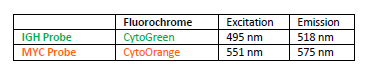
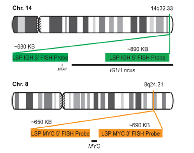
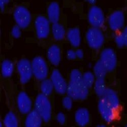
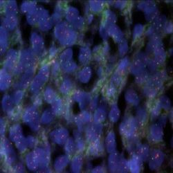
روش و پروتکل انجام تست فیش (FISH): این پروب برای بررسی و استفاده از تکنیک هیبریدازیسیون (FISH) بر روی بافت های پارافین قابل استفاده می باشد و پیشنهاد می شود برای استفاده از این پروب فیش از کیت آماده سازی Master diagnostica FISH Pretreatment و پروتکل های پیشنهادی آن استفاده نمایید.

توجه: پیشنهاد می شود که قبل از استفاده از ویال, آن را با کمک سمپلر مخلوط نموده و سپس استفاده کنید.

زمان و دمای دناتوره شدن: 5 دقیقه – 80 درجه سانتی گراد

زمان دمای هیبرید شدن: 43 درجه سانتی گراد – 16 ساعت
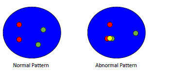
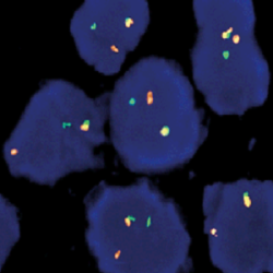
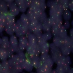
تفسیر نتایج:

به طور کلی جهت تفسیر نتایج بر روی این پروب فیش (FISH) پیشنهاد می شود که معیارهای ارزیابی پذیرفته شده که در سرتاسر جهان استفاده میشود را مورد بررسی قرار دهید.

لطفا کاتالوگ پروب را از همین صفحه دانلود کنید و اطلاعات بیشتر را مطالعه فرمایید.


دانلود کاتالوگ

.well.c2a3 .btn { padding: 11px 30px; font-size: 11pt; } .well.c2a3 .media-body { font-size: 11pt; } .well.c2a3 .wpdm_icon { height: 42px; width: auto; }

Reviews

There are no reviews yet.

Be the first to review “پروب فیش IGH-MYC Fusion/Translocation FISH Probe”

نشانی ایمیل شما منتشر نخواهد شد. بخش‌های موردنیاز علامت‌گذاری شده‌اند *

پیمایش به بالا