-
 آنتی بادیهای ایمونوهیستوشیمی
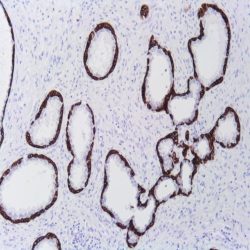
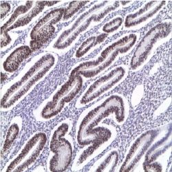
آنتی بادی Cytokeratin 5/6 (EP24/EP67)
.(Name: Mouse anti-human Cytokeratin5/6 Monoclonal Antibody (Clone EP24/EP67
Description and applications: Keratins are cytoplasmic intermediate filament proteins expressed by epithelial cells. CK5 is a type II cytokeratin. Loss-of-function mutations in the keratin 5 gene (KRT5) affected family members and in six unrelated patients with Dowling-Degos disease (DDD), an autosomal dominant genodermatosis. This suggests a crucial role for keratins in the organization of cell adhesion, melanosome uptake, organelle transport, and nuclear anchorage. CK5 labels myoepithelial cells of breast and prostate basal cells. CK5 and calretinin have been useful in different studies as immunohistochemical markers suggestive of mesothelioma, and their expression is analyzed for the histological differentiation with adenocarcinomas, especially when confronting metastatic tumors of unknown origin. The human type II Cytokeratin 6 (CK6; 56 kDa) is well known for its strong induction in stratified epithelia that feature an enhanced cell proliferation rate or abnormal differentiation during wound healing, in several diseases (e.g. psoriasis, actinic keratosis) and cancer. CK6 is expressed on stratified epithelia including oral mucosa, esophagus, basal layer of epidermis, the outer root sheath of hair follicles, and in glandular epithelia. CK6 is a marker of hyperproliferative and activated keratinocytes found in psoriasis.Anti-CK6 paired with the CK5 antibody is useful for differentiating mesothelioma (positive) from lung carcinoma (negative) or metastatic carcinoma (negative) in the pleura. An antibody against CK5/6 has also been used to distinguish usual ductal hyperplasia of the breast (strong staining) from solid papillary DCIS (negative).
Immunogen: A synthetic peptide.
-
 آنتی بادیهای ایمونوهیستوشیمی
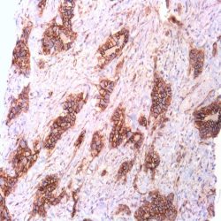
آنتی بادی BRST-2/GCDFP-15 (EP95)
Name: Rabbit anti-human Gross Cystic Disease Fluid Protein-15 (BRST-2/GCDFP-15) Monoclonal Antibody (Clone EP95).
Description and Applications: Gross cystic disease fluid protein (GCDFP-15), also called prolactininducible protein (PIP), is a single polypeptide chain with a versatile function in human reproductive and immunological systems. GCDFP-15 binds to CD4, exerts a potent inhibition on T lymphocyte apoptosis mediated by CD4/T-cell receptor (TCR) activation, and carries a fibronectinspecific aspartyl protease activity. It is up regulated by prolactin and androgens, while it is down regulated by estrogen. In normal adult tissues, GCDFP-15 expression was found in all apocrine, lacrimal, ceruminous, and Moll’s glands and in numerous serous cells of the submandibular, sublingual, and minor salivary glands. The serous cells of nasal and bronchial glands were also positive. It is used as a marker of apocrine differentiation. GCDFP-15 has been found in the cyst fluid of cystic breast disease and primary and metastatic breast cancer, and considered a highly specific marker for identification of breast cancer. GCDFP-15 expression has also been found in other cancer types including salivary glands, sweat glands, prostate, and lung.
COMPOSITION: Anti-human BRST-2/GCDFP-15 rabbit monoclonal antibody purified from serum and prepared in 10mM PBS, pH 7.4, with 0.2% BSA and 0.09% sodium azide.
IMMUNOGEN: A synthetic peptide corresponding to residues in human Gross Cystic Disease Fluid Protein15 (GCDFP-15) protein.
-
 آنتی بادیهای ایمونوهیستوشیمی
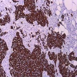
آنتی بادی Cytokeratin 34betaE12 (34BE12)
Name: Cytokeratin HMW Antibody clone 34BE12
Description and applications: The proteins generically known intermediate filaments measuring between 7 and 22 nm diameter (ie with a size between -5 to 7 nm like actin and 22 to 25 nm like tubulin), are part, together with the above mentioned, of the vertebrates cytoskeleton. This superfamily comprises six subfamilies of molecules with different tissue expression. Cytokeratins constitute two homology groups (I and II) and in humans are encoded by more than 49 different genes on chromosomes 17 (I) and 12 (II). The nomenclature coined in 1982 by Moll and Franke assigns ranges of 1 to 8 for type II cytokeratins (neutralor basic) and between 9 and 21 for type I (acidic). Normally, keratins are assembled in heterodimers I / II and are co-expressed in pairs specific to each tissue. This antibody recognizes keratins of 68, 58, 56.5 and 50 kD obtained from the skin stratum corneum. By immunohistochemistry, the antibody reacts with squamous epithelium, ductal epithelia and other similar structure. This antibody is used confirm carius types of tumors like breast, pancreatic, bile duct, salivary gland, prostate and transitional cell carcinoma.
Composition: anti-HMW-CK mouse monoclonal antibody obtained from supernatant culture and prediluted in a tris buffered solution pH 7.4 containing 0.375mM sodium azide solution as bacteriostatic and bactericidal.
Immunogen: Solubilized keratin extracted from human stratum corneum.
-
 آنتی بادیهای ایمونوهیستوشیمی
آنتی بادی Cytokeratin 20 (KS20.8)
.Name: CytoKeratin 20 Antibody Clone Ks20.8
Description and aplications: Keratins (also known as cytokeratins) are intermediate filament/forming proteins that represents the main cytoskeleton of epithelial cells. They classically are classified based on Moll cataloging which grouped the basic-to-neutral type II keratins as K1–K8 and the acidic type I keratins as K9– K19. An updated classification including 24 types of keratins was then proposed in order to enable other mammalian species keratins to be added. Keratin 20 / cytokeratin20 (CK20) is a Type-I keratin which is primarily expressed in gastric and intestinal epithelium, urothelium, and Merkel-cells. CK20 is expressed in adenocarcinomas of the colon, stomach, pancreas and the bile system. CK20 is also present in mucinous ovarian tumors, transitional-cell and Merkel cell carcinomas. Notably, the squamous cell carcinomas and adenocarcinomas of the breast, lung, and endometrium, nonmucinous tumors of the ovary, and small cell carcinomas lack in CK20.
Composition: anti human keratin 20 mouse monoclonal antibody obtained from supernatant culture and prediluted in a Tris buffered solution pH 7.4 containing 0.375mM sodium azide solution as bacteriostatic and bactericidal.
Immunogen: semi-purified human cytokeratin preparation.
-
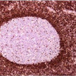
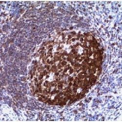 آنتی بادیهای ایمونوهیستوشیمی
آنتی بادی BOB-1 (SP92)
Name: BOB-1 (SP92)
Description and applications: The BOB.1, OBF.1, OCA-B (now called BOB.1 / OBF.1) is a 35 kD protein encoded by a gene located on chromosome 11q23.1. BOB.1 from “B cell binding Octamer protein 1 “is a specific coactivator of transcription factors Oct-1 and Oct-2 of B lymphocytes. BOB.1 mission is to increase transcriptional activity of such proteins. Interaction with transcription factor OCT2 octamer ATGCAAAT nucleotides is critical and decisive for the expression of immunoglobulin genes. It has been recently demonstrated that alteration in immunoglobulin transcription is correlated with the downregulation of the transcription factors Oct-2 and BOB.1 / OBF.1 on B lymphocytes. This antibody recognizes BOB.1 of human, murine and rat origin. In normal lymphoid tissue, BOB.1 is expressed mainly in the nuclei of germinal center B lymphocytes, with lower intensity in the follicular mantle and dispersed form in B lymphocytes localized in the interfolicular areas of the lymphoid tissue; for this reason it is useful in identifying B cell lymphomas. Variable intensity has been demonstrated in Hodgkin disease, classic type without statistical relationship with its different subtypes. The expression of specific activator proteins of B lymphocytes like “octamer-binding transcription factor 2” [Oct-2] and BOB.1 with pan-B lymphocyte markers (CD20 or CD79a ) are useful for the differential diagnosis of diffuse large B-cell lymphoma, nodular lymphocyte-predominant Hodgkin lymphoma (OCT-2 + / BOB.1 + / PAN-B +) with anaplastic lymphoma B (OCT-2 / BOB. 1- / PAN-B +). The antibody shows no cross-react with Oct-1 or Oct-2.
Composition: anti-human BOB-1 rabbit monoclonal antibody purified from serum and prepared in 10mM PBS, pH 7.4, with 0.2% BSA and 0.09% sodium azide
Intended use: Immunohistochemistry (IHC) on paraffin embedded tissues. Not tested on frozen tissues or Western-Blotting
-
 آنتی بادیهای ایمونوهیستوشیمی
آنتی بادی Beta Catenin-1 (EP35)
Name: Beta-catenin Antibody Clone EP35
Description and applications: Beta-Catenin is a key regulatory protein involved in cell adhesion and signal transduction through the Wnt pathway, and plays important roles in development, cellular proliferation, and differentiation. Mutations in the Beta-Catenin gene CTNNB1 leading to stabilization of Beta-Catenin in the cytoplasm and translocation to the nucleus have been implicated in various forms of tumor including familial adenomatous polyposis, fibromatosis, solitary fibrous tumors and endometrial carcinoma. A nuclear accumulation of Beta-Catenin in fibromatosis (desmoid tumor) in various locations including breast and mesentery is useful in the differentiation of this tumor from other fibroblast like lesions.
Composition: anti-human Beta-catenin rabbit monoclonal antibody purified and prepared in 10mM PBS, pH 7.4, with 0.2% BSA and 0.09% sodium azide.
-
 آنتی بادیهای ایمونوهیستوشیمی
آنتی بادی Bcl-2 (EP36)
Name: Rabbit Anti-Human Bcl-2 Monoclonal Antibody Clone EP36
Description and applications: The Bcl-2 family of proteins regulates apoptosis by controlling mitochondrial permeability and release of cytochrome c. Bcl-2 is an anti-apoptotic protein that resides in the outer mitochondrial wall and inhibits release of cytochrome c. Over-expression of Bcl-2 has been shown to promote cell survival by suppressing apoptosis. It has been documented that bcl-2 becomes deregulated in tumor cells as a result of translocation into the immunoglobulin heavy-chain locus and is therefore activated in B cell malignancies. Bcl-2 antibody is useful in differentiation of follicular lymphoma from reactive follicular proliferation (bcl-2 negative). In addition, bcl-2 expression has been shown to be correlated with disease prognosis in breast cancer, prostate cancer, ovarian cancer, endometrial cancer and colon cancer. The sensitivity of this antibody in diagnosis of follicular lymphoma Nucleolar dot-like staining is frequently seen with this antibody and it should not interfere with the staining interpretation.
Composition: Antibody to Bcl-2 is affinity purified and diluted in 10 mM Phosphate buffered saline (PBS), pH 7.2 containing 1% bovine serum albumin (BSA) and 0.09% sodium azide (NaN3).
Immunogen: A synthetic peptide corresponding to residues between BH3 and BH4 of human Bcl-2 protein
-
 آنتی بادیهای ایمونوهیستوشیمی
آنتی بادی Cytokeratin 19 (BA17)
(Name: Mouse anti-human Cytokeratin 19 Monoclonal Antibody (Clone BA17
Description and Applications: Cytokeratin 19 is a member of type I acidic subfamily of intermediate filaments. It is expressed in variou different human tissues except in liver. Keratin 19 is not expressed in hepatocytes, therefore, antibody to keratin 19 is useful in the identification of liver metastasis. It is expressed by the great majority of breast carcinomas and used as a marker for sentinel lymph node evaluation both in frozen tissues and formalin fixed, paraffin embedded samples.
COMPOSITION: Anti-human Cytokeratin 19 mouse monoclonal antibody purified from serum and prepared in 10mM PBS, pH 7.4, with 0.2% BSA and 0.09% sodium azide.
IMMUNOGEN: Detergent soluble extract of human mammary epithelium.
-
 آنتی بادیهای ایمونوهیستوشیمی
آنتی بادی Bcl-6 (LN22)
.(Name: Mouse anti-human BCL-6 Monoclonal Antibody (Clone LN22
Description and applications: Bcl-6 proto-oncogene product (a Kruppel-type zinc-finger protein) is mainly expressed in normal germinal center B cells and related lymphomas. Bcl-6 is involved in chromosome rearrangements at 3q27 in non-Hodgkin’s lymphomas and Bcl-6 rearrangements have been detected in 33%-45% of diffuse large B cell lymphomas. Bcl-6 has been detected immunohistochemically in follicular lymphomas, diffuse large B cell lymphomas, Burkitt’s lymphomas and in nodular lymphocyte predominant Hodgkin lymphoma.
Composition: anti-human BCL-6 mouse monoclonal antibody purified from ascites. Prepared in 10mM PBS, pH 7.4, with 0.2% BSA and 0.09% sodium azide.
Immunogen: Recombinant BCL-6 protein.
-
 آنتی بادیهای ایمونوهیستوشیمی
آنتی بادی BRST-3/TAG-72/CA72-4 (B72.3)
(Name: Mouse anti-human BRST-3/TAG-72/CA72-4 Monoclonal Antibody (Clone B72.3
Description and applications: The Signet B72.3 monoclonal antibody recognizes TAG-72 (Tumor Associated Glycoprotein-72), a high molecular weight (220-400 kDa) glycoprotein complex.
In mature human tissue it does not produce positive reaction or if anything is very weak except for the secretory endometrium.
This antibody specifically recognizes the glycoprotein TAG-72, one oncofetal tumor-associated antigen expressed in a variety of human adenocarcinomas (84% of invasive ductal breast carcinomas, 85-90% of adenocarcinomas of the colon, pancreas, stomach , esophagus, lung, ovary and endometrium). It is not expressed in leukemias, lymphomas, sarcomas, mesotheliomas, melanomas or benign tumors.Composition: anti-human BRST-3rabbit monoclonal antibody purified from serum and prepared in 10mM PBS, pH 7.4, with 0.2% BSA and 0.09% sodium azide.
Immunogen: A membrane-enriched fraction of a breast carcinoma metastatic to the liver.
-
 آنتی بادیهای ایمونوهیستوشیمی
آنتی بادی Androgen Receptor (SP107)
.(Name: Mouse anti-human Androgen Receptor Monoclonal Antibody (Clone SP107
Description and applications: The expression of AR is reportedly inversely correlated with histologic grade i.e. well differentiated prostate tumors show higher expression than the poorly differentiated tumors. In prostate cancer, AR has been proposed as a marker of hormone-responsiveness. Composition: anti-human androgen receptor (AR) mouse monoclonal antibody purified from ascites. Prepared in 10mM PBS, pH 7.4, with 0.2% BSA and 0.09% sodium azide.
Immunogen: A synthetic peptide from human AR.
-
 آنتی بادیهای ایمونوهیستوشیمی
آنتی بادی Desmin (D33)
(Name: Mouse anti-human Desmin Monoclonal Antibody (Clone D33
Description and applications: Desmin is an intermediate filament protein of both smooth and striated muscles Antibody to desmin reacts with striated (skeletal and cardiac) as well as smooth muscle cells. In skeletal and cardiac muscles, the staining is confined to the Z-bands giving a characteristic striated appearance. Anti-desmin antibody is useful in identification of tumors of myogenic origin. It reacts with leiomyosarcomas (smooth muscle) as well as rhabdomyosarcomas (striated muscle). The antibody does not stain epithelial or myoepithelial cells, endometrial stromal tumors or ovarian cancer, lymphoid tissue, osteocytes, chondrocytes or mesothelial cells.
Composition: anti-human Desmin mouse monoclonal antibody purified from ascites. Prepared in 10mM PBS, pH 7.4, with 0.2% BSA and 0.09% sodium azide.
Immunogen: Desmin from human muscle.
-
 آنتی بادیهای ایمونوهیستوشیمی
آنتی بادی DOG1 (Anoctamin-1) (SP31)
.(Name: Rabbit anti-human DOG-1 Monoclonal Antibody (Clone SP31
Description and aplications: Dog1 is a cell surface protein of unknown function selectively expressed in gastrointestinal stromal tumors (GIST). Among GIST cases with KIT mutations the Dog1 antibody identified 11% more cases than c-Kit antibody . Dog1 identifies vast majority of both c-Kit negative and PDGFRA mutated GIST cases that may still benefit from imatinib mesylate (Gleevac), an inhibitor of the kit tyrosin kinase. In addition, Dog1 immunoreactivity is seen in fewer cases of mesenchymal, epithelial tumours and melanomas when compared with c-Kit. The use of this highly sensitive and specific novel marker will increase the accuracy of GIST diagnosis.
Composition: anti-human DOG1 rabbit monoclonal antibody purified from ascites. Prepared in 10mM PBS, pH 7.4, with 0.2% BSA and 0.09% sodium azide.
Immunogen: Synthetic peptide of human DOG-1 protein.
-
 آنتی بادیهای ایمونوهیستوشیمی
آنتی بادی Epidermal Growth Factor Receptor (EP22)
.(Name: EGFR (Epidermal growth factor receptor) Antibody (Clone EP22
Description and applications: The epidermal growth factor receptor (EGFR) is a transmembrane glycoprotein of 170 kDa with tyrosine-kinase activity after binding to the epidermal growth factor (EGF) and is involved in cell growth and differentiation. Tyrosine(Tyr) residues Tyr 1068, Tyr 1148 and Tyr 1173 located at the carboxy (C) terminal portion of the EGFR, are the places of autophosphorylation upon binding with the EGF. Determining the level of expression of EGFR is necessary to establish a specific treatment with monoclonal antibodies (cetuximab). The EGFR is overexpressed in some tumors of the colon, esophagus, stomach, breast, CNS, bladder, lung, head and neck, uterine cervix, vulva, ovary and endometrium. EGFR is expressed in a high percentage of metastases of squamous cell carcinomas of different locations and therefore its expression has been considered as an indicator of poor prognosis in these tumors. The antibody detects both EGFR phosphorylated on Tyr1068 of the mature human isoform 1 (corresponding to Y1092 from the precursor form P00533-1/p170), and also unphosphorylated EGFR.
Composition: anti-human EGFR rabbit monoclonal antibody purified by protein affinity and prepared in 10mM PBS, pH 7.4, with 0.2% BSA and 0.09% sodium azide.
Immunogen: A synthetic phoshopeptide corresponding to residues Tyr1068 of human EGFR was used as immunogen.
-
 آنتی بادیهای ایمونوهیستوشیمی
آنتی بادی Epithelial Specific Antigen (BER-EP4)
.(Name: Epithelial Specific Antigen (Clone Ber-EP4
Description and aplications: This antibody reacts with two glycoproteins of 34 and 49 kD molecular weight present on the surface and in the cytoplasm of all epithelial cells except the surface layer of the squamous epithelium, hepatocytes and parietal cells of the stomach. This antibody is specific to HEA125 equivalent. The antibody shows a broad pattern of reactivity with human epithelial tissues like simple epithelium, basal layer of the stratified squamous non-keratinized epithelia and epidermis. In contrast to other known anti-epithelial antibodies, this antibody does not mark the mesothelial cells and only a small number of mesotheliomas stained positively. This antibody does not react with nervous, glial, muscle, mesenchymal or lymphoid tissue. Due to the labile nature of the epitope in tissues fixed in buffered formalin and embedded in paraffin, negative staining results should be considered with caution.
Composition: anti-Ber-EP4 mouse monoclonal antibody obtained from supernatant culture and prediluted in a tris buffered solution pH 7.4 containing 0.375mM sodium azide solution as bacteriostatic and bactericidal.
Immunogen: MCF-7 human breast carcinoma cell line.
-
 آنتی بادیهای ایمونوهیستوشیمی
آنتی بادی ERCC1 (SP68)
.(Name: ERCC1 Antibody (Clone SP68
Description and applications: Nucleotide Excision Repair (NER) is a versatile mechanism capable of recognizing and eliminating damaged DNA, most commonly caused by UV radiations. ERCC1 is one of the three endonucleases that catalyzes this process. The ERCC1 gene is located on the chromosomal region 19q13.3-q13.2 and encodes a protein of 297 amino acids and about 32.5 kDa molecular weight. Although there are only few immunohistochemical studies with antibodies against ERCC1, it seems that tumors with extensive and intense immunohistochemical expression of this molecule have a worse prognosis and are resistant to platinum-based therapy. These tumors include non-small cell lung adenocarcinomas, gastrointestinal tract adenocarcinomas, breast, urinary bladder and adrenal cortex carcinomas, and advanced stage genital tract carcinomas.
Composition: anti-ERCC1 rabbit monoclonal antibody obtained from supernatant culture and prediluted in a tris buffered solution pH 7.4 containing 0.375mM sodium azide solution as bacteriostatic and bactericidal.
Immunogen: Recombinant human ERCC1 protein.
-
 آنتی بادیهای ایمونوهیستوشیمی
آنتی بادی Estrogen Receptor (SP1)
Name: Rabbit Anti-Human Estrogen Receptor (ER) Monoclonal Antibody clone SP1
Description and aplications: This rabbit monoclonal antibody (clone SP1) specifically reacts with the human alpha isoform of the estrogen receptors not having been tested in other species as mouse, rat and hamsters were it shows reactivity.This antibody is useful for immunohistochemical identification of estrogen receptor in normal and neoplastic cells estrogen dependent. However, the description of various types of benign and malignant neoplasms positive for estrogen receptors as meningiomas, lung and stomach adenocarcinoma, and above all in breast cancer, the amount of estrogen receptors expression in the tissue is an important parameter in predicting the prognosis of the disease and its response to endocrine therapy.
Presentation: ready-to-use
Composition: anti-ER rabbit monoclonal antibody obtained from supernatant culture and prediluted in a tris buffered solution pH 7.4 containing 0.375mM sodium azide solution as bacteriostatic and bactericidal.
Immunogen: Synthetic peptide derived from the C-terminus of human estrogen receptor alpha
Species reactivity: In vitro diagnostics in humans. Not tested in other species
-
 آنتی بادیهای ایمونوهیستوشیمی
آنتی بادی FACTOR VIII (Polyclonal)
Name: Factor VIII Polyclonal Antibody
Description and applications: Factor VIII related antigen or von Willebrand factor is a multimeric glycoprotein, which binds to platelet glycoprotein Ib, glycoprotein IIb/IIIa, collagen and heparin. It is synthesized by endothelial cells and stored in the Weibel-Palade granules. It mediates platelet adhesion to injured vessel walls and serves as a carrier and stabilizer for coagulation factor VIII. von Willebrand factor is one of the most useful markers to identify endothelial (or megakaryocytic) lineage of neoplasms. As not all endothelial cells synthesize / store) this molecule, about 30% of tumors of vascular origin fail to stain for factor VIII related antigen, regardless of whether they are benign or malignant. Staining for factor VIII related antigen has also been used to measure angiogenesis, an indicator of tumor recurrence. This antibody has helped to establish the endothelial nature of some lesions of disputed histogenesis, e.g. Kaposi’s sarcoma and cardiac myxoma.
Composition: anti-human Factor VIII (FVIII) rabbit polyclonal antibody purified from ascites. Prepared in 10mM PBS, pH 7.4, with 0.2% BSA and 0.09% sodium azide.
-
 آنتی بادیهای ایمونوهیستوشیمی
آنتی بادی Factor XIIIa (EP292)
.(Name: Factor XIIIa (FXIIIa) Antibody (Clone EP292
Description and applications: Factor XIII (plasma transglutaminase, fibrin stabilizing factor) is a glycoprotein that circulates in blood as a tetramer, consisting of two A and two B subunits. Subunit A of factor XIII is an un-glycosylated 730-residue peptide with a molecular mass of 83 kD. It is the last enzyme generated in the blood coagulation cascade and is the zymogen for fibrinoligase, a transglutaminase that forms intramolecular gamma-glutamyl-epsilon-lysine crosslinking between fibrin molecules and thus stabilizes blood clots.
Factor XIII A is present in plasma, platelets, and monocytes, as well as macrophages and bone marrow precursors of these cell types and selective expression in subsets of stromal inflammatory cells. Factor XIII A is a marker for alternatively activated macrophages, while absence of Factor XIII A in monocyte-derived
macrophages is an indicator of their activated state. In addition, Factor XIII A is useful in distinguishing malignant fibrous histiocytoma (positive) from soft tissue tumor (negative).
Composition: anti-human FXIIIa rabbit monoclonal antibody purified from serum and prepared in 10mM PBS, pH 7.4, with 0.2% BSA and 0.09% sodium azide.
Immunogen: A synthetic peptide corresponding to residues of human Factor XIII A protein.
-
 آنتی بادیهای ایمونوهیستوشیمی
آنتی بادی FLI1 (G146-222)
Name: Fli-1 Antibody-Clone G146-222
Description and applications: The FLI-1 gene encodes the transcription factor of the same name (FLI-1 “Friend leukemia integration 1 transcription factor”) that has a fundamental role in embryonic development of blood vessels. Expression of Fli-1 appears as the first reliable marker of endothelial differentiation. In tumors such as Ewing sarcoma / primitive neuroectodermal tumor (ES / PNET), FLI-1 plays a pathogenic role and more than 90% of ES / PNET have the translocation t (11; 22) (q24, q12) producing the EWS fusion gene located on chromosome 22 with the Fli-1 gene located on chromosome 11. In other tumors, nuclear positivity for FLI-1 has been detected in 95% of benign (hemangiomas) and malignant (hemangioendothelioma, angiosarcoma, Kaposi’s sarcomas) vascular tumors, in different lymphomas, including lymphomas of precursor cells (lymphoblastic leukemia / lymphoblastic lymphoma), T cell lymphoma, of anaplastic large cell lymphomas, T angioinmmunoblastic lymphoma, enteropathy-associated T-cell lymphoma, all stages of mycosis fungoides with variable intensity and in 50% of peripheral T-cell lymphoma NOS. It was recently reported in melanomas.
Composition: anti-Fli1 mouse monoclonal antibody obtained from supernatant culture and prediluted in a tris buffered solution pH 7.4 containing 0.375mM sodium azide solution as bacteriostatic and bactericidal.
برای تماس کلیک کنید
سما تشخیص آریا ، عرضه کننده متنوع ترین و کامل ترین محصولات کیت های آزمایشگاهی