-
 آنتی بادیهای ایمونوهیستوشیمی
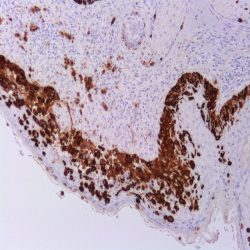
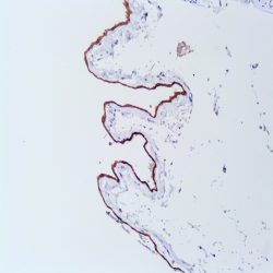
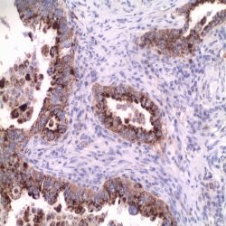
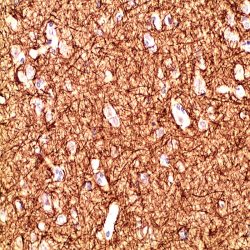
آنتی بادی MELAN A/MART-1 (EP43)
Name: Melan A/Mart-1 Antibody Clone EP43
Description and applications: MART-1, also known as Melan-A,is a melanocyte lineage-specific protein (MART-1; melanoma antigen recognized by T cells 1) recognized by the T lymphocytes of patients with established malignancy. Antibody to MART-1 labels both normal melanocyte and diseased cell with melanocyte differentiation. It is useful for diagnosis of tumors with melanocyte differentiation, especially metastatic melanoma. Identification of MART-1 also opens possibilities for the development of immunotherapies for patients with melanoma.
Composition: Anti-human Melan A/Mart-1 rabbit monoclonal antibody purified from serum and prepared in 10mM PBS, pH 7.4, with 0.2% BSA and 0.09% sodium azide INTENDED USE Immunohistochemistry (IHC) on paraffin embedded tissues. Not tested on frozen
tissues or Western-Blotting. -
 آنتی بادیهای ایمونوهیستوشیمی
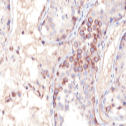
آنتی بادی Melanoma-Associated Antigen-1 (MAGE-1) (6C1)
Name: Melanoma-Associated Antigen-1 (MAGE-1)Antibody Clone 6C1
Description and applications: Human malignant neoplasms carry rejection antigens that are recognized by the patients’ autologous,
tumor-directed and specific, cytolytic, CD8+ T lymphocyte clones (CTL). An important group of antigens is coded by the MAGE family of genes. It was identified that melanomas and primary glial brain tumors express common melanoma associated antigens (MAAs). Because MAGE-1 is expressed on a significant proportion of human neoplasms of various histological types (melanoma, brain tumors of glial origin, neuroblastoma, non-small cell lung cancer, breast, gastric, colorectal, ovarian, renal cell carcinomas) and not on normal tissues, the encoded antigen may serve as a marker of early detection and target for cancer immunotherapy.Composition: Anti-human MAGE-1 mouse monoclonal antibody purified from serum and prepared in 10mM PBS, pH 7.4, with 0.2% BSA and 0.09% sodium azide.
-
 آنتی بادیهای ایمونوهیستوشیمی
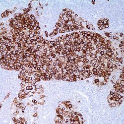
آنتی بادی MELANOMA (HMB45)
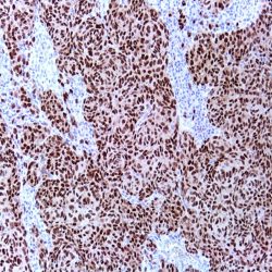
Name:Melanoma Antibody Clone HMB-45
Description and applications:This antibody gives a qualitative assessment of malignant melanoma, which can be extremely difficult
to diagnose. Metastatic amelanotic melanoma can often be confused with a variety of poorly diferentiated carcinomas, large cell lymphomas, and sarcomas. It is also difficult to differentiate melanoma from spindle cell carcinomas and various types of mesenchymal neoplasms. Melanoma antibody stains fetal and neonatal melanocytes, junctional and blue nevus cells, and malignant melanocytes. Typically, a keratin negative, vimentin rich neoplasm, that immunoreacts with antibody to S-100 protein and this melanoma antibody is, with rare exception, a melanoma.As a marker of melanosomes, careful should be taken as malanofages can also be stained.Composition: Anti-human Melanoma mouse monoclonal antibody purified from serum and prepared in 10mM PBS, pH 7.4, with 0.2% BSA and 0.09% sodium azide.
-
 آنتی بادیهای ایمونوهیستوشیمی
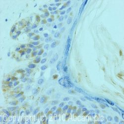
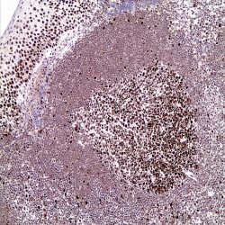
آنتی بادی Melanoma Associated Antigen (KBA.62)
Name: anti-Melanoma Associated Antigen Antibody Clone KBA.62
Description and applications: Anti-KBA.62 (Melanoma Associated Antigen) is a novel antimelanoma antibody. Studies thus far have shown a similar sensitivity to melanocytic proliferations as that seen with S-100 protein staining, which is somewhat higher than that seen with HMB-45. This has been con#rmed by one study on a series of 215 sentinel lymph nodes. Moreover, anti-KBA.62 identi#ed 6 patients (3%) who had con#rmed sentinel lymph node metastasis but stained negative for HMB-45. In this setting, the resolution appears to be higher than S- 100 protein in that the staining pattern (membranous) is quite distinct. Interestingly, most cases of desmoplastic and spindle cell melanomas show strongly positive results with anti-KBA.62, unlike that seen with other melanocyte markers. It should be noted that anti-KBA.62 will label occasional endothelial cells which can serve as an internal positive control. A small percentage of well differentiated squamous cell carcinomas of the skin (and lung) have also been noted to stain with this antibody; however, the poorly-differentiated forms of carcinoma do not, thus resolving a greater practical problem in differential diagnosis. Therefore, anti-KBA.62 is a useful additional marker for melanoma, specifically in desmoplastic/spindle cell cases and in the context of micro-metastasis in sentinel lymph node.
Composition: anti-human KBA.62 mouse monoclonal antibody purified from serum and prepared in 10mM PBS, pH 7.4, with 0.2% BSA and 0.09% sodium azide.
-
 آنتی بادیهای ایمونوهیستوشیمی
آنتی بادی Mesothelin (EP140)
Name: Mesothelin Antibody Clone EP140
Description and aplications: Mesothelin gene encodes a 69 kDa precursor protein that progress to a protein of 40-kDa bound to a glycosylphosphatidylinositol (GPI), the mature mesothelin, present on the cell surface. Its biological function is not well understood, but recent studies have shown it forms a strong and specific complex with the MUC16, a union that has been suggested as the basis of ovarian cancer metastasis. Mesothelin is present on the surface of normal mesothelium that coats the pleura, peritoneum and pericardium. Mesothelin is overexpressed in patients with epithelial ovarian carcinoma and malignant mesotheliomas and may be useful to distinguish mesothelioma from other solid tumors. Its overexpression has been observed in other tumors too, including pancreatic carcinomas and cholangiocarcinoma.
Composition: anti-human Mesothelin rabbit monoclonal antibody obtained from supernatant culture and prediluted in a tris buffered solution pH 7.4 containing 0.375mM sodium azide solution as bacteriostatic and bactericidal.
Immunogen: A synthetic peptide corresponding to residues at the C-terminus of human Mesothelin protein
-
 آنتی بادیهای ایمونوهیستوشیمی
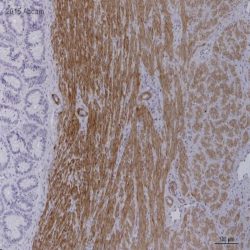
آنتی بادی Smooth muscle myosin, heavy chain (EP166)
Name: Myosin, heavy chain Antibody-Clone EP1663
Description and applications: Myosin heavy chain 11 (MYH11) is a smooth muscle myosin belonging to the myosin heavy chain family. It is a subunit of a hexameric protein that consists of two heavy chain subunits and two pairs of non-identical light chain subunits. Myosin heavy chain functions as a major contractile protein, converting chemical energy into mechanical energy through the hydrolysis of ATP. An aberration in this protein is associated with acute myeloid leukemia of the M4Eo subtype. MYH labels smooth muscle cells and myoepithelial cells in various tissues. The immunoreactivity in glial cells of the cerebellum and spermatocytes in the testis is also observed. MYH has been a useful marker for myoepithelial cell as well as smooth muscle cell differentiation. MYH together with p63 are one of the most useful antibodies in establishing the invasive character of breast lesions.
Composition: Anti-human Smooth muscle myosin, heavy chain rabbit monoclonal antibody purified from serum and prepared in 10mM PBS, pH 7.4, with 0.2% BSA and 0.09% sodium azide.
-
 آنتی بادیهای ایمونوهیستوشیمی
آنتی بادی MITF (Microphthalmia-Associated Transcription Factor) (C5+D5)
Name: MITF (Microphthalmia-Associated Transcription Factor) Antibody Clone C5+D5
Description and applications: This antibody reacts with human microphthalmia transcription factor (MITF). The MITF gene (microphthalmia transcription factor, MITF) encodes a nuclear helix-loop-helix transcription factor involved in pigmentation and mast and bone cell development. The MITF gene is essential for the normal development of melanocytes and is responsible for the regulation of the expression of other genes related to the melanocytic lineage: tyrosinase genes, SILV/PMEL17/GP100 (encoding HMB45), MLANA/MART1 (coding for Melan A), TRP-1, and TRP-2. Mutation of the MITF gene in humans causes Waardenburg syndrome IIA and Tietz syndrome, characterized by pigmentation disorders and deafness, the latter due to melanocyte deficiency in the inner ear. There are two known isoforms of MITF that differ in 66 amino acid residues from the amino terminus. The shortest isoform of MITF is melanocyte-specific. MITF-M is expressed in both normal and malignant melanocytes. The other isoforms, MITF-A, MITF-C, and MITF-H, differ structurally from MITF-M at the amino terminus and are expressed in osteoclasts, mast and heart cells, and in B16 melanoma cell lines (although not in other melanoma cell lines). The anti-MITF antibody reacts against the melanocytic and non melanocytic isoforms of the MITF gene, and although it does not distinguish between benign and malignant melanocytic lesions, it shows high sensitivity and specificity as a histopathological marker of melanocytes and melanocyte-derived neoplasms (melanomas). Using MITF and Melan A in the same panel of melanoma markers, the sensitivity (95%) and specificity (100%) values obtained are higher than those obtained using the S100 protein and HMB45, even in cytological samples. MITF, Melan-A, and tyrosinase are sensitive markers for the diagnosis of epithelioid melanomas. Low levels of Melan-A and MITF have been shown to correlate with a poorer prognosis. MITF has demonstrated 100% sensitivity and specificity to detect invasive malignancies of melanocytic origin and to study sentinel lymph nodes.
Composition: Anti-human MITF mouse monoclonal antibody purified from serum and prepared in 10mM PBS, pH 7.4, with 0.2% BSA and 0.09% sodium azide.
-
 آنتی بادیهای ایمونوهیستوشیمی
آنتی بادی MLH1 (BS29)
Name: MLH1 Antibody Clone BS29
Description and aplications: MLH1 is a mismatch repair gene that is deficient in a high proportion of patients with microsatellite instability (MSI-H). This finding is associated with the autosomal dominant condition known as Hereditary Non-Polyposis Colon Cancer (HNPCC). The anti-MLH1 antibody is useful in screening patients and families for this condition. Colon cancers that are microsatellite unstable have a better prognosis than their microsatellite stable counterparts. Along with PMS2, MSH2 and MSH6, MLH1 antibody is helpful in diagnosis of MSI. An IHC study conducted by Mayo clinic on 535 cases with MSI-high, 90% of the tumors showed loss of MLH1, MSH2 and/or MSH6 expression, while 70% of the remaining cases showed isolated loss of PMS2 expression. Endometrial carcinomas are the most common non-colorectal cancers occur in HNPCC. The most common IHC abnormality in endometrial carcinomas with MSI was concurrent loss of MLH1/PMS2. Adding of PMS2 and MSH6 to MLH1 and MSH2 antibodies increased sensitivity for diagnosis of MSI. Tumors with low-level MSI show unfavourable pathological characteristics compared to tumours with no and tumours with high-level MSI.
Composition: anti-human MLH1 mouse monoclonal antibody obtained from supernatant culture and prediluted in a tris buffered solution pH 7.4 containing 0.375mM sodium azide solution as bacteriostatic and bactericidal.
-
 آنتی بادیهای ایمونوهیستوشیمی
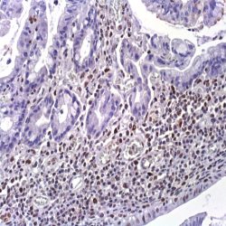
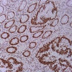
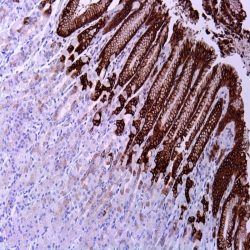
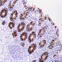
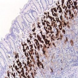
آنتی بادی Napsin A (BS10)
Name: Napsin A Antibody-Clone BS10
Description and aplications: Napsin is a pepsin-like aspartic proteinase, in the A1 group of the AA class of proteinases. There are two closely related napsins, napsin A and napsin B. Napsin A is expressed as a single chain protein with the molecular weight of approximately 38 kDa. Immunohistochemical studies revealed high expression levels of napsin A in human lung and kidney but low expression in spleen. Napsin A is expressed in type II pneumocytes and in adenocarcinomas of lung. The high specificity expression of napsin A in adenocarcinomas of lung is useful to distinguish primary lung adenocarcinomas from adenocarcinomas of other organs. Napsin A is also useful for the study of renal papillary adenocarcinomas and clear cell adenocarcinomas as it might be positive in high percentage of the cases. Conversely it is expressed by less than 5% of papillary carcinomas of the thyroid.
Composition: anti-Napsin A mouse monoclonal antibody obtained from supernatant culture and prediluted in a tris buffered solution pH 7.4 containing 0.375mM sodium azide solution as bacteriostatic and bactericidal.
-
 آنتی بادیهای ایمونوهیستوشیمی
آنتی بادی MPCyV Large T-Antigen (CM2B4)
Name:MPCyV Large T-Antigen Antibody Clone CM2B4
Description and applications: Merkel cells are round neuroendocrine cells found in skin where they form synaptic complexes with
somatosensory afferent fibres. These complexes are responsible for fine touch and produce various neuroactive substances that act as fast-acting skin neurotransmitters or neuromodulators. In pathological conditions, Merkel cells can proliferate and generate a rare but very aggressive form of skin cancer known as Merkel cell carcinoma (CCM). Around 80% of CMMs are caused by the recently described Merkel cell polyomavirus (MCPyV or MCV), which integrates into the genome of Merkel cells and proliferates clonally. The MCPyV virus is a nonencapsulated DNA virus consisting of two early units (two T antigens, one large and one small) and three late units (VP1, 2, and 3). The large T antigen is the MCPyV’s functional nuclear protein, which has a molecular mass of 125 kDa and is expressed in tumour cells, where it binds to cell cycle suppressor molecules, degrades and sequesters them, promoting their S phase. The large T antigen has natural truncated mutations that also produce functionally active proteins of smaller size. The CM2B4 antibody, which shows no cross-staining with other types of polyomavirus and recognises the MCPyV large T antigen, has been synthesized against a peptide derived from exon 2 of the mentioned antigen’s locus, for which it is highly specific, as well as for its 57kT isoforms; however, it may not detect some of its minor truncated forms. CM2B4, assessed along with a panel of markers including cytokeratins 20 and 7, p63, TTF1, neuronspecific enolase, S100 protein, HMB45, and CD45, facilitates the diagnosis of approximately 80% of classical Merkel cell carcinoma cases. However, most CCM cases with eccrine, squamous, rhabdomyoblastic, leiomyosarcomatous, or neuroblastic divergent differentiation are MCPyV negative and are associated with a worse prognosis of the disease. In comparison with MCPyV level detection using PCR, the antibody has a sensitivity of 95% and a specificity of 83% since, as mentioned above, MCPyV negative CCM cases have been observed, as well as occasional positivity of uncertain meaning in mature lymphocytes, sweat glands, and vascular endothelium.Composition: Anti-human MPCyV Large T-Antigen mouse monoclonal antibody purified from serum and prepared in 10mM PBS, pH 7.4, with 0.2% BSA and 0.09% sodium azide.
-
 آنتی بادیهای ایمونوهیستوشیمی
آنتی بادی Nestin (10C2)
Name: Mouse anti-human Nestin Monoclonal Antibody clone 10c2
Description and applications: Nestin is a class VI intermediate filament (IF) protein. It has been reported that nestin expression is significantly increased in melanoma and correlated with more advanced stages of the disease. An immunohistochemical analysis identified nestinpositive cells in 84% of primary melanoma and 83% of metastatic melanoma. Nestin immunoreactivity was also observed in melanoma cells in all cases of HMB45-negative amelanotic and melanotic, nondesmoplastic melanoma. Nestin expression has been reported in tumors of the CNS, including astrocytoma, ependymoma, oligodendroglioma, glioblastoma, and primitive neuroectodermal tumors. Nestin has been detected in human gliomas, including low and high grade, but its expression is observed more frequently in high grade than in low grade gliomas. Nestin overexpression is seen in carcinomas, including prostatic adenocarcinoma, pancreatic ductal carcinoma, thyroid carcinoma, and in mesenchymal tumors such as GIST and DFSP. Among the breast carcinoma subtypes, Nestin is highly expressed in basal breast cancer subtype (ERα-/PR-/Her2-) but not in the Her2 subtype (ERα-/PR-/Her2+) or luminal epithelial phenotype (ERα+/PR+). Only cytoplasmic staining is considered positive, whereas any nuclear staining is considered as background artifact. In normal skin, nestin is observed in endothelial cells and the bulge area of hair follicles.
Composition: Anti-human Nestin mouse monoclonal antibody purified from serum and prepared in 10mM PBS, pH 7.4, with 0.2% BSA and 0.09% sodium azide
Intend use : Immunohistochemistry (IHC) on paraffin embedded tissues. Not tested on frozen tissues or Western-Blotting
-
 آنتی بادیهای ایمونوهیستوشیمی
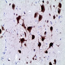
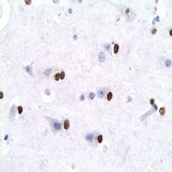
آنتی بادی NeuN Protein (A60)
Name: Neuronal Nuclear Protein (NeuN)-clone A60
Description and aplications: NeuN antibody (NEUronal Nuclei; clone A60) specifically recognizes the DNA-binding, neuron-specific protein NeuN, which is present in most CNS and PNS neuronal cell types of all vertebrates. NeuN protein distributions are apparently restricted to neuronal nuclei, perikarya and some proximal neuronal processes in both fetal and adult brain although, some neurons fail to be recognized by NeuN at all ages: INL retinal cells, Cajal-Retzius cells, Purkinje cells, inferior olivary and dentate nucleus neurons, and sympathetic ganglion cells are examples. Immunohistochemically detectable NeuN protein first appears at developmental timepoints that correspond with the withdrawal of the neuron from the cell cycle and/or with the initiation of terminal differentiation of the neuron. Immunoreactivity appears around E9.5 in the mouse neural tube and is extensive throughout the developing nervous system by E12.5. Strong nuclear staining suggests a nuclear regulatory protein function; however, no evidence currently exists as to whether the NeuN protein antigen has a function in the distal cytoplasm or whether it is merely synthesized there before being transported back into the nucleus. No difference between protein isolated from purified nuclei and whole brain extract on immunoblots has been found. The A60 antibody is a neuronal widely used tumor marker. NeuN is expressed in the neuronal component gangliogliomas , ganglioneuromas , Lhermitte -Duclos disease ( gangliocytoma with cerebellar dysplasia) , neurocitomas , in well-differentiated neurons and the oligodendrocytes like component present in dysembryoplastic neuroepithelial tumors . The expression in over 60% of the cells in glial tumors has been referenced in specific cases (1 oligodendroglioma , 3 glioblastomas and one clear cell ependymoma from over 600 cases studied ) . NeuN is often negative in the component neuronal ganglion cell tumors in particular when it is morphologically differentiated . This antibody cross-reacts with other species (mouse, rat, pig , etc.).
Composition: anti- Neuronal Nuclear Protein (NeuN) mouse monoclonal antibody obtained from supernatant culture and prediluted in a tris buffered solution pH 7.4 containing 0.375mM sodium azide solution as bacteriostatic and bactericidal. The quantity of the active antibody was not determined.
Immunogen: Purified cell nuclei from mouse brain
-
 آنتی بادیهای ایمونوهیستوشیمی
آنتی بادی Neurofilament 70/200 kDa (2F11)
Name: Neurofilament 70/200 kDa Antibody-Clone 2F11
Description and aplication: AntiNeurofilament antibody stains an antigen localized in a number of neural, neuroendocrine, and endocrine tumors. Neuromas, ganglioneuromas, gangliogliomas, ganglioneuroblastomas and neuroblastomas stain positively for neurofilament. Neurofilaments are also present in paragangliomas and adrenal and extraadrenal pheochromocytomas. Carcinoids, neuroendocrine carcinomas of the skin, and oat cell carcinomas of the lung also express neurofilament.
Composition:Anti-human Neurofilament mouse monoclonal antibody purified from serum and prepared in 10mM PBS, pH 7.4, with 0.2% BSA and 0.09% sodium azide.
-
 آنتی بادیهای ایمونوهیستوشیمی
آنتی بادی MSH2 (FE11)
Name: MSH2 (MutS Protein Homolog 2) Mouse] Antibody Clone FE11
Description and applications: MSH2 is involved in DNA repair as a mismatch repair protein, and mutations of MSH2 are found in approximately 50% of inherited non polyposis colorectal carcinoma (HNPCC) (Lynch syndrome) cases. HNPCC is an autosomal, dominantly inherited disease associated with marked increase in cancer susceptibility. It is characterized by a familial predisposition to early onset colorectal carcinoma and extra-colonic cancers of the gastrointestinal, urological and female reproductive tracts.
Composition: anti-human MSH2 mouse monoclonal antibody purified from ascites fluid by Protein A chromatography. Prepared in 10mM PBS, pH 7.4, with 0.2% BSA and 0.09% sodium azide.
-
 آنتی بادیهای ایمونوهیستوشیمی
آنتی بادی MSH6 (EP49)
Name: MSH6 Antibody (Clone EP49)
Description and aplications: The MutS homologue 6 protein (MSH6) is a member of the MutS homolog family required in the DNA match repair system.Carriers of the mismatch repair gene mutations have a high lifetime risk of developing Hereditary Non-Polyposis Colon Cancer (HNPCC) and several other cancers including endometrial cancer due to microsatellite instability (MSI) caused by accumulation of DNA replication errors in proliferating cells. MSH6 antibody is useful for screening and diagnosis of patients with MSI. The level of MSI has been reported to be associated with prognosis in colon cancer. Adding of PMS2 and MSH6 to MLH1 and MSH2 antibodies increased sensitivity for diagnosis of MSI. Tumors with low-level MSI show unfavourable pathological characteristics compared to tumours with no and tumours with high-level MSI.
Composition: anti-human MSH6 rabbit monoclonal antibody purified from ascites. Prepared in 10mM PBS, pH 7.4, with 0.2% BSA and 0.09% sodium azide.
-
 آنتی بادیهای ایمونوهیستوشیمی
آنتی بادی Oct3/4 (C-10)
Name: Mouse Anti-OCT 3/4 Monoclonal Antibody
Description and aplications: OCT-3 (officially POU5F1, also known as OCT4, OCT 3/4, OTF3 or OTF4) is a nuclear transcription factor involved in the initiation, maintenance and differentiation of germ cells and stem cells during embryonic development. The antibody recognizes a protein of 18kDa molecular weight encoded by the gene POU5F1 located on chromosome 6p21.3 which is essential in the development of the blastocyst. Three isoforms of OCT-3-called OCT-3A, OCT-3B and OCT3B1 are generated by alternative splicing. Isoforms A and B1 are expressed only in germ cell tumors while OCT3B isoform may also be expressed in tumors of somatic origin. This antibody is useful for the identification of transcription factor OCT-3/4 present in normal tissues (embryonic testis) and neoplastic ones. Gonadoblastomas, intratubular germ cell tumors, testicular seminoma, ovarian dysgerminomas, extragonadal germinomas and embryonal carcinoma show nuclear positivity with different degrees of intensity. By contrast the yolk sac tumors, spermatocytic seminomas, choriocarcinoma, testicular and ovarian teratomas are negative. Its expression has also been demonstrated in several tumor lines of somatic origin and linked to poor prognosis
Composition: anti-OCT3/4 mouse monoclonal antibody obtained from supernatant culture and prediluted in a tris buffered solution pH 7.4 containing 0.375mM sodium azide solution as bacteriostatic and bactericidal.
Immunogen: amino acids 1-134 of Oct-3/4 of human origin
-
 آنتی بادیهای ایمونوهیستوشیمی
آنتی بادی Mucin 5AC (MUC5AC) (MD-19 also known as MRQ-19)
Name: Mucin 5AC (MUC5AC) Antibody (Clone MUC5AC, MD-19 also known as MRQ-19)
Description and applications: Mucins are glycoproteins of high molecular weight linked to oligosaccharides in the threonine or serine wastes by glycosidic bonds produced by different epithelial cells. In humans, at least 14 genes coding for mucins have been identified (MUC1, MUC2, MUC3, MUC4, MUC5AC, MUC5B, MUC6, MUC7, MUC8, MUC9, MUC11, MUC12, MUC13 and MUC16). Mucins are classified as follows: Membrane-bound mucins (MUC1, MUC3, MUC4, MUC12 and MUC16), secreted mucins forming gel-like secretion (MUC2, MUC5AC, MUC5B, and MUC6) and secreted soluble mucin (MUC7). Mucins are the main component of the mucous membrane that protects the gastric mucosa from the chemical and mechanical insults. This antibody reacts with the mucin 5AC. MUC5AC isthe glycoprotein secreted forming gel-like secretions expressed by the gastric epithelium and the respiratory tract. The distribution of the expression of the mucins is heterogeneous MUC1 (stomach and pancreas), MUC2 (small and large intestine), MUC4 (stomach), MUC5AC (stomach) and MUC6 (stomach, small intestine and pancreas). MUC5AC is expressed in apical rim in normal cells and also in the lateral edges of the cytoplasmic membrane and intracytoplasmically in carcinomas of different tissues. MUC5AC is expressed in normal gastric epithelium in the cardia, fundus and pyloric antrum. Barrett’s esophagus shows positivity for this mucin; MUC5AC is not expressed in the small intestine, large intestine nor pancreas. In neoplastic tissues, it is expressed in pancreatic, colon and appendix adenocarcinomas, extramammary Paget’s disease Colloid carcinomas of the breast nor cholangiocarcinomas in general do not express MUC5AC. The mucin expression pattern in tumors has been associated with the tumor location, the biological insult and the infiltration capacity of some neoplasias.
Composition: Anti-human Mucin 5AC mouse monoclonal antibody purified from serum and prepared in 10mM PBS, pH 7.4, with 0.2% BSA and 0.09% sodium azide.
-
 آنتی بادیهای ایمونوهیستوشیمی
آنتی بادی Mucin 2 (Ccp 58)
Name: Mucin 2 (MUC-2) Antibody (Clone Ccp58)
Description and applications: Secreted epithelial mucins are large macromolecules which exhibit extreme polydispersity. Mucin 2 is the major intestinal mucin. O-glycans are attached to MUC2 in a potentially diverse arrangement, which is crucial for their interaction with endogeneous and exogeneous lectins. MUC2 is expressed in glandular epithelium (goblet cells) of normal small and large intestine, Barrett’s esophagus and breast glandular epithelium; it is not expressed in stomach or pancreas. In neoplastic tissues the pattern of mucins expression in tumours has been associated with tumor location, the biological aggressiveness and the infiltration capacity of certain malignancies. MUC2 is expressed by colon and appendix signet ring adenocarcinomas, breast colloid carcinoma, Paget’s disease of the breast and prostate carcinoma. It is not expresses by endometrial carcinomas, the non-mucinous breast or pancreatobiliary carcinomas.
Composition: anti-human MUC-2 mouse monoclonal antibody purified from serum and prepared in 10mM PBS, pH 7.4, with 0.2% BSA and 0.09% sodium azide.
-
 آنتی بادیهای ایمونوهیستوشیمی
آنتی بادی Mucin 6 (MD-20 also known as MRQ-20)
Name: Anti-Mucin 6 (MUC6) Antibody (Clone MRQ-20)
Description and aplications: Mucins are high molecular weight glycoproteins produced by epithelial cells. In humans, at least 14 genes encoding mucin has been identified (MUC1, MUC2, MUC3, MUC4, MUC5AC, MUC5B, MUC6, MUC7, MUC8, MUC9, MUC11, MUC12, MUC13 and MUC16). Mucins are classified as membrane associated (MUC1, MUC3, MUC4, MUC12 and MUC16), secreted mucins forming gels (MUC2, MUC5AC, MUC5B, and MUC6), or soluble secreted mucin (MUC7). Mucins are the major component of the mucus layer which protects against chemical and mechanical gastric mucosa aggression. MUC6 is expressed in the normal gastric epithelium of the pyloric antrum, in Brüner duodenal glands, small intestine, and pancreas; MUC6 is not expressed in ducts or acini of normal breasts or Barrett’s esophagus. In neoplastic tissues MUC6 is expressed in colloidal breast carcinomas. Breast carcinomas, pancreatic adenocarcinomas and cholangiocarcinomas do not express MUC6. The pattern of mucin expression in tumors has been associated with tumor location, the biological aggressiveness, and the infiltration capacity of certain malignancies.
Composition: anti-MUC-6 mouse monoclonal antibody obtained from supernatant culture and prediluted in a tris buffered solution pH 7.4 containing 0.375mM sodium azide solution as bacteriostatic and bactericidal.
-
 آنتی بادیهای ایمونوهیستوشیمی
آنتی بادی Olig 2 Protein (211F1.1)
Name: Olig 2 Protein Antibody-Clone 211F1.1
Description and applications: Olig2, a basic helix–loop–helix transcription factor, is involved in oligodendroglial specification. Olig2 expression has been reported in most glial tumors, such as oligodendrogliomas and astrocytomas. Although more than half of glioblastomas are positive for Olig2, expression is very weak in terms of both percentage of labeled cells and intensity. No Olig2 expression has been found in the non-glial tumors including neuroepithelial tumors, ependymomas, subependymomas, medulloblastomas, and nonneuroepithelial tumors, such as CNS lymphomas, meningiomas, schwannomas, atypical teratoid/rhabdoid tumor, and haemangioblastomas. Compared to the strong staining seen in glioma samples, a weak expression is observed in nontumoral brain tissue (gliosis). In order to characterize cellular subtypes that constitute astrocytomas, oligoastrocytomas and oligodendrogliomas, double labeling of Olig2 and GFAP has been performed which identified two phenotypically distinct tumor populations. The first is Olig2+/GFAP– which has an oligodendroglial morphology, corresponding to pure oligodendrogliomas that contain only oligodendroglial cells; the second is Olig2–/GFAP+ which has an astrocytic phenotype, including not only oligoastrocytomas, but also WHO astrocytomas. Depending on proportion and spatial clustering of the two phenotypically distinct tumor populations, the tumor (Olig2-/GFAP+) is classified either as an astrocytoma when both populations are intermingled with a dominance of GFAP+ cells or oligoastrocytoma when there is some degree of spatial clustering of the GFAP+ cells.
Composition: anti-human Olig 2 protein mouse monoclonal antibody purified from serum and prepared in 10mM PBS, pH 7.4, with 0.2% BSA and 0.09% sodium azide.
برای تماس کلیک کنید
سما تشخیص آریا ، عرضه کننده متنوع ترین و کامل ترین محصولات کیت های آزمایشگاهی