-
 آنتی بادیهای ایمونوهیستوشیمی
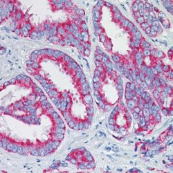
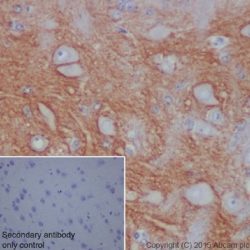
آنتی بادی Cdk4 (DCS-35)
Name: Anti-human CDK4 Mouse Monoclonal Antibody clone DCS-35
Description and aplications: Cell cycle progression is controlled in part by a family of cyclin proteins and cyclin dependent kinases (Cdks). Cdk proteins work in concert with the cyclins to phosphorylate key substrates involved in each phase of cell cycle progression.
Another family of proteins, Cdk inhibitors, also plays a role in regulating the cell cycle by binding to cyclin-Cdk complexes and modulating their activity. Several Cdk proteins have been identified, including Cdk2-Cdk8, PCTAIRE-1-PCTAIRE-3, PITALRE and PITSLRE. Cdk4, in complex with D-type cyclins, is thought to regulate cell growth during the G1 phase of the cell cycle. This association with a D-type cyclin upregulates Cdk4 activity, whereas binding to the Cdk inhibitor p16 downregulates Cdk4 activity. Activation of the Cdk4-cyclin complexes requires phosphorylation on a single threonyl residue of Cdk4, catalyzed by a Cdk-activating protein (CAK).Composition: Anti-human CDK4 mouse monoclonal antibody purified from serum and prepared in 10mM PBS, pH 7.4, with 0.2% BSA and 0.09% sodium azide
Intended use: Immunohistochemistry (IHC) on paraffin embedded tissues. Not tested on frozen tissues or Western-Blotting
-
 آنتی بادیهای ایمونوهیستوشیمی
آنتی بادی CD95 (EP208)
Name: CD95 Monoclonal Antibody clone EP208
Description and aplications: The CD95 (Fas) protein is a cell surface receptor belonging to the tumor necrosis factor (TNF) family that transduces death signaling on engagement by multimeric Fas ligand (CD95L), of which there are eight in its membrane–bound form or in its soluble form resulting from cleavage by a putative metalloproteinase. CD95 is a widely expressed protein. CD95-mediated apoptosis is an essential mechanism for the maintenance of normal tissue homeostasis, and disruption of this death pathway has been associated with a wide range of human diseases, including autoimmune diseases, lymphoproliferative disorders and malignancies. The Fas death system also plays important roles in various apoptosis conditions such as those evoked by irradiation, chemotherapeutic
agents and viral infections. The expression of CD95 serves as a prognostic marker in predicting the outcome of disease progression and treatment in many types of tumors.Composition: Anti-human CD95 rabbit monoclonal antibody purified from serum and prepared in 10mM PBS, pH 7.4, with 0.2% BSA and 0.09% sodium azide
-
 آنتی بادیهای ایمونوهیستوشیمی
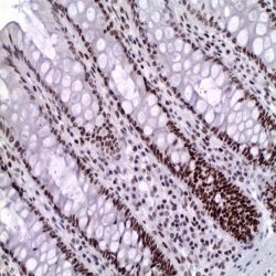
آنتی بادی CNPasa (11-5B)
Name: CNPase Monoclonal Antibody clone 11-5B
Description and aplications: This monoclonal antibody recognises two proteins with a molecular mass of 46 kD (CNP1) and 48 kD (CNP2), identified as CNPase isoforms. CNPase (2’,3’-cyclic nucleotide 3′-phosphodiesterase) is expressed at high levels in oligodendrocytes of the central nervous system and in Schwann cells of the peripheral nervous system and it is virtually absent in other cell types. Since CNPase is expressed early in postnatal development, this antibody is useful for the identification of oligodendrocytes and Schwann cells. CNPase activity is decreased in demyelinating diseases such as multiple sclerosis. The CNPase gene is located on chromosome 17q21, very close to the familial breast cancer gene BRCA1. This antibody shows cross-reactivity with monkey, cow, sheep, goat, pig, cat, dog, rabbit, rat, and mouse. It does not cross-react with guinea pig and chicken.
Composition: Anti-human CNPase mouse monoclonal antibody purified from serum and prepared in 10mM PBS, pH 7.4, with 0.2% BSA and 0.09% sodium azide
Intended use: Immunohistochemistry (IHC) on paraffin embedded tissues. Not tested on frozen tissues or Western-Blotting
-
 آنتی بادیهای ایمونوهیستوشیمی
آنتی بادی Cytokeratin 17 (EP98)
Name: Cytokeratin 17 Monoclonal Antibody clone EP98
Description and aplications: The widely known intermediate filaments proteins, which measure between 7 and 22 nm diameter (that is, a size between the one of the actin -5-7 nm and the tubulin -22-25 nm-), along with the previous ones, belong to the cytoskeleton of the vertebrates. This superfamily is made up of six subfamilies of molecules with different tissue expressions. Cytokeratins make up the homology I and II in humans. They are encoded in more than 49 different genes in the chromosomes 17 (I) and 12 (II). The nomenclature adopted in 1982 by Moll and Franke assigns the ranks 1 to 8 for the type II cytokeratins (neutral or basic) and between 9 and 21 for the type I (acid). Nowadays, an analogue nomenclature has been defined to name the hair keratin with theaddition of the Ha and Hb letters to separate the ones of the group I from group II. Structurally speaking, cytokeratins share with the rest of intermediate filaments a central axis of 310 amino acids made up of four α-helices domains (1A, 1B, 2A and 2B) highly preserved that define the type of intermediate filament that they will make up after its packing, separated by three non-helix binding sites (L1, L12 and L2) and two extreme domains highly different in size and sequence (head-1- and tail-2-), each of them with constant (E1/E2), variable (V1/V2) and homology regions (H1/H2), the last ones are characteristic of the type II keratins and absent of the type I. In variable domains, they have the most immunogenic properties and he main differences between each type of keratin. Usually, keratins assemble in heterodimers I/II and they co-express in pairs specifically in each tissue. Cytokeratin 17 of 46 KDa of molecular mass is a type I intermediate filament with an isoelectric point of 5.1 encoded by the gene of the same name located in the chromosome 17. Cytokeratin 17 is expressed in the basal and suprabasal cells of complex epithelium of the skin and skin adnexa. Furthermore, it is expressed in the basal cells of the pseudostratified epithelium of the trachea, larynx and bronchus. This antibody reacts with myoepithelial cells of the salivary and sweat glands. This antibody is useful to diagnose epithelial neoplasias that express cytokeratin 17 such as basal cell carcinomas, squamous lung cancers, adenoid cystic and mucoepidermoid salivary gland carcinomas, mesotheliomas, cholangiocarcinomas and numerous lung adenocarcinomas (>50%), colorectal adenocarcinomas (<50%), pancreatic adenocarcinomas (30%). Cytokeratin 17 is not detected in gastric adenocarcinomas, mucinous colorectal adenocarcinomas, hepatocellular carcinomas and Merkel-cell carcinomas.
Composition: Anti-human Cytokeratin 17 rabbit monoclonal antibody purified from serum and prepared in 10mM PBS, pH 7.4, with 0.2% BSA and 0.09% sodium azide
-
 آنتی بادیهای ایمونوهیستوشیمی
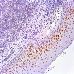
آنتی بادی ISLET 1 (EP283)
Name: Islet 1 Monoclonal Antibody clone EP283
Description and aplications: Islet-1, Insulin gene enhancer protein ISL-1, is a protein that in humans is encoded by the ISL1 gene. This gene encodes a transcription factor containing two N-terminal LIM domains and one C-terminal homeo domain. The encoded protein plays an important role in the embryogenesis of pancreatic islets of Langerhans. ISL1 has been shown to interact with Estrogen Receptor alpha. Islet-1 produces a strong nuclear staining in the islets of normal pancreas and tumor cells of the pancreatic neuroendocrine tumors. Islet-1 has been found to be a reliable marker of pancreatic endocrine tumors and metastasis. It shows a comparable sensitivity and specificity as CDX2 as a marker of ileal and appendiceal neuroendocrine tumors and their metastasis. A panel of Islet-1, CDX2 and TTF1 may be useful in differentiation well-differentiated endocrine carcinomas of unknown origin.
Composition: Anti-human Islet 1 rabbit monoclonal antibody purified from culture supernatant, filtered, sterilized and prepared in 10mM PBS, pH 7.4, with 0.2% BSA and 0.09% sodium azide
-
 آنتی بادیهای ایمونوهیستوشیمی
آنتی بادی Mucin 1 (ZM35 also known as MRQ-17)
Name: Mouse anti-human Mucin 1 Monoclonal Antibody clone ZM35
Description and aplications: Mucins are high molecular weight glycoproteins which constitute the major component of the mucus layer that protects the gastric epithelium from chemical and mechanical aggressions. In humans, at least 14 mucin genes have been identified that code for the mucin proteins. They are designated as MUC1, MUC2, MUC3, MUC4, MUC5AC, MUC5B, MUC6, MUC7, MUC8, MUC9, MUC11, MUC12, MUC13, and MUC16. The heterogeneous pattern of mucin expression, including the expression of the intestinal mucin MUC2, may provide new insights into the differentiation pathways of gastric carcinoma. Comparative studies showed that: (1) mucin expression is associated with tumor type (MUC5AC with dffuse and infiltrative carcinomas and MUC2 with mucinous carcinomas) but not with the clinicobiological behavior of the tumors; and (2) mucin expression is associated with tumor location (MUC5AC with antrum carcinomas and MUC2 with cardia carcinomas).
Composition: Anti-human Mucin 1 mouse monoclonal antibody purified from serum and prepared in 10mM PBS, pH 7.4, with 0.2% BSA and 0.09% sodium azide
Intended use: Immunohistochemistry (IHC) on paraffin embedded tissues. Not tested on frozen tissues or Western-Blotting
-
 آنتی بادیهای ایمونوهیستوشیمی
آنتی بادی PCNA (PC10)
Name: PCNA (Proliferating Cell Nuclear Antigen) Monoclonal Antibody clone PC10
Description and aplications: Expression of proliferating cell nuclear antigen (PCNA) or cyclin or polymerase delta auxiliary protein is elevated in the nucleus during late G1 phase immediately before the onset of DNA synthesis, becoming maximal during S-phase and declining during G2 and M phases. Its level correlates directly with rates of cellular proliferation and DNA synthesis. PCNA/cyclin may act as an auxiliary protein of DNA polymerase-delta to play a fundamental role in the initiation of cell proliferation The level of PCNA expression is directly related to the rate of cell proliferation and DNA synthesis. PCNA / cyclin could act as an auxiliary protein of DNA polymerase delta, which plays an essential role in the initiation of cell proliferation.This antibody is useful tool for studying the proliferating cells in normal tissues and its potential aberrant expression in neoplasms
Composition: anti-human PCNA mouse monoclonal antibody purified from ascites fluid by Protein A chromatography. Prepared in 10mM PBS, pH 7.4, with 0.2% BSA and 0.09% sodium azide
Immunogen: Recombinant rat PCNA protein
-
 آنتی بادیهای ایمونوهیستوشیمی
آنتی بادی PD-1 (NAT105)
Name:PD-1 Antibody clone NAT105
Description and aplications: Programmed death-1 (PD-1) is a member of the CD28 family of receptors that includes CD28, cytotoxic T-lymphocyteassociated antigen 4 (CTLA-4), inducible costimulator (ICOS), and B- and T-lymphocyte attenuator. These receptors play a role in the cellular immune response. For example, CD28 serves as a costimulatory receptor that enhances T-cell activation, whereas CTLA-4 serves as an inhibitor of T-cell activation. PD-1 also has an inhibitory function on T cells and B cells, and is important in peripheral tolerance. There are at least 2 ligands for PD-1, PD-L1, and PD-L2, which are expressed on a range of cells.
CD28 is constitutively expressed on most or all CD4+ T cells and approximately 50% of CD8+ T cells, whereas CTLA-4 is not expressed on resting T cells. PD-1 is also expressed on activated T cells, B cells, and myeloid cells. Iwai and coworkers studied the microanatomic distribution of PD-1 in human tonsil and found that PD-1 is expressed on most T cells and a small subset of B cells in the light zone of germinal centers, but not elsewhere in the tonsil. On that basis, it was postulated that PD-1 may play a role in the process of clonal selection of centrocytes, which occurs in this subanatomic site in germinal centers.Composition: anti-human PD-1 mouse monoclonal antibody purified from ascites. Prepared in 10mM PBS, pH 7.4, with 0.2% BSA and 0.09% sodium azide
-
 آنتی بادیهای ایمونوهیستوشیمی
آنتی بادی PD-L1 (CAL10)
Name: PD-L1 Monoclonal Antibody clone CAL10
Description and applications:Programmed death ligand 1 (PD-L1, also known as CD274) inhibits tumor-reactive T cells via binding to the programmed death-1 (PD-1) receptor, rendering tumor cells resistant to CD8+ T cell-mediated lysis. Studies have shown that the inhibitory receptor PD-1 is expressed on tumor-infiltrating lymphocytes (TIL) while PD-L1 is expressed on tumor cells. Assessment of PD-L1 expression in combination with CD8+ TIL density may be a useful predictive metric in multiple cancers, including stage III NSCLC, hormone receptor negative breast cancer and sentinel lymph node melanoma. Clinical trials utilizing humanized chimeric antibodies that block inhibitory checkpoints, such as anti-PD-1 and anti-PD-L1, have demonstrated delayed tumor growth and increased survival. While identification of PD-L1 overexpression by IHC is not yet standardized, it has become increasingly important to identify these tumors, as a directed therapy may improve clinical outcomes in these patients. In cutaneous melanoma, the targeting of PD1/PD-L1 has provided meaningful clinical benefit for patients in just the past 5-10 years. The use of IHC for protein identification, along with novel therapies, such as checkpoint inhibitors and vaccines, are generating new options for the treatment of cancer patients. The PD-L1 [CAL10] clone does not cross react with PDL2.
Composition: Anti-human PD-L1 rabbit monoclonal antibody purified from serum and prepared in 10mM PBS, pH 7.4, with 0.2% BSA and 0.09% sodium azide
Intended use: Immunohistochemistry (IHC) on paraffin embedded tissues. Not tested on frozen tissues or Western-Blotting
Immunogen: Peptide corresponding to the region within human PD-L1.
-
 آنتی بادیهای ایمونوهیستوشیمی
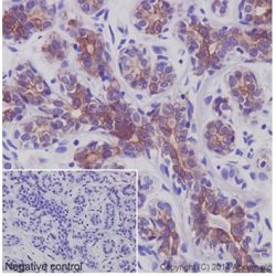
آنتی بادی PDX1 (EP139)
Name: PDX1 Monoclonal Antibody clone EP139
Description and applications: The homeobox 1 duodenal protein (PDX1) is a transcription factor that is required for the embryonic development of the pancreas and the maturation of the β-cells of the islets of Langerhans. Moreover, PDX1 activates somatostatin, glucokinase, islet amyloid polypeptide, and glucose transporter type 2 gene transcription. The PDX1 protein is encoded by the PDX1 gene, located in the chromosomal region 13q12.2. Clinically, mutations in the PDX1 gene have been shown to cause pancreatic agenesis and hereditary maturity onset diabetes mellitus of the young (MODY); likewise, the PDX-1 factor plays a major role in the pathogenesis of non-insulindependent (type II) diabetes mellitus. The PDX1 protein is initially expressed in the intestinal region of the embryo and then in the primitive pancreatic epithelium, where it allows its proliferation, branching, and differentiation. In the adult, this transcription factor is selectively expressed in endocrine cells, such as pancreatic beta cells, duodenum’s Brunner’s gland cells, and endocrine cells of the stomach’s pyloric region. Within the pancreas, PDX1 can also be observed in a subset of centroacinar and pancreatic duct exocrine cells, as well as in the beta cells of the islets of Langerhans. Increased expression of PDX1 has been reported in pancreatic (both exocrine and neuroendocrine), colon, and prostate tumours, suggesting that PDX1 may act as a biomarker in patients with these malignant tumours. However, further studies are needed to prove their specificity.
Composition: Anti-human PDX1 rabbit monoclonal antibody purified from serum and prepared in 10mM PBS, pH 7.4, with 0.2% BSA and 0.09% sodium azide
Intended use: Immunohistochemistry (IHC) on paraffin embedded tissues. Not tested on frozen tissues or Western-Blotting
-
 آنتی بادیهای ایمونوهیستوشیمی
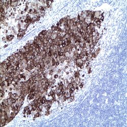
آنتی بادی Perforin 1 (5B10)
Name: Perforin Monoclonal Antibody clone 5B10
Composition: anti-Perforin mouse monoclonal antibody obtained from supernatant culture and prediluted in a tris buffered solution pH 7.4 containing 0.375mM sodium azide solution as bacteriostatic and bactericidal.
Immunogen: Recombinant protein encoding C-terminal region of human perforin
-
 آنتی بادیهای ایمونوهیستوشیمی
آنتی بادی PIN Cocktail (AMACR/p504s+p63) (13H4+4A4)
Name: Anti-AMACR/p504s+Anti-p63 human cocktail (PIN cocktail) clone 13H4+4A4
Description and aplications: The main use of the combination of both antibodies in a single reagent is simultaneously immunostain prostate glands in order to detect whether or not neoplastic. In the case of prostate adenocarcinoma, neoplastic glands must present cytoplasmic staining produced by expression of p504s and absence of nuclear staining as these glands lack basal cells and thus are negative for p63. Normal or hyperplastic glands must present nuclear staining in the basal cell layer. The Due to the different pattern of staining of the antibodies included in the cocktail, the detection could be realised with a polyvalent HRP detection systems. Nevertheless, as the antibodies are developed in two different host, a dual detection system can be used.
Composition: anti-human AMACR/p50s rabbit monoclonal antibody + anti-human p63 mouse monoclonal antibody prepared in 10mM PBS, pH 7.4, with 0.2% BSA and 0.09% sodium azide
Immunogen: Human AMACR polypeptide + aminoacids 1-250 of the N-terminal portion of the human p63 protein
-
 آنتی بادیهای ایمونوهیستوشیمی
آنتی بادی PMS2 (EP51)
Name: PMS2 Monoclonal Antibody clone EP51
Description and aplications: PMS2, a mismatch repair endonuclease, is a member of a family of genes involved in DNA mismatch repair. Carriers of the mismatch repair gene mutations have a high lifetime risk of developing Hereditary Non-Polyposis Colon Cancer (HNPCC) and several other cancers including endometrial cancer due to microsatellite instability (MSI) caused by accumulation of DNA replication errors in proliferating cells. Along with MLH1, MSH2 and MSH6, PMS2 antibody is helpful in diagnosis of MSI. An IHC study conducted by Mayo clinic on 535 cases with MSI-high, 90% of the tumors showed loss of MLH1, MSH2 and/or MSH6 expression, while 70% of the remaining cases showed isolated loss of PMS2 expression. The loss of PMS2 was associated with young age of diagnosis and right-sided location but not with a striking family history of cancer. Endometrial carcinomas are the most common noncolorectal cancers occur in HNPCC. The most common IHC abnormality in endometrial carcinomas with MSI was concurrent loss of MLH1/PMS2. Adding of PMS2 and MSH6 to MLH1 and MSH2 antibodies increased sensitivity for diagnosis of MSI. Tumors with low-level MSI show unfavourable pathological characteristics compared to tumours with no and tumours with highlevel MSI.
Composition: anti-human PMS2 rabbit monoclonal antibody purified from ascites. Prepared in 10mM PBS, pH 7.4, with 0.2% BSA and 0.09% sodium azide
Intended use : Immunohistochemistry (IHC) on paraffin embedded tissues. Not tested on frozen tissues or Western-Blotting
Immunogen: A synthetic peptide corresponding to residues in human PMS2 protein
-
 آنتی بادیهای ایمونوهیستوشیمی
آنتی بادی PNL2 (PNL2)
Name: PNL2 Monoclonal Antibody clone PNL2
Description and applications: PNL2, initially developed as a human somatostatin receptor marker, is a novel monoclonal antibody directed against an antigen (not well affiliated with melanocytes) that is chemically resistant to fixation and allows immunostaining after melanin bleaching and treatment with decalcifying agents. On a wide range of normal and neoplastic human tissues and compared to other similar markers such as HMB45, Melan A, MiTF, CD63, MAGE1, MAGE3, or tyrosinase, this antibody has revealed high specificity for melanocytes and for various benign and malignant melanocytic lesions derived from them. When the anti-PNL2 antibody is used on normal tissues, it gives a strong cytoplasmic staining of skin and oral mucosae melanocytes, as well as of granulocytes and osteoclast-like multinucleated giant cells when used at high concentration. In benign melanocytic lesions, PNL2 labels intraepidermal nevi, whereas intradermal nevi and the dermal component of compound nevi are basically negative although scattered nevus cells in the papillary dermis are usually positive. In primary melanomas, irrespective of their histologic type, PNL2 usually labels more than 70% of the neoplastic cells. In this context and within a broader panel of antibodies, PNL2 may contribute to the differential diagnosis between atypical nevus lesions and melanomas, although this antibody does not label desmoplastic melanomas. Moreover, the vast majority of metastatic melanomas are positive against PNL2 and, although the proportion of labelled cells is occasionally lower than in the primary tumour, it is always greater than that obtained with the commonly used markers for these tumours (HMB45 and Melan A). Like Melan A and HMB-45, PNL2 also stains most of the clear cell sarcoma cells, and a few cells in angiomyolipomas and lymphangioleiomyomatosis. Other non-melanocytic lesions positive for this antibody include PEComas and melanotic schwannoma. In isolated cases of chronic myeloid leukaemia, the PNL2 antibody stains neutrophils and myelocites, while blast cells are negative.
Composition: Anti-human PNL2 mouse monoclonal antibody purified from serum and prepared in 10mM PBS, pH 7.4, with 0.2% BSA and 0.09% sodium azide
-
 آنتی بادیهای ایمونوهیستوشیمی
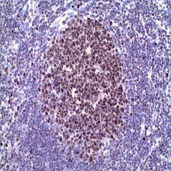
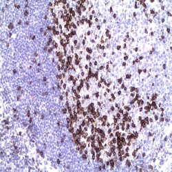
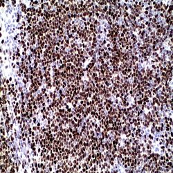
آنتی بادی SOX-11 (MD-58 also known as MRQ-58)
Name: SOX-11 Antibody clone MD-58 also known as MRQ-58
Description and applications:Mantle cell lymphoma (MCL) accounts for 5% to 10% of mature Bcell neoplasms and is an aggressive disease genetically characterized by overexpression of cyclin D1 (CCND1) due to the specific translocation t(11;14)(q13;q32). It is necessary that MCL be distinguished from potential morphologic mimics, including chronic lymphocytic leukemia/ small lymphocytic lymphoma (CLL/SLL), follicular lymphoma (FL), and marginal zone lymphoma (MZL) based on immunohistochemical (IHC) staining for CD5, CD23, and CD10. MCL and CLL both express CD5 but MCL, in contrast to CLL, generally lacks CD23 expression by IHC. FL lacks expression of both CD5 and CD23 but most often expresses CD10, whereas MZL is typically negative for all 3 antigens. Cyclin D1 overexpression is thus the hallmark of MCL even though approximately 5%-10% of MCLs lack Cyclin D1 expression and may be misdiagnosed by overreliance on CyclinD1 IHC. The recognition of cyclin D1-negative MCL is difficult because it may resemble other small B-cell lymphomas morphologically and phenotypically. Although the clinical information on cyclin D1negative MCL is limited, published data indicate that the behaviour of the variant is as aggressive as that of conventional MCL. On the other hand, patients with small B-cell lymphomas mimicking MCL have a significantly better outcome than those with true MCL. It is, therefore, important to find reliable biomarkers that may allow the identification of cyclin D1-negative MCL in clinical practice. SOX-11, the SRY (sex-determining region Y)-box11 gene, a transcription factor, normally is expressed in the developing human central nervous system, medulloblastoma, and glioma. In a series study, SOX- 11 expression was investigated in 54 cyclin D1positive MCL and 209 other lymphoid neoplasms. Interestingly, nearly all MCL were strongly positive for anti-SOX-11 (50/54, 93%), with a nuclear pattern. The staining was intense and relatively homogeneous in most of the cells. Compared to anti-cyclin D1 staining, anti-SOX-11 reactivity was stronger and more homogeneous. Five T-cell and B-cell lymphoblastic leukemia/lymphomas showed strong SOX-11 nuclear expression. One case of classic Hodgkin lymphoma, two of eight BL and two of three T-cell prolymphocytic leukemias were also positive. SOX-11 protein expression was examined by immunohistochemistry in the 12 cyclin D1-negative MCL, and all of them showed strong nuclear positive staining similar to that occurring in conventional cyclin D1-positive MCL. The expression of SOX-11 in reactive tonsil, lymph node and spleen specimens was studied. No nuclear expression was observed in any lymphocyte compartment. Only cytoplasmic staining was seen in cells from reactive germinal centers. Therefore, the authors confirmed prior reports that SOX-11 nuclear expression was a specific marker for MCL, including cyclin D1-negative MCL with typical morphology. Their study indicates that SOX-11 IHC is of value in further defining pathologic features of CD5+ DLBCL. Routine use of anti-SOX-11 in cases of suspected CD5+ DLBCL might help identify additional cases of cyclin D1-negative blastoid MCL. In summary, nuclear protein expression of SOX-11 is highly associated with both cyclin D1-positve and negative MCL. The detection of this transcription factor is a useful biomarker for identifying true cyclin D1- negative MCL. SOX-11 IHC is of value in further defining pathologic features of CD5+ DLBCL. Routine use of anti-SOX-11 in cases of suspected CD5+ DLBCL might help identify additional cases of cyclin D1- negative blastoid MCL. SOX-11 can also be detected in some BL, LBL and T-PLL, although the different morphological and phenotypic features of these malignancies allow easy recognition of the cases of cyclin D1-negative MCL
Composition:Anti-human SOX-11 mouse monoclonal antibody purified from serum and prepared in 10mM PBS, pH 7.4, with 0.2% BSA and 0.09% sodium azide
Intended use: Immunohistochemistry (IHC) on paraffin embedded tissues. Not tested on frozen tissues or Western-Blotting
-
 آنتی بادیهای ایمونوهیستوشیمی
آنتی بادی L1CAM (Neural Cell Adhesion Molecule L1) (BSR3)
Name: L1CAM Antibody clone BSR3
Description and applications:L1CAM (L1 cell adhesion molecule protein) is a cell adhesion molecule with an important role in the development of the nervous system. The L1, neural cell adhesion molecule (L1CAM) plays an important role in axon growth, fasciculation, neural migration and in mediating neuronal differentiation. L1 protein is expressed to tissues arising from neuroectoderm. L1CAM plays also an important role in the malignancy of human tumors and according to several studies, L1CAM positive carcinomas have a bad prognosis. L1CAM is overexpressed in many human carcinomas but it is useful especially in endometrium carcinoma diagnostic.
Composition : Anti-human L1CAM rabbit monoclonal antibody purified from culture supernatant, filtered, sterilized and prepared in 10mM PBS, pH 7.4, with 0.2% BSA and 0.09% sodium azide
Intended use : Immunohistochemistry (IHC) on paraffin embedded tissues. Not tested on frozen tissues or Western-Blotting
-
 آنتی بادیهای ایمونوهیستوشیمی
آنتی بادی p21/Waf-1 (DCS-60.2)
Name: p21/Waf-1 (DCS-60.2)
Description and aplications: p21WAF1/Cip1/Sdi1/Pic1 is a tumor suppressor protein. Expression of p21WAF1 is induced by wild type, but not mutant, p53 suppressor protein. The p21WAF1 protein binds to cyclin/CDK complexes and inhibits their kinase activity thereby stopping cell cycle progression. It also binds to PCNA (proliferating cell nuclear antigen) and blocks DNA replication but not the DNA repair process.
Composition: anti-human p21 mouse monoclonal antibody purified from ascites. Prepared in 10mM PBS, pH 7.4, with 0.2% BSA and 0.09% sodium azide
Intended use : Immunohistochemistry (IHC) on paraffin embedded tissues. Not tested on frozen tissues or Western-Blotting
Visualization: Nuclear
-
 آنتی بادیهای ایمونوهیستوشیمی
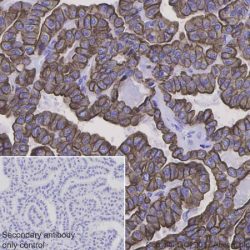
آنتی بادی Cytokeratin 7 (OVTL 12/30)
Name: Cytokeratin 7 Antibody clone OV-TL 12/30
Description and aplications: Keratins (also known as cytokeratins) are intermediate filament/forming proteins that represents the main cytoskeleton of epithelial cells. They classically are classified based on Moll cataloging which grouped the basic-to-neutral type II keratins as K1–K8 and the acidic type I keratins as K9– K19. An updated classification including 24 types of keratins was then proposed in order to enable other mammalian species keratins to be added. This monoclonal antibody reacts specifically with the cytokeratin 7, immunostaining of the producing a protein band of 54 kD in the cytoskeleton of cell lines and uni-dimensional immunoblots. This antibody is useful to distinguish between different types of normal glandular epithelia as it marks the epithelia of lung and breast, being negative for colon and prostate. We have not observed any cross reaction with other cytokeratins and overall this antibody does not react with stratified squamous epithelium. In liver, hepatocytes are negative and the epithelial cells of the bile ducts are positive. This antibody reacts with numerous both benign and malignant epithelial lesions. Cytokeratin 7 is expressed in specific subtypes of ovarian adenocarcinomas, breast and lung carcinomas while the gastrointestinal tract, except stomach, are negative. Carcinomas arising from transitional epithelium also express cytokeratin, whereas prostate cancer is generally negative.
Composition: anti human keratin 7 mouse monoclonal antibody obtained from supernatant culture and prediluted in a Tris buffered solution pH 7.4 containing 0.375mM sodium azide solution as bacteriostatic and bactericidal.
Intended use : Immunohistochemistry (IHC) on paraffin embedded tissues. Not tested on frozen tissues or Western-Blotting
Immunogen: OTN II ovarian carcinoma cell line
-
 آنتی بادیهای ایمونوهیستوشیمی
آنتی بادی Cytokeratin 8 (EP17)
Name: Cytokeratin 8 antibody clone EP17
Description and aplications: Cytokeratin 8 (CK8) is an intermediate filament protein produced early in embryogenesis. It is the only type-II CK occurring in many simple epithelial in respiratory, gastrointestinal, male and female reproductive tract and thyroid. CK8 is often co-expressed with Cytokeratin 18. CK8/18 is the major keratin pair in simple-type epithelia, as found in the liver, pancreas, and intestine. CK8 antibody is used to detect adenocarcinomas with simple epithelium origin. The difference in staining pattern is useful to distinguish duct (peripheral staining) from lobular (perinuclear staining) breast carcinoma
Composition: anti-human cytokeratin 8 rabbit monoclonal antibody purified from ascites. Prepared in 10mM PBS, pH 7.4, with 0.2% BSA and 0.09% sodium azide
Immunogen: A synthetic peptide corresponding on the C-terminus
-
 آنتی بادیهای ایمونوهیستوشیمی
آنتی بادی Cytokeratin 8/18 (B22.1&B23.1)
Name : Cytokeratin 8/18 antibody clone B22.1/B23.1
Description and applications:Cytokeratins 8&18 can be found in most simple epithelium,e.g. thyroid,female breast, gastrointestinal tract, and respiratory tract. Adenocarcinomas and most non-keratinizing squamous carcinomas will stain, but keratinizing squamous carcinomas will not. This antibody is used when attempting to demonstrate the presence of Paget cells; there is very little keratin 18 in the normal epidermis so this will only stain Paget cells. This approach facilitates the interpretation using immunostains and is more sensitive than mucin histochemistry.
Composition:Anti-human Cytokeratin 8/18 cocktail of two mouse monoclonal antibodies purified from serum and prepared in 10mM PBS, pH 7.4, with 0.2% BSA and 0.09% sodium azide
Intended use : Immunohistochemistry (IHC) on paraffin embedded tissues. Not tested on frozen tissues or Western-Blotting
برای تماس کلیک کنید
سما تشخیص آریا ، عرضه کننده متنوع ترین و کامل ترین محصولات کیت های آزمایشگاهی