برای تماس کلیک کنید

سما تشخیص آریا ، عرضه کننده متنوع ترین و کامل ترین محصولات کیت های آزمایشگاهی

آنتی‌ بادی PNL2 (PNL2)

Name: PNL2 Monoclonal Antibody clone PNL2

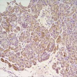
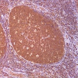
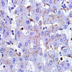
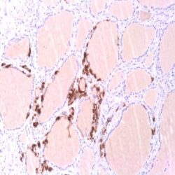
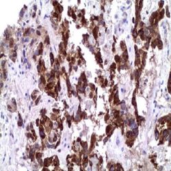
Description and applications: PNL2, initially developed as a human somatostatin receptor marker, is a novel monoclonal antibody directed against an antigen (not well affiliated with melanocytes) that is chemically resistant to fixation and allows immunostaining after melanin bleaching and treatment with decalcifying agents. On a wide range of normal and neoplastic human tissues and compared to other similar markers such as HMB45, Melan A, MiTF, CD63, MAGE1, MAGE3, or tyrosinase, this antibody has revealed high specificity for melanocytes and for various benign and malignant melanocytic lesions derived from them. When the anti-PNL2 antibody is used on normal tissues, it gives a strong cytoplasmic staining of skin and oral mucosae melanocytes, as well as of granulocytes and osteoclast-like multinucleated giant cells when used at high concentration. In benign melanocytic lesions, PNL2 labels intraepidermal nevi, whereas intradermal nevi and the dermal component of compound nevi are basically negative although scattered nevus cells in the papillary dermis are usually positive. In primary melanomas, irrespective of their histologic type, PNL2 usually labels more than 70% of the neoplastic cells. In this context and within a broader panel of antibodies, PNL2 may contribute to the differential diagnosis between atypical nevus lesions and melanomas, although this antibody does not label desmoplastic melanomas. Moreover, the vast majority of metastatic melanomas are positive against PNL2 and, although the proportion of labelled cells is occasionally lower than in the primary tumour, it is always greater than that obtained with the commonly used markers for these tumours (HMB45 and Melan A). Like Melan A and HMB-45, PNL2 also stains most of the clear cell sarcoma cells, and a few cells in angiomyolipomas and lymphangioleiomyomatosis. Other non-melanocytic lesions positive for this antibody include PEComas and melanotic schwannoma. In isolated cases of chronic myeloid leukaemia, the PNL2 antibody stains neutrophils and myelocites, while blast cells are negative.

Composition: Anti-human PNL2 mouse monoclonal antibody purified from serum and prepared in 10mM PBS, pH 7.4, with 0.2% BSA and 0.09% sodium azide

حجم موجود: 3 / 7 / 12 میلی‌لیتر

مورد استفاده: ایمونوهیستوشیمی روی بافت پارافین. بر روی بافتهای فروزن یا وسترن بلات تست نشده است.

کلون: PNL2

ایزوتایپ: IgG1

کنترل مثبت IHC: پوست نرمال یا بخشی از بافت ملانوما

قابلیت دیده شدن: سیتوپلاسمی

روش پیشنهادی IHC:

  • برش تهیه شده با ضخامت μm4 باید روی لام شارژ شده در طول شب و در دمای 60 درجه سانتی گراد خشک شود.
  • دپارافینه کردن، رهیدراته کردن و استفاده از حرارت جهت بازیابی اپی توپ : بافت را با استفاده از بافر EDTA pH 8 برای بیست دقیقه در دمای 95 درجه سانتی گراد بجوشانید. سپس 5-3 بار با استفاده از آب مقطر یا دیونیزه شده شسته و سپس بیست دقیقه در دمای اتاق خنک کنید.
  • بلاک پروکسیدازهای درونی : بلاک با استفاده از محلول پروکسیداز به مدت ده دقیقه
  • آنتی بادی اولیه: برای 10 دقیقه در انکوبه کنید پروتکل ممکن است بسته به تهیه نمونه و کاربرد خاص متفاوت باشد. شرایط استاندارد باتوجه به آزمایشگاه و کاربر متفاوت می باشد.
  • برای رنگ آمیزی ، می توانید از کیت Master Polymer Plus Detection یا کیتهای دیتکشن سایر کمپانی ها استفاده کنید.
  • رنگ میزی نهایی با هماتوکسیلین و فیکس نهایی اسلاید.

شرایط نگهداری و پایداری : حداکثر 18 ماه؛ در دمای 2-8 درجه سانتیگراد نگهداری شود. فریز نشود.


این آنتی‌بادی متعلق به پنل‌‌‌‌‌‌‌‌ تشخیصی DERMATOPATHOLOGY است.


Download Cataloge

Icon

PNL2 (PNL2) 95.45 KB 7 downloads

...
.well.c2a3 .btn { padding: 11px 30px; font-size: 11pt; } .well.c2a3 .media-body { font-size: 11pt; } .well.c2a3 .wpdm_icon { height: 42px; width: auto; }

Reviews

There are no reviews yet.

Be the first to review “آنتی‌ بادی PNL2 (PNL2)”

Your email address will not be published. Required fields are marked *

Scroll to Top