برای تماس کلیک کنید

Welcome to Sama Tashkhis Aria Official Website

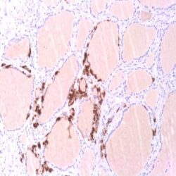
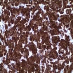
آنتی‌ بادی PD-L1 (CAL10)

Name: PD-L1 Monoclonal Antibody clone CAL10

Description and applications:Programmed death ligand 1 (PD-L1, also known as CD274) inhibits tumor-reactive T cells via binding to the programmed death-1 (PD-1) receptor, rendering tumor cells resistant to CD8+ T cell-mediated lysis. Studies have shown that the inhibitory receptor PD-1 is expressed on tumor-infiltrating lymphocytes (TIL) while PD-L1 is expressed on tumor cells. Assessment of PD-L1 expression in combination with CD8+ TIL density may be a useful predictive metric in multiple cancers, including stage III NSCLC, hormone receptor negative breast cancer and sentinel lymph node melanoma. Clinical trials utilizing humanized chimeric antibodies that block inhibitory checkpoints, such as anti-PD-1 and anti-PD-L1, have demonstrated delayed tumor growth and increased survival. While identification of PD-L1 overexpression by IHC is not yet standardized, it has become increasingly  important to identify these tumors, as a directed therapy may improve clinical outcomes in these patients. In cutaneous melanoma, the targeting of PD1/PD-L1 has provided meaningful clinical benefit for patients in just the past 5-10 years. The use of IHC for protein identification, along with novel therapies, such as checkpoint inhibitors and vaccines, are generating new options for the treatment of cancer patients. The PD-L1 [CAL10] clone does not cross react with PDL2.

Composition: Anti-human PD-L1 rabbit monoclonal antibody purified from serum and prepared in 10mM PBS, pH 7.4, with 0.2% BSA and 0.09% sodium azide

Intended use: Immunohistochemistry (IHC) on paraffin embedded tissues. Not tested on frozen tissues or Western-Blotting

Immunogen: Peptide corresponding to the region within human PD-L1.

حجم موجود: 3 / 7 / 12 میلی‌لیتر

مورد استفاده: ایمونوهیستوشیمی روی بافت پارافین. بر روی بافتهای فروزن یا وسترن بلات تست نشده است.

کلون: CAL10

ایزوتایپ: IgG

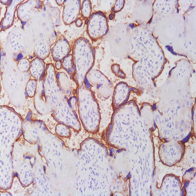
کنترل مثبت IHC: آدنوکارسینومای ریه، جفت یا لوزه

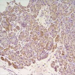
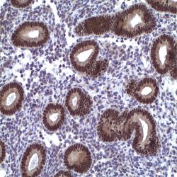
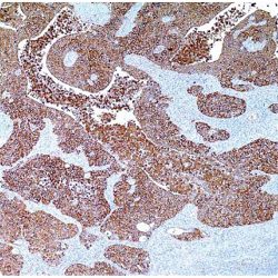
قابلیت دیده شدن: غشایی و سیتوپلاسمی

روش پیشنهادی IHC:

  • برش تهیه شده با ضخامت μm4 باید روی لام شارژ شده در طول شب و در دمای 60 درجه سانتی گراد خشک شود.
  • دپارافینه کردن، رهیدراته کردن و استفاده از حرارت جهت بازیابی اپی توپ : بافت را با استفاده از بافر EDTA pH 8 برای بیست دقیقه در دمای 95 درجه سانتی گراد بجوشانید. سپس 5-3 بار با استفاده از آب مقطر یا دیونیزه شده شسته و سپس بیست دقیقه در دمای اتاق خنک کنید.
  • بلاک پروکسیدازهای درونی : بلاک با استفاده از محلول پروکسیداز به مدت ده دقیقه
  • آنتی بادی اولیه: برای 10 دقیقه در انکوبه کنید پروتکل ممکن است بسته به تهیه نمونه و کاربرد خاص متفاوت باشد. شرایط استاندارد باتوجه به آزمایشگاه و کاربر متفاوت می باشد.
  • برای رنگ آمیزی ، می توانید از کیت Master Polymer Plus Detection یا کیتهای دیتکشن سایر کمپانی ها استفاده کنید.
  • رنگ میزی نهایی با هماتوکسیلین و فیکس نهایی اسلاید.

شرایط نگهداری و پایداری : حداکثر 18 ماه؛ در دمای 2-8 درجه سانتیگراد نگهداری شود. فریز نشود.


این آنتی‌بادی متعلق به پنل‌‌‌‌‌‌های تشخیصی DERMATOPATHOLOGY ،PLEUROPULMONARY و UROGENITAL است.


Download Cataloge

Icon

PD-L1 (CAL10) 95.56 KB 55 downloads

...
.well.c2a3 .btn { padding: 11px 30px; font-size: 11pt; } .well.c2a3 .media-body { font-size: 11pt; } .well.c2a3 .wpdm_icon { height: 42px; width: auto; }

Reviews

There are no reviews yet.

Be the first to review “آنتی‌ بادی PD-L1 (CAL10)”

Your email address will not be published. Required fields are marked *

Scroll to Top