برای تماس کلیک کنید

سما تشخیص آریا ، عرضه کننده متنوع ترین و کامل ترین محصولات کیت های آزمایشگاهی

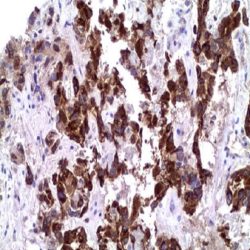
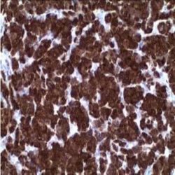
Name: Arginase-1 Antibody (Clone EP261)

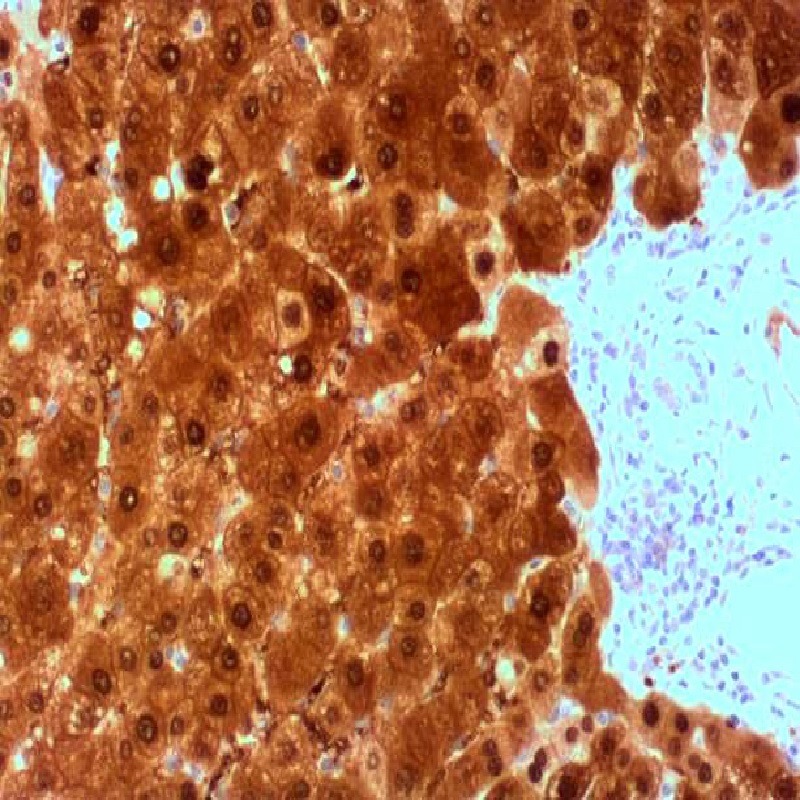
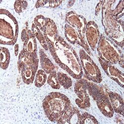
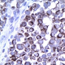
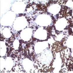
Description and applications: Arginase is a manganese metalloenzyme that catalyzes the hydrolysis of arginine to generate ornithine and urea. Arginiase I and II are isoenzymes which differ in subcellular localization, regulation, and possibly function. Arginase I is a cytosolic enzyme, which is expressed mainly in the liver as part of the urea cycle, whereas arginase II is a mitochondrial protein found in a variety of tissues. Antibody to ARG-1 labels hepatocytes in normal tissues and granulocytes in peripheral blood. ARG-1 is a sensitive and specific marker for identification of hepatocellular carcinoma. This antibody is very useful in distinguishing between: 1) liver metastases of various adenocarcinomas and hepatocellular carcinoma (HCC), which can be a realdiagnostic challenge, especially in small biopsies or material from fine needle aspiration ( FNA) and 2) the distinction between different histological variants of HCC and cholangiocarcinoma. Specifically, the ARG-1 antibody is key in the diagnosis of scirrhous hepatocellular carcinoma, where specific markers for adenocarcinomas are generally positive while HepPar-1 in some cases it may be negative. In addition, several studies have shown that ARG-1 antibody is more sensitive than HepPar-1 for immunohistochemical diagnosis of hepatocellular carcinoma while the second is usually negative in poorly differentiated hepatocellular carcinomas and can be positive in adenocarcinomas of pancreatic, gastric, colic origin or even cholangiocarcinoma. However, isolated cases of pancreatic adenocarcinomas and cholangiocarcinomas presented focal staining against ARG-1. In this line, it should be considered that the ARG-1 antibody does not allow differential diagnosis between benign, dysplastic and malignant hepatocyte lesions.

Composition: anti-human ARG-1 rabbit monoclonal antibody purified from serum and prepared in 10mM PBS, pH 7.4, with 0.2% BSA and 0.09% sodium azide.


حجم موجود3 / 7 / 12 میلی‌لیتر

مورد استفادهایمونوهیستوشیمی روی بافت پارافین. بر روی بافتهای فروزن یا وسترن بلات تست نشده است.

کلون: EP261

ایزوتایپ: Rabbit IgG

کنترل مثبت IHC: کبد

قابلیت دیده شدن: سیتوپلاسمی و هسته ای

روش پیشنهادی IHC:

  • برش تهیه شده با ضخامت μm4 باید روی لام شارژ شده در طول شب و در دمای 60 درجه سانتی گراد خشک شود.
  • دپارافینه کردن، رهیدراته کردن و استفاده از حرارت جهت بازیابی اپی توپ : بافت را با استفاده از بافر EDTA pH 8 برای بیست دقیقه در دمای 95 درجه سانتی گراد بجوشانید. سپس 5-3 بار با استفاده از آب مقطر یا دیونیزه شده شسته و سپس بیست دقیقه در دمای اتاق خنک کنید.
  • بلاک پروکسیدازهای درونی : بلاک با استفاده از محلول پروکسیداز به مدت ده دقیقه
  • آنتی بادی اولیه: برای 10 دقیقه در انکوبه کنید پروتکل ممکن است بسته به تهیه نمونه و کاربرد خاص متفاوت باشد. شرایط استاندارد باتوجه به آزمایشگاه و کاربر متفاوت می باشد.
  • برای رنگ آمیزی ، می توانید از کیت Master Polymer Plus Detection یا کیتهای دیتکشن سایر کمپانی ها استفاده کنید.
  • رنگ میزی نهایی با هماتوکسیلین و فیکس نهایی اسلاید.

شرایط نگهداری و پایداری : حداکثر 18 ماه؛ در دمای 2-8 درجه سانتیگراد نگهداری شود. فریز نشود.


این آنتی‌بادی متعلق به پنل تشخیصی DIGESTIVE است.


Download Cataloge

Icon

ARG-1 (EP261) 433.94 KB 14 downloads

...
.well.c2a3 .btn { padding: 11px 30px; font-size: 11pt; } .well.c2a3 .media-body { font-size: 11pt; } .well.c2a3 .wpdm_icon { height: 42px; width: auto; }

Reviews

There are no reviews yet.

Be the first to review “آنتی­ بادی ARG-1 (EP261)”

Your email address will not be published. Required fields are marked *

Scroll to Top