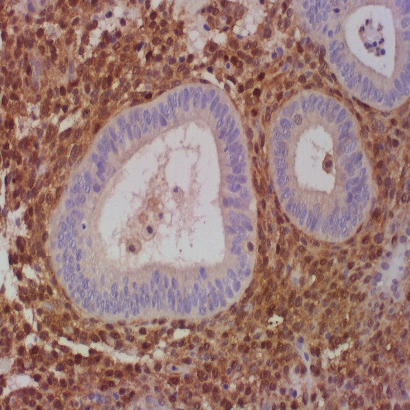
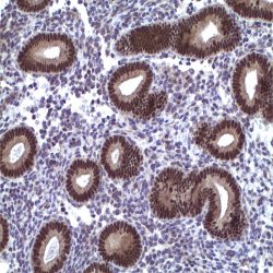
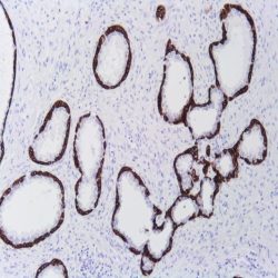
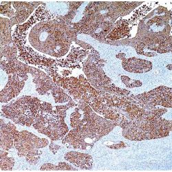
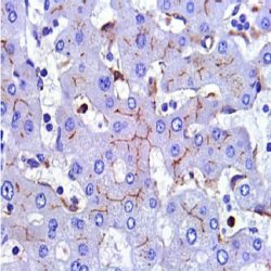
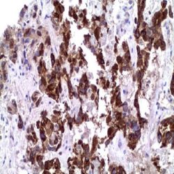
Name: PTEN Antibody (Clone 6H2.1)
Description and aplications: The antibody recognizes a protein fraction of 403 amino acids encoded by the PTEN gene located on chromosome 10q23.3 region. PTEN protein acts as a dual specificity phosphatase. On one hand, it dephosphorylates the phosphorylated proteins in tyrosine, serine, and threonine residues and on the other hand, it acts as a lipid phosphatase removing the phosphate group of the D3 position of the inositol ring in phosphatidylinositol-3,4,5-trisphosphate, phosphatidylinositol-3,4-diphosphate, phosphatidylinositol-3-phosphate, and inositol 1,3,4,5- tetrakisfosfato. This last feature is critical to act as a tumor suppressor through the negative regulation of the signaling pathway PI3K-AKT / PKB. This latter controls cell cycle progression, the tissue development, cell migration, and inhibition of apoptosis. In this signaling pathway, PTEN antagonistic effect on PI3K-AKT / PKB also maintains the stability of p53. PTEN also controls other cell cycle proteins such as p27, p130, myc, and cyclin D1. PTEN gene mutations induce alterations in the functioning of cells in a manner that the loss of PTEN protein expression is associated with tumor genesis, cancer progression, and cancer-resistance to different
treatments. Due to its role in cell growth, the vast majority of normal tissues express high amount of the PTEN protein. The loss of PTEN protein expression was observed in some malignancies associated with abnormalities in the PTEN gene. These abnormalities
can be monoallelic (gliomas and carcinomas of breast, prostate, lung, and colon) or biallelic (endometrial carcinomas, prostate and breast cancer, or glioblastoma multiforme). There are also some genetic diseases with alterations of germinal type in the PTEN gene like Cowden syndrome, Bannayan-Riley-Ruvalcaba syndrome, Lhermitte-Duclos disease and a subgroup of patients with Proteus syndrome. Somatic mutations of PTEN with loss of protein expression present variable incidences although they are often observed in sporadic endometrial carcinomas and in glioblastoma multiforme. These mutations are usually related to the histological grade of the tumor and are considered poor prognostic factors. Loss of PTEN expression is also a prognostic factor in gastrointestinal stromal tumors (GIST), tumors of the salivary glands, intrahepatic cholangiocarcinoma, gallbladder carcinomas, etc. In addition to being a prognostic factor, PTEN antibody immunostaining acts as a predictive indicator of the response to therapy using chemotherapeutic agents aimed at molecular targets. Controversial results obtained with the PTEN antibody are apparently related to the antibody clone used and/or tissue fixation issues. The only clone with reproducible results in the diagnosis of paraffin embedded samples, especially in endometrial adenocarcinomas and their precursor lesions, is the 6H2.1. However, superior results are always obtained in excisional biopsy or curettage samples in comparison with immunostaining of hysterectomies samples, where fixing is usually worse. In this sense and to mitigate immunostaining loss it is recommended to use freshly obtained serial sections of the sample.
Composition: anti-PTEN mouse monoclonal antibody obtained from supernatant culture and prediluted in a tris buffered solution pH 7.4 containing 0.375mM sodium azide solution as bacteriostatic and bactericidal.
Reviews
There are no reviews yet.