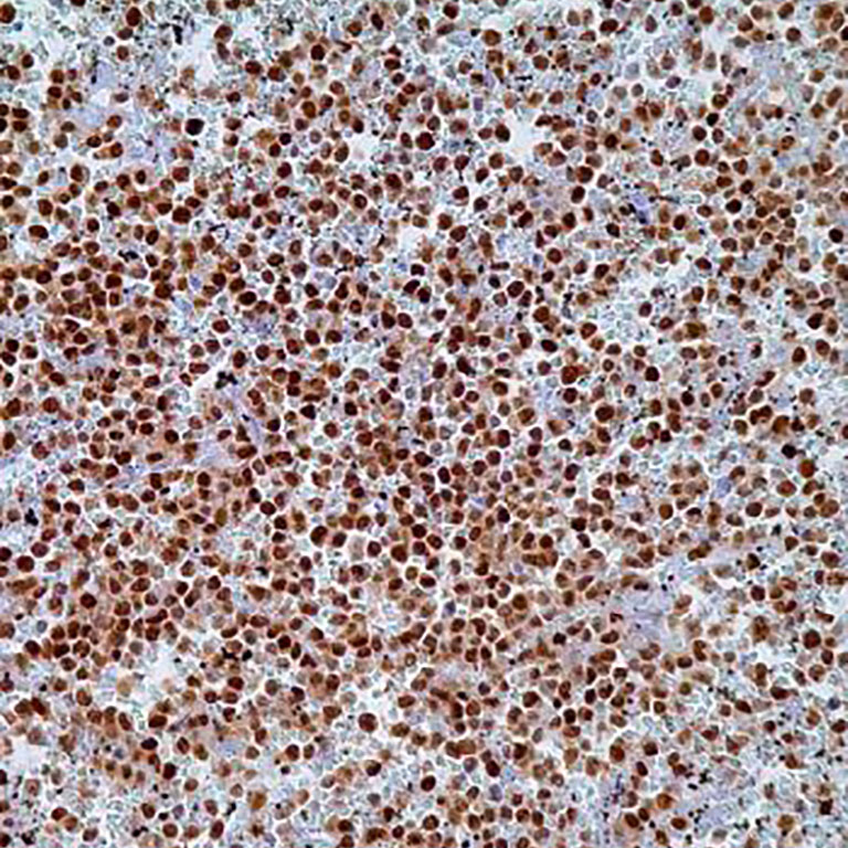
Name: T-Box/TBX21 Antibody (Clone 4B10)
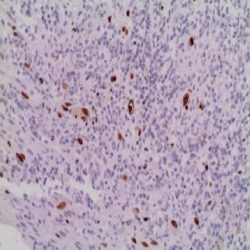
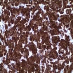
Description and applications: The T-bet transcription factor (more correctly called T-box TBX21 transcription factor), expressed in T cells, is one of the members of the T-box transcription factor family specific to Th1 cells. It is encoded by a gene located in the chromosomal region 17q21.32. The T-bet factor was originally isolated from nuclear extracts of resting and PMA/ionomycin-activated AE7 lymphoid cells; it is expressed in low levels in resting cells, and in increased levels in stimulated cells. T-bet regulates the secretion of IFN-γ by CD4 Th1 cells (although this mediator is also produced by NK cells and CD8+ cytotoxic T cells, in the latter case, independently of the presence of T-bet). Similarly, and under the right conditions, T-bet also converts effector Th2 cells into the opposing Th1 subset. In mice carrying T-bet gene deletions, lymphocytes differentiate into a predetermined stage of Th2 cells and are protected against lesions equivalent torheumatoid arthritis, which highlights the crucial role that this transcription factor plays in the induction of a Th1 response. For this reason, it is now known that an adequate level of T-bet activation contributes to a decrease of the excessive Th2 response found in bronchial hyperreactivity, oedema, and eosinophil granulocyte infiltration underlying bronchial asthma. In normal tissue, T-bet is expressed selectively and more intensely in Th1 cells, although at a lower level it is also found in NK cells and CD8+ cytotoxic T cells, and its expression level is increased in response to signals mediated by the T cell-specific receptor (TCR) and IL-12. In pathological situations, T-bet, in addition to being involved in the pathogenic mechanisms of some T cell lymphoproliferative processes, also regulates the response in some autoimmune diseases such as Crohn’s disease and type I diabetes mellitus. Finally, the presence of genetic variations in T-bet is associated with a greater susceptibility to bronchial asthma with sinonasal polyposis development and intolerance to aspirin. The implication of T-bet in the pathological development of B cells, as well as the development of some of the neoplastic lesions that are derived from them, has not yet been sufficiently analysed. In neoplasms, the T-bet transcription factor is expressed in all lymphomas originating from mature T cells, especially in peripheral T lymphomas, and is consistently negative in lymphomas derived from T cell precursors (lymphoblastic lymphomas). Likewise, most nasal NK/T lymphomas express nuclear T-bet regardless of the presence of Epstein-Barr virus or the presence of TCR receptor clonal rearrangement. Among B cell-derived neoplasms that most frequently express T-bet are marginal zone lymphomas, both extranodal and splenic, B lymphoblastic leukaemia, and hairy-cell leukaemia, in which T-bet positivity, along with TRAP, DBA.44, CD123, and Annexin-1 positivity, confirm diagnosis. Moreover, this antibody against T-bet has the advantage of being one of the few antibodies that work consistently in spine biopsies with hairy-cell leukaemia infiltration. Other lymphoproliferative lesions that may occasionally express T-bet are B cell chronic lymphocytic leukaemia, diffuse large B cell lymphoma, and classical Hodgkin lymphoma. Likewise, positivity has been described in some adenocarcinomas. In the differential diagnosis between lymphocytic colitis and coeliac disease, predominance of T-betpositive T cells, both in the epithelium and in the lamina propria, supports the latter diagnosis, while positive cells for both T-bet and GATA3 are more characteristic of lymphocytic colitis.
Composition: Anti-human T-Box TBX21 mouse monoclonal antibody purified from serum and prepared in 10mM PBS, pH 7.4, with 0.2% BSA and 0.09% sodium azide.
Immunogen:Recombinant mouse T-bet protein.
Reviews
There are no reviews yet.